吉林大学学报(医学版) ›› 2024, Vol. 50 ›› Issue (3): 728-738.doi: 10.13481/j.1671-587X.20240317

• 基础研究 • 上一篇    

miR-487a通过靶向调控TIA1对胃癌肿瘤相关巨噬细胞M2型极化的抑制作用

曲颜1,戴霖1,王彪1,阮笃激1,钟裕昌1,杨雪峰1,2()   

  1. 1.遵义医科大学第二附属医院胃肠外科,贵州 遵义 563006
    2.遵义医科大学附属医院胃肠外科,贵州 遵义 563000
  • 收稿日期:2023-07-11 出版日期:2024-05-28 发布日期:2024-07-01
  • 通讯作者: 杨雪峰 E-mail:yangxuefeng1978@126.com
  • 作者简介:曲 颜(1984-),男,山东省临沂市人,主治医师,医学硕士,主要从事消化道肿瘤发病机制方面的研究。
  • 基金资助:
    贵州省科技厅科技计划项目(黔科合基础〔2020〕1Y335)

Inhibitory effect of miR-487a on M2-type polarization of gastric cancer tumor-associated macrophages by targeting TIA1

Yan QU1,Lin DAI1,Biao WANG1,Duji RUAN1,Yuchang ZHONG1,Xuefeng YANG1,2()   

  1. 1.Department of Gastrointestinal Surgery,Second Affiliated Hospital,Zunyi Medical University,Zunyi 563006,China
    2.Department of Gastrointestinal Surgery,Affiliated Hospital,Zunyi Medical University,Zunyi 563000,China
  • Received:2023-07-11 Online:2024-05-28 Published:2024-07-01
  • Contact: Xuefeng YANG E-mail:yangxuefeng1978@126.com

摘要:

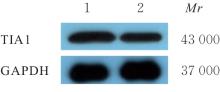
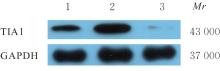
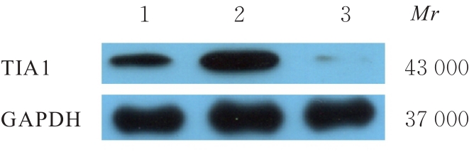
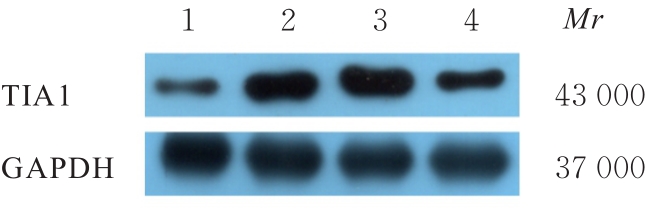
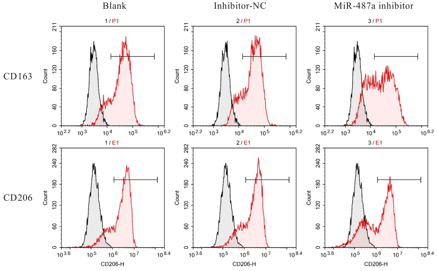
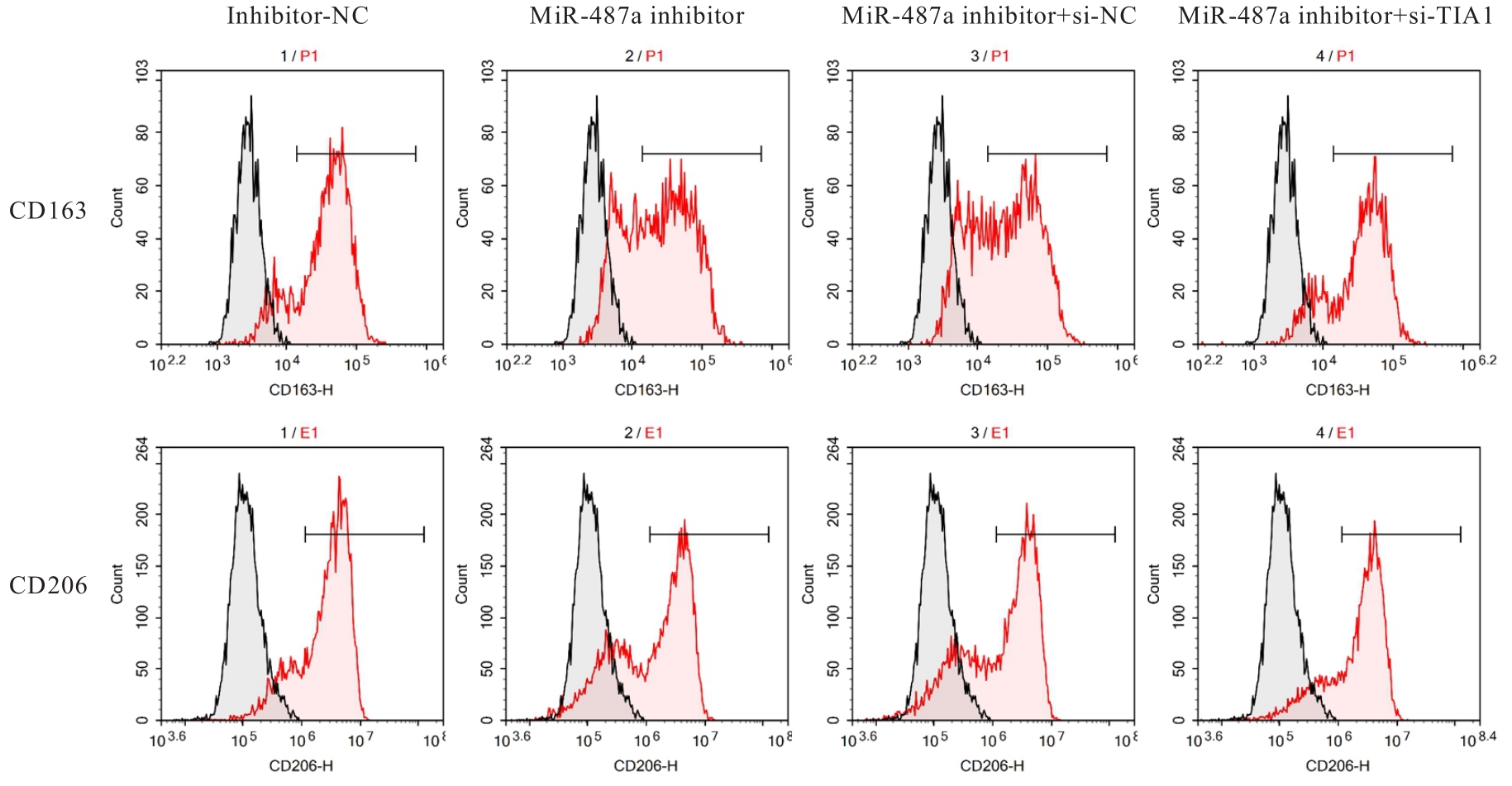
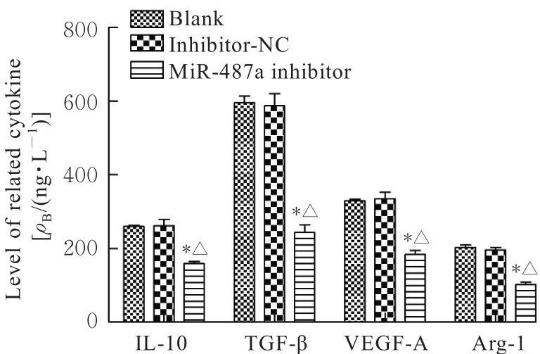
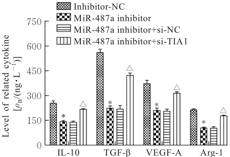
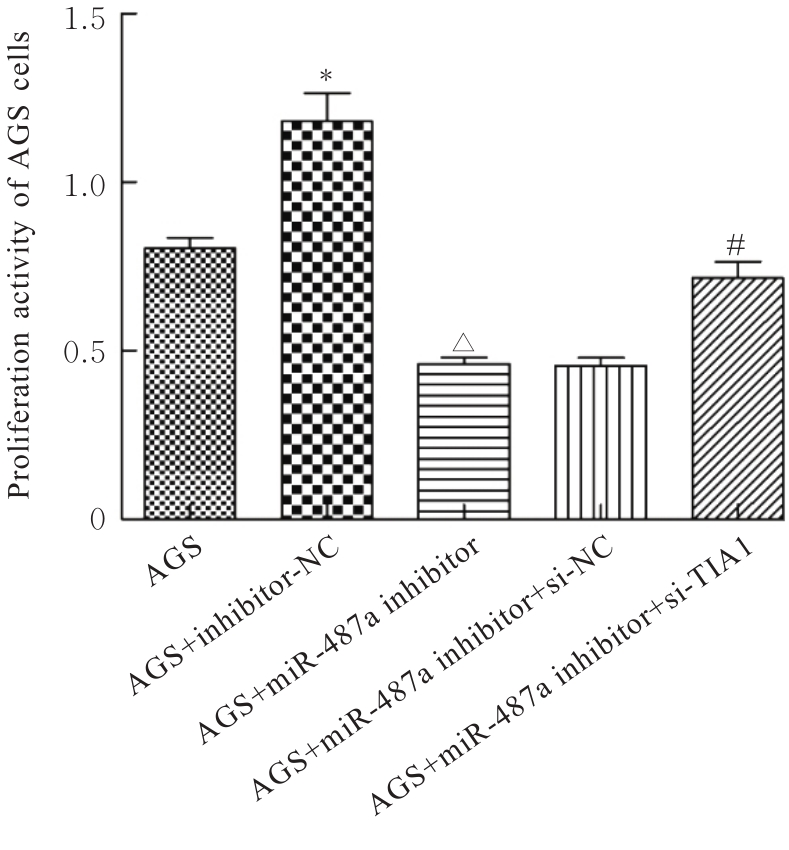
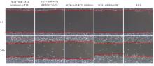
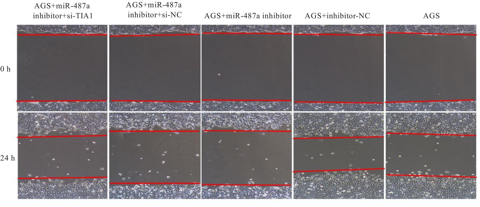
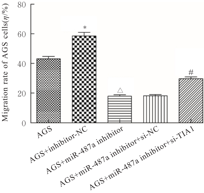
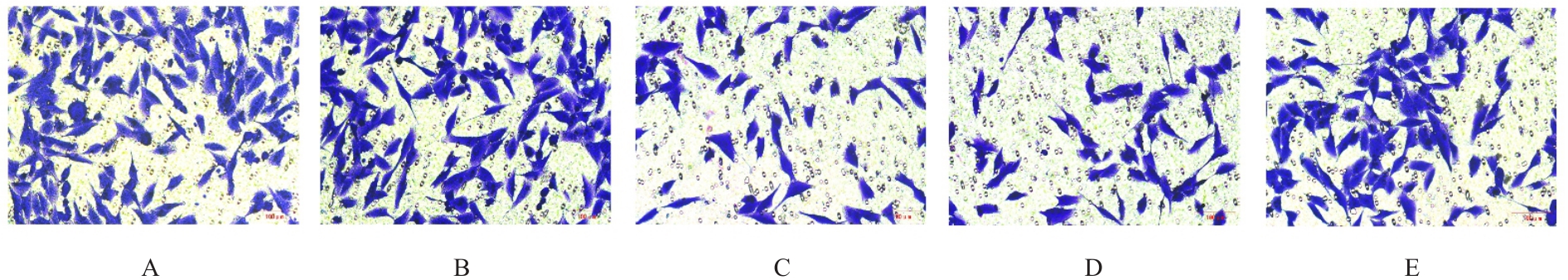
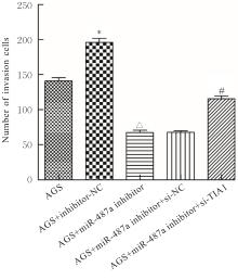
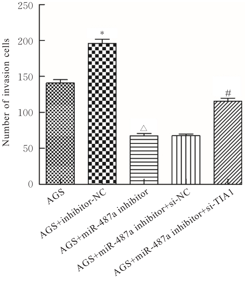
目的 探讨微小RNA(miR)-487a对胃癌肿瘤相关巨噬细胞(TAMs)M2型极化的抑制作用,并阐明其对胃癌AGS细胞增殖、侵袭和迁移的影响。 方法 分离和培养原发性胃癌患者胃癌组织TAMs及癌旁组织来源的正常巨噬细胞(NTMs),体外诱导人单核细胞THP-1分化为TAMs,将分化得到的M0、M1和M2型巨噬细胞经条件培养基(CM)刺激培养24 h,分别获取TAMs、M1-TAMs和M2-TAMs。转染TAMs,分为空白组、inhibitor-NC组、miR-487a inhibitor组、miR-487a inhibitor+si-NC组和miR-487a inhibitor+si-TIA1组,采用实时荧光定量PCR(RT-qPCR)法和Western blotting法验证转染效率。将M2-TAMs与AGS细胞共培养,分为AGS组、AGS+inhibitor-NC组、AGS+miR-487a inhibitor 组、 AGS+miR-487a inhibitor +si-NC 组和 AGS+miR-487a inhibitor+si-TIA1组,RT-qPCR法检测胃癌组织TAMs和癌旁组织NTMs及各组TAMs中miR-487a和T淋巴细胞胞浆内抗原-1(TIA1)mRNA表达水平,Western blotting法检测胃癌组织TAMs和癌旁组织NTMs及各组TAMs中TIA1蛋白表达水平,流式细胞术检测各组TAMs中CD206和CD163水平,酶联免疫吸附试验(ELISA)法检测各组TAMs培养上清中白细胞介素10(IL-10)、转化生长因子β(TGF-β)、血管内皮生长因子A(VEGF-A)和精氨酸酶1(Arg-1)水平,CCK-8法检测各组AGS细胞增殖活性,细胞划痕实验检测各组AGS细胞迁移率,Transwell实验检测各组AGS细胞侵袭细胞数。 结果 RT-qPCR法,与癌旁组织NTMs比较,胃癌组织TAMs中miR-487a表达水平明显升高(P<0.01),TIA1 mRNA表达水平明显降低(P<0.01);与TAMs比较,M1-TAMs中miR-487a表达水平明显降低(P<0.01),TIA1 mRNA表达水平明显升高(P<0.01);M2-TAMs中miR-487a表达水平明显升高(P<0.01),TIA1 mRNA表达水平明显降低(P<0.01);转染后,与空白组和inhibitor-NC组比较,miR-487a inhibitor组细胞中miR-487a表达水平明显降低(P<0.01),提示细胞转染成功。Western blotting法,与癌旁组织NTMs比较,胃癌组织TAMs中TIA1蛋白表达水平明显降低(P<0.01);与TAMs比较,M1-TAMs中TIA1蛋白表达水平明显升高(P<0.01),M2-TAMs中TIA1蛋白表达水平明显降低(P<0.01);共转染后,与inhibitor-NC组比较,miR-487a inhibitor组细胞中TIA1蛋白表达水平明显升高(P<0.01);与miR-487a inhibitor+si-NC组比较,miR-487a inhibitor+si-TIA1组细胞中TIA1蛋白表达水平明显降低(P<0.01)。流式细胞术,与空白组和inhibitor-NC组比较,miR-487a inhibitor组细胞中CD206和CD163水平明显降低(P<0.01);共转染后,与inhibitor-NC组比较,miR-487a inhibitor组细胞中CD206和CD163水平均明显降低(P<0.01);与miR-487a inhibitor+si-NC组比较,miR-487a inhibitor+si-TIA1组细胞中CD206和CD163水平均明显升高(P<0.01)。ELISA法,与空白组和inhibitor-NC组比较,miR-487a inhibitor组TAMs细胞培养上清中IL-10、TGF-β、VEGF-A和Arg-1水平均明显降低(P<0.01);共转染后,与inhibitor-NC组比较,miR-487a inhibitor组TAMs细胞培养上清中IL-10、TGF-β、VEGF-A和Arg-1水平均明显降低(P<0.01);与miR-487a inhibitor+si-NC组比较,miR-487a inhibitor+si-TIA1组TAMs细胞培养上清中IL-10、TGF-β、VEGF-A和Arg-1水平均明显升高(P<0.01)。CCK-8法,与AGS组比较,AGS+inhibitor-NC组细胞增殖活性明显升高(P<0.01);与AGS+inhibitor-NC组比较,AGS+miR-487a inhibitor组细胞增殖活性明显降低(P<0.01);与AGS+miR-487a inhibitor+si-NC组比较,AGS+miR-487a inhibitor +si-TIA1组细胞增殖活性明显升高(P<0.01)。细胞划痕实验,与AGS组比较,AGS+inhibitor-NC组AGS细胞迁移率明显升高(P<0.05);与AGS+inhibitor-NC组比较,AGS+miR-487a inhibitor组AGS细胞迁移率明显降低(P<0.01);与AGS+miR-487a inhibitor+si-NC组比较,AGS+miR-487a inhibitor+si-TIA1组AGS细胞迁移率明显升高(P<0.05)。Transwell实验,与AGS组比较,AGS+inhibitor-NC组AGS细胞侵袭细胞数明显升高(P<0.01);与AGS+inhibitor-NC组比较,AGS+miR-487a inhibitor组AGS细胞侵袭细胞数明显降低(P<0.01);与AGS+miR-487a inhibitor+si-NC组比较,AGS+miR-487a inhibitor+si-TIA1组AGS细胞侵袭细胞数明显升高(P<0.01)。 结论 沉默miR-487a表达可通过靶向上调TIA1抑制胃癌肿瘤相关巨噬细胞M2型极化,并抑制胃癌细胞增殖、迁移和侵袭。

关键词: 胃肿瘤, 微小RNA-487a, T淋巴细胞内抗原1, 肿瘤相关巨噬细胞, M2型极化

Abstract:

Objective To discuss the inhibitory effect of microRNA-487a (miR-487a) on the M2 polarization of tumor-associated macrophages (TAMs) in gastric cancer, and to clarify its effect on the proliferation, invasion, and migration of the gastric cancer AGS cells. Methods The TAMs from gastric cancer tissue and adjacent normal tissue macrophages (NTMs) from adjacent tissue of the primary gastric cancer patients were isolated and cultured. The human monocyte THP-1 cells were induced in vitro to differentiate into TAMs, and the differentiated M0, M1, and M2 macrophages were cultured for 24 h by conditioned medium (CM) to obtain the TAMs, M1-TAMs, and M2-TAMs respectively. The TAMs were transfected and then divided into blank group, inhibitor-NC group, miR-487a inhibitor group, miR-487a inhibitor+si-NC group, and miR-487a inhibitor+si-TIA1 group. The transfection efficiencies of the cells in various groups were detected by real-time fluorescence quantitative PCR (RT-qPCR) and Western blotting methods. The M2-TAMs were co-cultured with the AGS cells, and divided into AGS group, AGS+inhibitor-NC group, AGS+miR-487a inhibitor group, AGS+miR-487a inhibitor+si-NC group, and AGS+miR-487a inhibitor+si-TIA1 group. RT-qPCR method was used to detect the expression levels of miR-487a and lymphocyte intracytoplasmic antigen-1 (TIA1) mRNA in TAMs from gastric cancer tissue and NTMs from adjacent normal tissue in various groups; Western blotting method was used to detect the expression level of TIA1 protein in TAMs from gastric cancer tissue and NTMs from adjacent normal tissue and TAMs in various groups; flow cytometry was used to detect the levels of CD206 and CD163 in TAMs in various groups; enzyme-linked immunosorbent assay (ELISA) was used to detect the levels of interleukin-10 (IL-10), transforming growth factor-beta (TGF-β), vascular endothelial growth factor A (VEGF-A), and arginase-1 (Arg-1) in culture supernatant of the TAMs cells; CCK-8 assay was used to detect the proliferative activity of the AGS cells in various groups; wound healing assay was used to detect the migration rates of the AGS cells in various groups; Transwell assay was used to detect the number of invasion AGS cells in various groups. Results The RT-qPCR results shoued that compared with NTMs from adjacent tissue, the expression level of miR-487a in the TAMs from gastric cancer tissue was significantly increased (P<0.01) and the expression level of TIA1 mRNA was significantly decreased (P<0.01). Compared with TAMs, the expression level of miR-487a in M1-TAMs was significantly decreased (P<0.01), and the expression level of TIA1 mRNA was increased (P<0.01); the expression level of miR-487a in M2-TAMs was significantly increased (P<0.01), and the expression level of TIA1 mRNA was decreased (P<0.01). After transfection, compared with blank group and inhibitor-NC group, the expression level of miR-487a in the cells in miR-487a inhibitor group was significantly decreased (P<0.01), indicating successful transfection. The Western blotting results showed that compared with NTMs from adjacent normal tissue, the expression level of TIA1 protein in TAMs from gastric cancer tissue was decreased (P<0.01); compared with TAMs, the expression level of T1A1 protein in M1-TAMs was significantly increased (P<0.01), and the expression of TIA1 protein in M2-TAMs was significantly decreased (P<0.01); after co-transfection, compared with inhibitor-NC group, the expression level of TIA1 protein in the cells in miR-487a inhibitor group was significantly increased (P<0.01); compared with miR-487a inhibitor+si-NC group, the expression level of TIA1 protein in the cells in miR-487a inhibitor+si-TIA1 group was significantly decreased (P<0.01). The flow cytometry results showed that compared with blank group and inhibitor-NC group, the levels of CD206 and CD163 in the cells in miR-487a inhibitor group were significantly decreased (P<0.01); after co-transfection, compared with inhibitor-NC group, the levels of CD206 and CD163 in the cells in miR-487a inhibitor group were significantly decreased (P<0.01); compared with miR-487a inhibitor+si-NC group, the levels of CD206 and CD163 in the cells in miR-487a inhibitor+si-TIA1 group were significantly increased (P<0.01). The ELISA results showed that compared with blank group and inhibitor-NC group, the levels of IL-10, TGF-β, VEGF-A, and Arg-1 in culture supernatant of the TAMs in miR-487a inhibitor group were significantly decreased (P<0.01); after co-transfection, compared with inhibitor-NC group, the levels of IL-10, TGF-β, VEGF-A, and Arg-1 in culture supernatant of the TAMs in miR-487a inhibitor group were significantly decreased (P<0.01); compared with miR-487a inhibitor+si-NC group, the levels of IL-10, TGF-β, VEGF-A, and Arg-1 in culture supernatant of the TAMs in miR-487a inhibitor+si-TIA1 group were significantly increased (P<0.01). The CCK-8 assay results showed that compared with AGS group, the proliferation activity of the cells in AGS+inhibitor-NC group was significantly increased (P<0.01); compared with AGS+inhibitor-NC group, the proliferation activity of the cells in AGS+miR-487a inhibitor group was significantly decreased (P<0.01); compared with AGS+miR-487a inhibitor+si-NC group, the proliferation activity of the cells in AGS+miR-487a inhibitor+si-TIA1 group was significantly increased (P<0.01). The wound healing assay results showed that compared with AGS group, the migration rate of the cells in AGS+inhibitor-NC group was significantly (P<0.05); compared with AGS+inhibitor-NC group, the migration rate of the cells in AGS+miR-487a inhibitor group was significantly decreased (P<0.01); compared with AGS+miR-487a inhibitor+si-NC group, the migration rate of the cells in AGS+miR-487a inhibitor+si-TIA1 group was significantly increased (P<0.05). The Transwell assay results showed that compared with AGS group, the number of invasion AGS cells in AGS + inhibitor-NC group was significantly increased (P<0.01); compared with AGS + inhibitor-NC group, the number of invasion AGS cells in AGS+miR-487a inhibitor group was significantly decreased (P<0.01); compared with AGS+miR-487a inhibitor+si-NC group, the number of invasion AGS cells in AGS+miR-487a inhibitor+si-TIA1 group was significantly increased (P<0.01). Conclusion Silencing the miR-487a expression can inhibit the M2 polarization of the gastric cancer-associated macrophages by targeted upregulation of TIA1, and suppress the proliferation, migration, and invasion of the gastric cancer cells.

Key words: Gastric neoplasm, MicroRNA-487a, T-lymphocyte-restricted intracellular antigen-1, Tumor-associated macrophage, M2 polarization

中图分类号: 

  • R735.2