吉林大学学报(医学版) ›› 2022, Vol. 48 ›› Issue (6): 1555-1565.doi: 10.13481/j.1671-587X.20220622

• 临床研究 • 上一篇    下一篇

miR-431-3p对胃癌细胞增殖和凋亡的影响及其靶向调控CTDP1基因表达机制

谢先顺1,王伟1(),蒋海兵2   

  1. 1.南华大学衡阳医学院附属第二医院血液肿瘤内科,湖南 衡阳 421001
    2.南华大学衡阳医学院附属第二医院消化内科,湖南 衡阳 421001
  • 收稿日期:2022-02-23 出版日期:2022-11-28 发布日期:2022-12-07
  • 通讯作者: 王伟 E-mail:wangwei5918@126.com
  • 作者简介:谢先顺(1989-),男,湖南省衡阳市人,主治医师,医学硕士,主要从事恶性肿瘤防治方面的研究。
  • 基金资助:
    湖南省卫计委科研项目(202103032022);湖南省衡阳市科技局指导性项目(2020jh042698)

Effect of miR-431-3p on proliferation and apoptosis of gastric cancer cells and its mechanism of targeted regulation of CTDP1 gene expression

Xianshun XIE1,Wei WANG1(),Haibing JIANG2   

  1. 1.Department of Oncology,Second Affiliated Hospital,Hengyang Medical School,University of South China,Hengyang 421001,China
    2.Department of Gastroenterology,Second Affiliated Hospital,Hengyang Medical School,University of South China,Hengyang 421001,China
  • Received:2022-02-23 Online:2022-11-28 Published:2022-12-07
  • Contact: Wei WANG E-mail:wangwei5918@126.com

摘要:

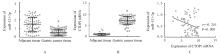
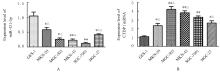
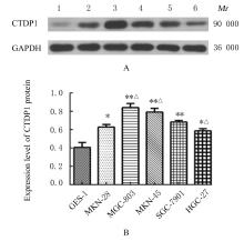
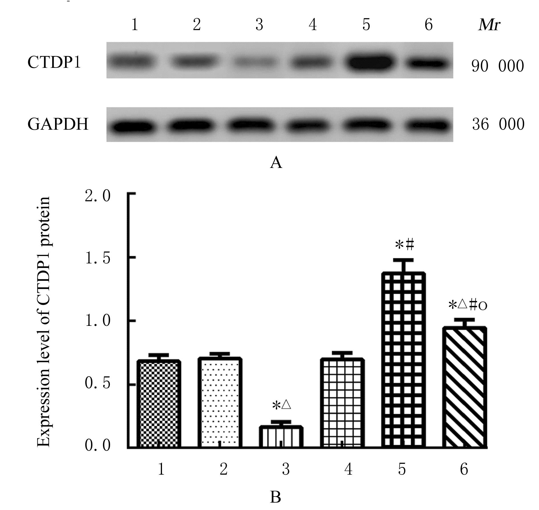
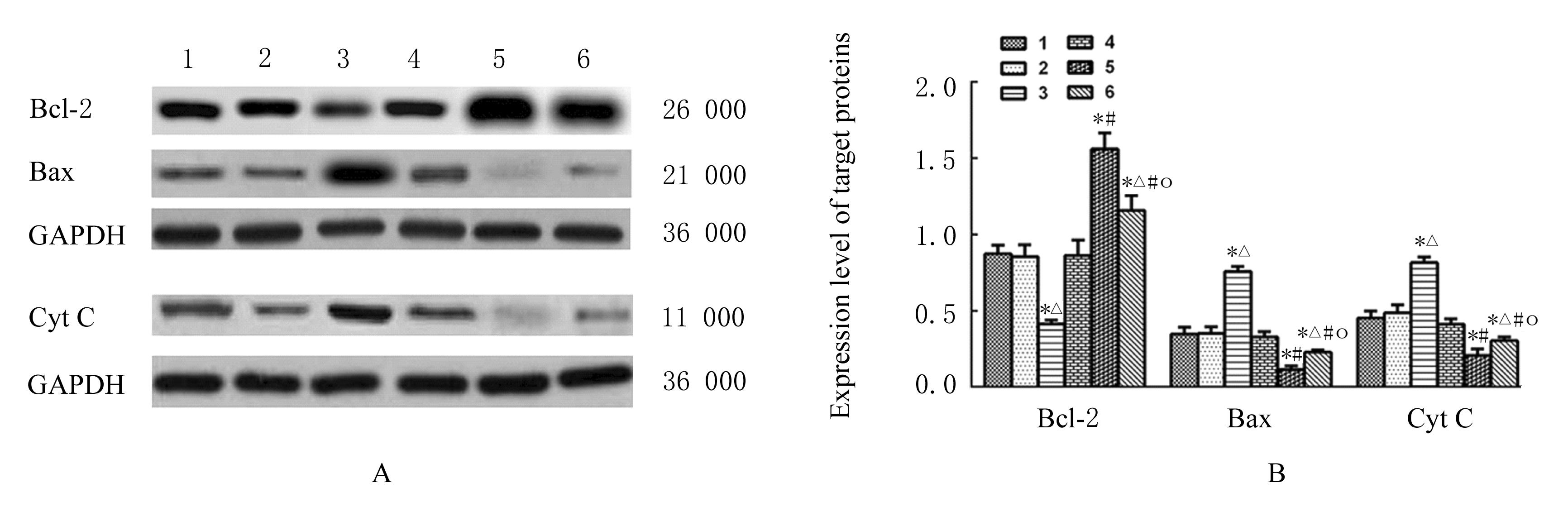
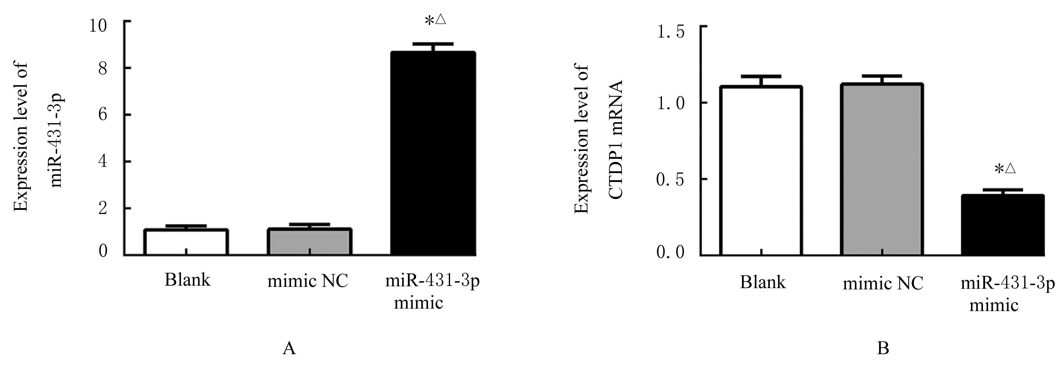
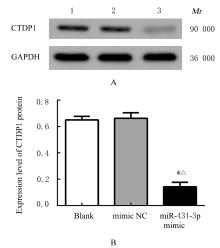
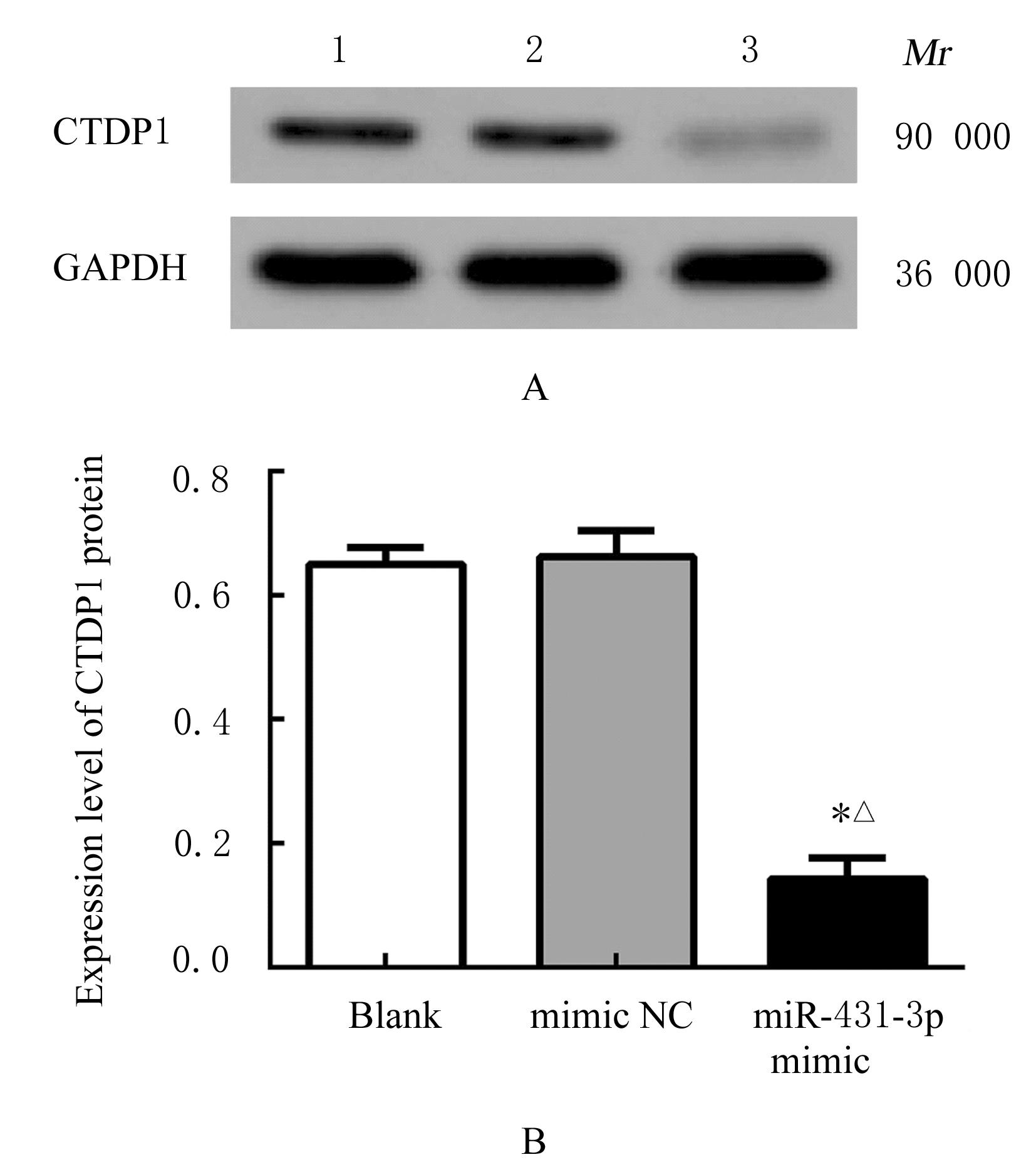
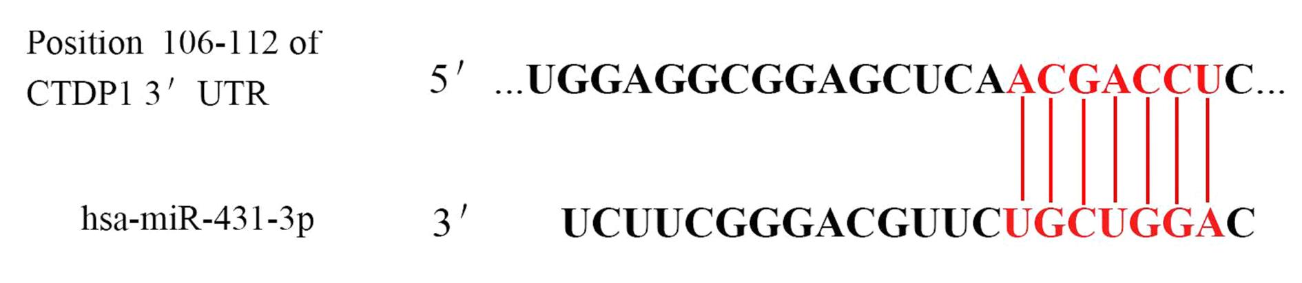
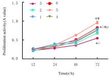
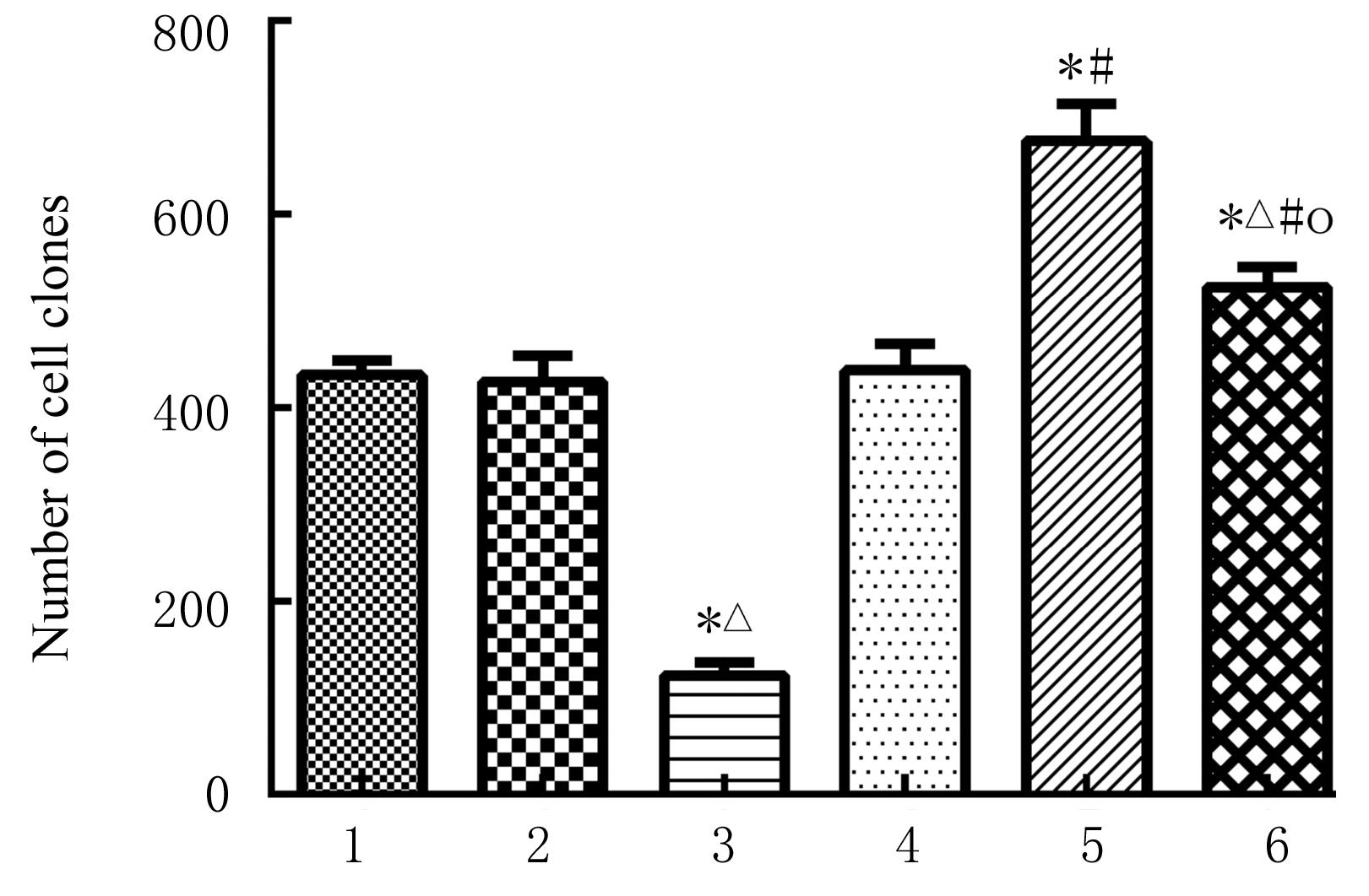
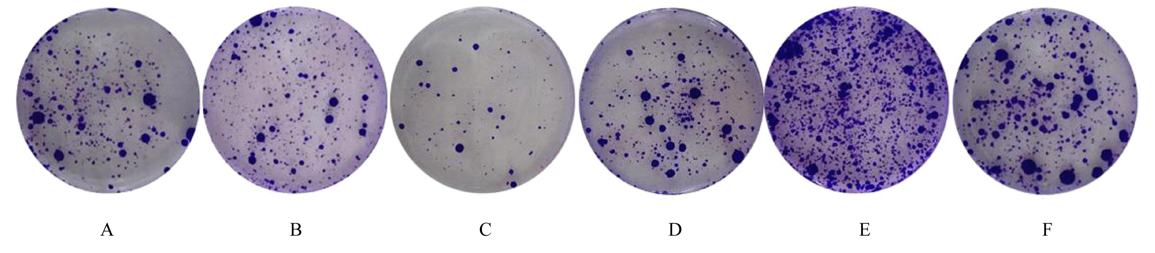
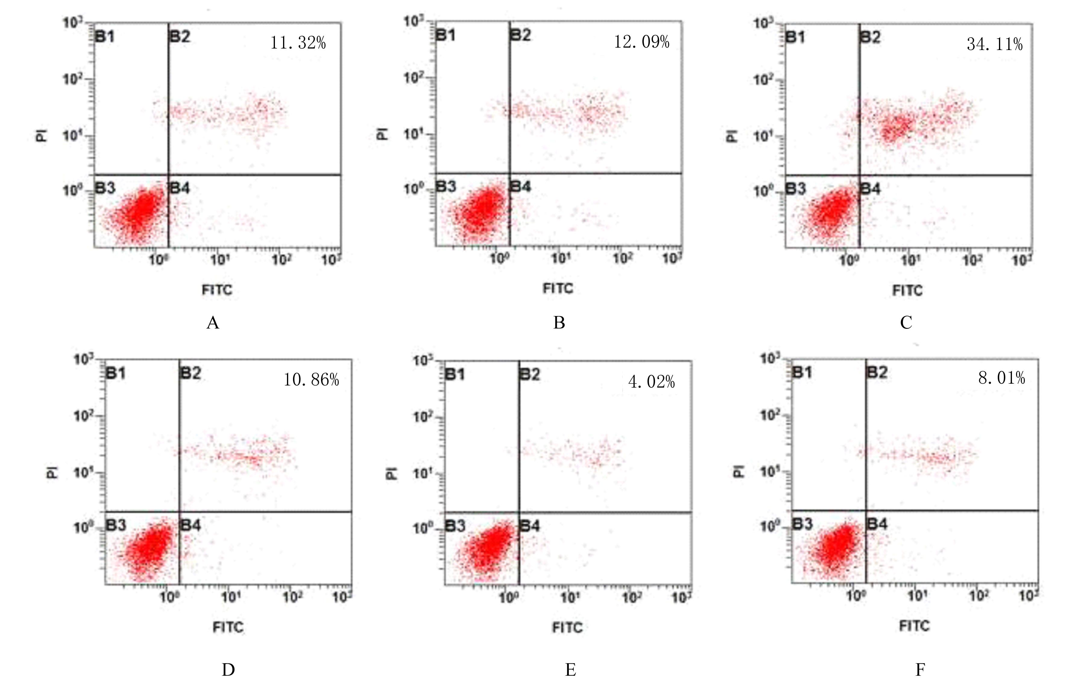
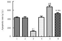
目的 探讨miR-431-3p对胃癌细胞增殖和凋亡的影响,阐明其可能的分子机制。 方法 取经病理诊断确诊为胃癌的68例患者的胃癌组织及其癌旁组织。采用实时荧光定量PCR(RT-qPCR)法检测胃癌组织、癌旁组织和人正常胃黏膜上皮GES-1细胞、人胃癌细胞(MKN-28、MGC-803、MKN-45、SGC-7901和HGC-27)中miR-431-3p及羧基末端结构域磷酸酶1(CTDP1)mRNA表达水平,Western blotting法检测上述细胞中CTDP1蛋白和各组SGC-7901细胞中细胞色素C(Cyt C)、B细胞淋巴瘤2(Bcl-2)和Bcl-2相关X蛋白(Bax)蛋白表达水平。采用Pearson相关分析法分析miR-431-3p与CTDP1 mRNA表达水平的相关性,双荧光素酶报告基因实验检测miR-431-3p和CTDP1的靶向关系。将miR-431-3p mimic和CTDP1过表达慢病毒分别或同时转染至SGC-7901细胞中,将细胞分为空白组、空载过表达(mimic NC)组、miR-431-3p过表达(miR-431-3p mimic)组、空载慢病毒(Vector)组、CTDP1过表达慢病毒(CTDP1过表达)组和miR-431-3p mimic+CTDP1过表达(共转染)组。MTT法检测各组SGC-7901细胞增殖活性,克隆形成实验检测各组SGC-7901细胞单克隆形成数,流式细胞术检测各组SGC-7901细胞凋亡率。 结果 与癌旁组织比较,胃癌组织中miR-431-3p表达水平降低(P<0.01),CTDP1 mRNA表达水平升高(P<0.01),并且二者表达水平呈负相关关系(r=-0.316,P=0.009)。与GES-1细胞比较,其他5种胃癌细胞中miR-431-3p表达水平均降低(P<0.01),CTDP1 mRNA和蛋白表达水平均升高(P<0.05)。双荧光素酶报告系统,miR-431-3p靶向调控CTDP1表达。与空白组和mimic NC组比较,miR-431-3p组SGC-7901细胞中CTDP1和Bcl-2蛋白表达水平、细胞增殖活性和克隆形成数降低(P<0.05),细胞凋亡率和细胞中Cyt C及Bax蛋白表达水平升高(P<0.05)。与空白组和Vector组比较,CTDP1过表达组SGC-7901细胞中CTDP1和Bcl-2蛋白表达水平、细胞增殖活性及克隆形成数升高(P<0.05);细胞凋亡率和细胞中Cyt C及Bax蛋白表达水平降低(P<0.05)。与空白组和miR-431-3p组比较,共转染组SGC-7901细胞中CTDP1和Bcl-2蛋白表达水平、细胞增殖活性及克隆形成数水平升高(P<0.05),细胞凋亡率和细胞中Cyt C及Bax蛋白表达水平降低(P<0.05)。 结论 miR-431-3p过表达能抑制人胃癌细胞增殖,并促进细胞凋亡,可能是与靶向下调CTDP1基因表达有关。

关键词: 胃肿瘤, miR-431-3p, 羧基末端结构域磷酸酶1, 细胞增殖, 细胞凋亡

Abstract:

Methods The gastric cancer tissue and the adjacent tissue of 68 patients with gastric cancer confirmed by pathological diagnosis were collected.The expression levels of miR-431-3p mRNA and carboxy-terminal domain phosphatase 1 (CTDP1) mRNA in gastric cancer tissue, adjacent tissue, human normal gastric mucosal epithelial GES-1 cells and human gastric cancer cells (MKN-28, MGC-803, MKN-45, SGC-7901, HGC-27 cells )were detected by real-time fluorescence quantitative PCR (RT-qPCR) method;the expression levels of CTDP1 protein in the above cells and the expression levels of cytochrome C(Cyt C),B cell lymphoma-2 (Bcl-2), and Bcl-2 associated X protein (Bax) in the SGC-7901 cells in various groups were detected by Western blotting method; the correlation between miR-431-3p and CTDP1 mRNA expression level was analyzed by Pearson correlation analysis;the targeting relationship between miR-431-3p and CTDP1 was detected by dual luciferase reporter gene experiment; the miR-431-3p mimic and CTDP1 over-expression lentivirus were transfected into the SGC-7901 cells separately or at the same time.The cells were divided into blank group, vector over-expression(mimic NC) group,miR-431-3p over-expression (miR-431-3p mimic) group,vector lentivirus(vector) group, CTDP1 over-expression lentivirus (CTDP over-expression)group and miR-431-3p mimic+CTDP1 over-expression (co-transfection) group.The proliferation activities of the cells in various groups were detected by MTT assay; the clone formation numbers of the SGC-7901 cells in various groups were detected by clone formation assay;the apoptotic rates of the SGC-7901 cells in various groups were detected by flow cytometry. Results Compared with the adjacent tissue, the expression level of miR-431-3p in the gastric cancer tissue was decreased (P<0.01),and the expression level of CTDP1 mRNA was increased (P<0.01), and there was a negative correlation between them (r=-0.316, P=0.009). Compared with the GES-1 cells, the expression levels of miR-431-3p in the other five kinds of gastric cancer cells were decreased (P<0.01), and the expression levels of CTDP1 mRNA and protein were increased (P<0.05). The results of dual luciferase reporter system showed that miR-431-3p targetedly regulated the expression of CTDP1. Compared with blank group and mimic NC group, the expression levels of CTDP1 and Bcl-2 proteins, proliferation activity,and number of clone formation of the SGC-7901 cells in miR-431-3p group were decreased (P<0.05), while the apoptotic rate, expression levels of Cyt C and Bax proteins were increased (P<0.05). Compared with blank group and vector group, the expression levels of CTDP1 and Bcl-2 proteins, proliferation activity, and number of clone formation of the SGC-7901 cells in CTDP1 over-expression group were increased (P<0.05), while the apoptotic rate, the expression levels of Cyt C and Bax proteins were decreased (P<0.05). Compared with blank group and miR-431-3p group, the expression levels of CTDP1 and Bcl-2 proteins, proliferation activity,and number of clone formation of the SGC-7901 cells in co-transfection group were increased (P<0.05), while the apoptotic rate, expression levels of Cyt C and Bax proteins were decreased (P<0.05). Conclusion Over-expression of miR-431-3p can inhibit the proliferation and promot the apoptosis of the human gastric cancer cells, and its mechanism may be related to targeted down-regulation of the CTDP1 gene expression. Objective To discuss the effect of miR-431-3p on the proliferation and apoptosis of the gastric cancer cells,and to elucidate its possible molecular mechanism.

Key words: Stomach neoplasm, MiR-431-3p, Carboxyl terminal domain phosphatase 1, Cell proliferation, Apoptosis

中图分类号: 

  • R735.2