吉林大学学报(医学版) ›› 2025, Vol. 51 ›› Issue (2): 403-411.doi: 10.13481/j.1671-587X.20250214

• 基础研究 • 上一篇    

高浓度葡萄糖对体外Raw264.7巨噬细胞极化状态的影响

胡晓霞1,李亚龙2,杨东亮3,拉巴泽仁,刘欣跃2()   

  1. 1.兰州大学第二临床医学院老年病科,甘肃 兰州 730030
    2.兰州大学第二临床医学院检验医学 中心,甘肃 兰州 730030
    3.西北民族大学生物医学研究中心 生物工程与技术国家民委重点实验室,甘肃 兰州 730030
  • 收稿日期:2024-03-13 接受日期:2024-06-20 出版日期:2025-03-28 发布日期:2025-04-22
  • 通讯作者: 刘欣跃 E-mail:liuxy@lzu.edu.cn
  • 作者简介:胡晓霞(1976-),女,甘肃省临夏市人,在读博士研究生,主要从事糖尿病急慢性并发症方面的研究。
  • 基金资助:
    甘肃省科技厅自然科学基金项目(23JRRA0958);兰州大学第二医院(第二临床医学院)“萃英科技创新”计划项目(CY2023-MS-B12)

Effect of high glucose on polarization of Raw264.7 macrophages in vitro

Xiaoxia HU1,Yalong LI2,Dongliang YANG3,Bazeren LA,Xinyue LIU2()   

  1. 1.Department of Geriatrics,Second Clinical Medical School,Lanzhou University,Lanzhou 730030,China
    2.Laboratory Medicine Center,Second Clinical Medical School,Lanzhou University,Lanzhou 730030,China
    3.Key Laboratory of Bioengineering and Biotechnology,National Ethnic Affairs Commission,Biomedical Research Center,Northwest Minzu University,Lanzhou 730030,China
  • Received:2024-03-13 Accepted:2024-06-20 Online:2025-03-28 Published:2025-04-22
  • Contact: Xinyue LIU E-mail:liuxy@lzu.edu.cn

摘要:

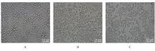
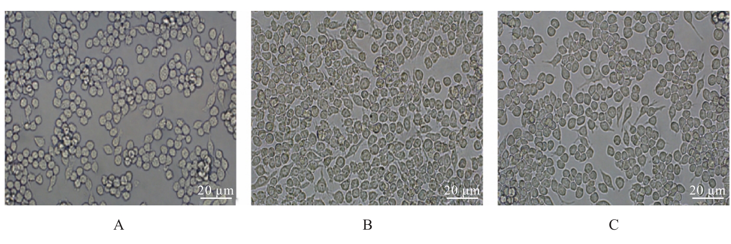
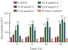
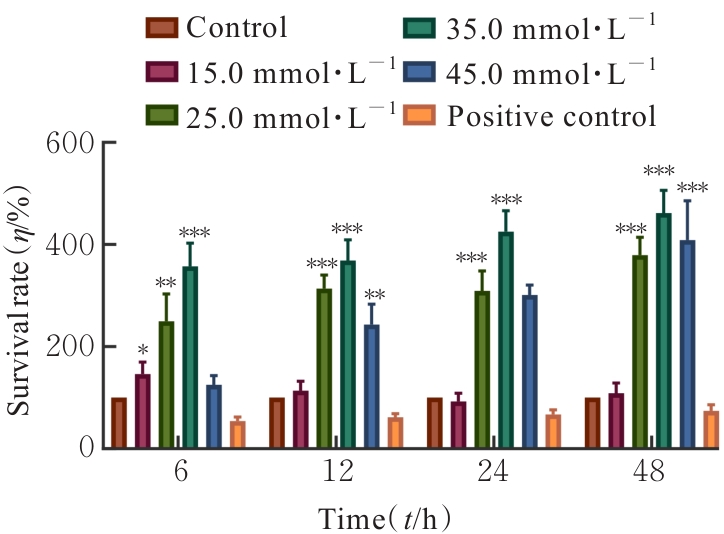
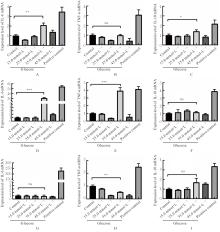
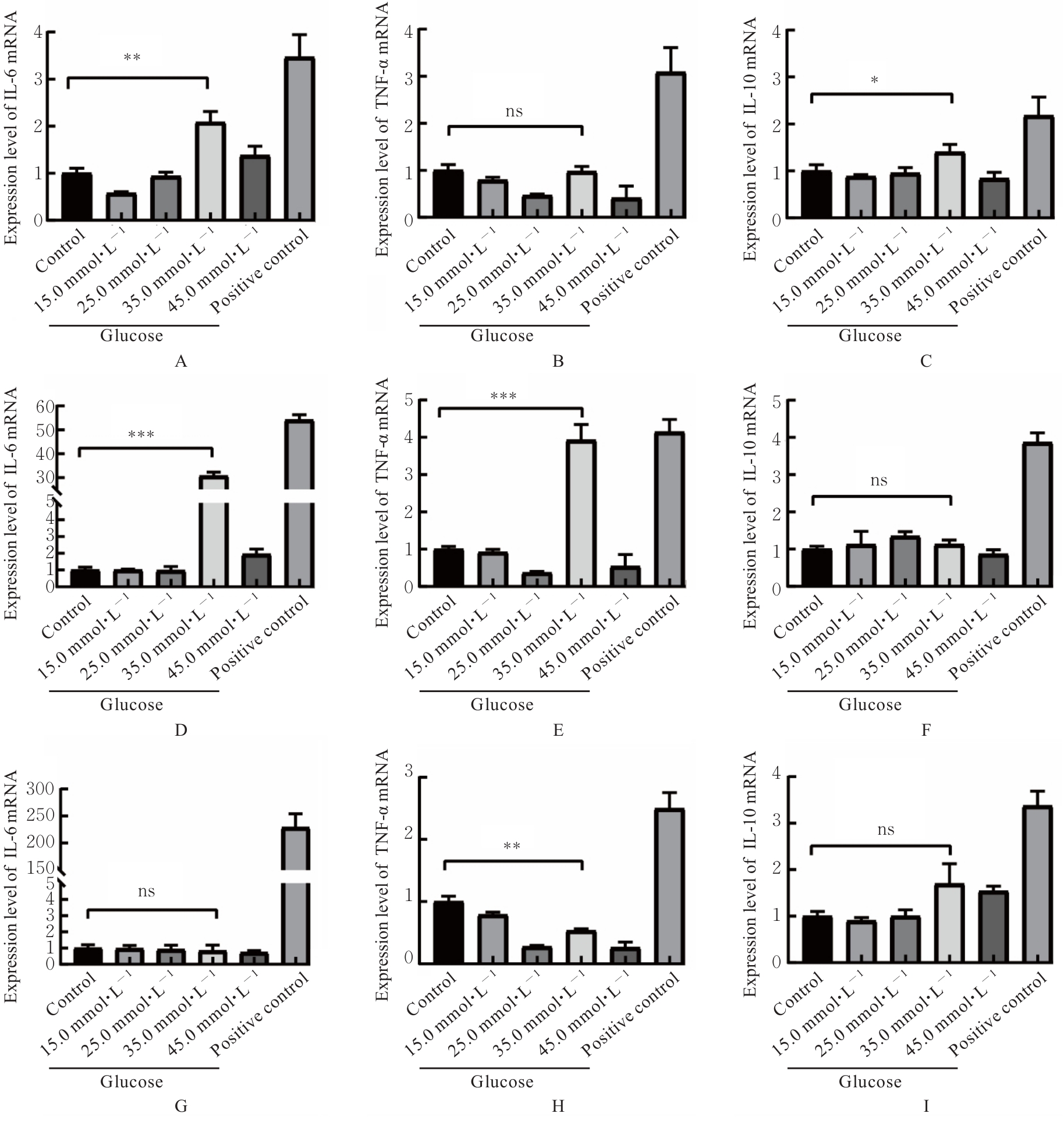
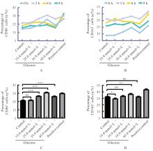
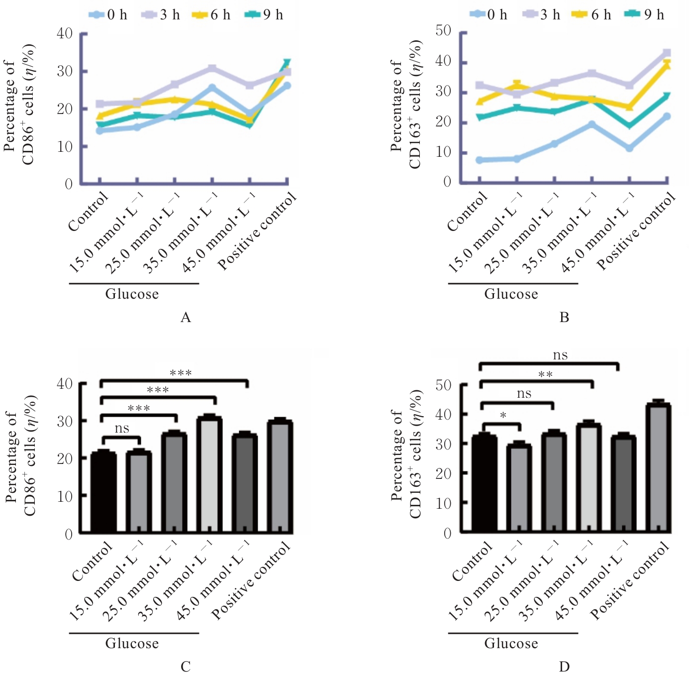
目的 探讨不同浓度葡萄糖对体外Raw264.7巨噬细胞极化的诱导作用。 方法 将DMEM培养基培养的Raw264.7细胞分为对照组(5.5 mmol·L-1葡萄糖)、不同浓度高糖组(15.0、25.0、35.0和45.0 mmol·L-1葡萄糖)和阳性对照组[脂多糖(LPS)],分别培养3、6和9 h,观察各组细胞形态,细胞计数试剂盒8(CCK-8)法检测各组细胞存活率,实时荧光定量PCR(RT-qPCR)法检测各组细胞中白细胞介素6(IL-6)、肿瘤坏死因子α(TNF-α)和白细胞介素10(IL-10)mRNA表达水平,酶联免疫吸附试验(ELISA)法检测各组细胞上清中IL-6、TNF-α和IL-10水平,流式细胞仪检测各组细胞M1和M2型巨噬细胞标志物CD86+及CD163+细胞百分比。 结果 对照组Raw264.7细胞贴壁生长,形态以圆形为主;35.0 mmol·L-1高糖组和阳性对照组细胞拉长、伪足形成,呈现炎症性改变。与对照组比较,作用6、12、24和48 h后不同浓度高糖组细胞存活率均升高(P<0.05)。与对照组比较,作用3 h后,35.0 mmol·L-1高糖组细胞中IL-6和IL-10 mRNA表达水平升高(P<0.05或P<0.01),细胞上清中IL-6、TNF-α和IL-10水平无明显变化,差异无统计学意义(P>0.05);作用6 h后,35.0 mmol·L-1高糖组细胞中TNF-α mRNA表达水平升高(P<0.001),细胞上清中IL-6、TNF-α和IL-10水平明显升高(P<0.05或P<0.001);作用3 h后,35.0 mmol·L-1高糖组巨噬细胞极化标志物CD86+和CD163+细胞百分比明显升高(P<0.01或P<0.001)。 结论 一定高浓度葡萄糖可诱导体外Raw264.7巨噬细胞向M1亚型极化。

关键词: Raw264.7细胞, 葡萄糖, 炎症, 巨噬细胞极化, 炎症细胞因子

Abstract:

Objective To investigate the induction effect of different concentrations of glucose on the polarization of Raw264.7 macrophages in vitro. Methods The Raw264.7 cells cultured in DMEM medium were divided into control group (5.5 mmol·L-1 glucose), different high-glucose groups (15.0, 25.0, 35.0, and 45.0 mmol·L-1 glucose), and positive control group [lipopolysaccharide(LPS)]. The cells were cultured for 3, 6, and 9 h, respectively. Cell morphology was observed in various groups. Cell counting kit-8 (CCK-8) method was used to assess the survival rates of cells in various groups. Real-time fluorescence quantitative PCR (RT-qPCR) method was used to detect the expression levels of interleukin-6 (IL-6), tumor necrosis factor-α (TNF-α), and interleukin-10 (IL-10) mRNA in cells in various groups. Enzyme-linked immunosorbent assay (ELISA) was used to measure the levels of IL-6, TNF-α, and IL-10 in the cell supernatants in various groups. Flow cytometry was used to determine the percentages of M1 and M2 macrophage markers, CD86+ and CD163+ cells, in various groups. Results In control group, the Raw264.7 cells adhered to the culture dish, with a predominantly round morphology. The cells in 35.0 mmol·L-1 high-glucose group and positive control group showed elongated shapes and pseudopodium formation, indicating inflammatory changes. Compared with control group, the survival rates in different high-glucose groups at 6, 12, 24, and 48 h after treatment were increased (P<0.05). Compared with control group, after 3 h of treatment, the expression levels of IL-6 and IL-10 mRNA in the cells in 35.0 mmol·L-1 high-glucose group were increased (P<0.05 or P<0.01), while no significant changes were observed in IL-6, TNF-α, and IL-10 levels in the cell supernatants (P>0.05); after 6 h of treatment, the expression level of TNF-α mRNA in the cells in 35.0 mmol·L-1 high-glucose group was increased (P<0.001), and the levels of IL-6, TNF-α, and IL-10 in the cell supernatants were significantly increased (P<0.05 or P<0.001); after 3 h of treatment, the percentages of macrophage markers CD86+ cells and CD163+ cells in 35.0 mmol·L-1 high-glucose group were significantly increased (P<0.01 or P<0.001). Conclusion A certain high concentration of glucose may induce the polarization of Raw264.7 macrophages towards the M1 subtype in vitro.

Key words: Raw264.7 cells, Glucose, Inflammation, Macrophage polarization, Inflammatory cytokine

中图分类号: 

  • R392.11