吉林大学学报(医学版) ›› 2025, Vol. 51 ›› Issue (3): 663-671.doi: 10.13481/j.1671-587X.20250311

• 基础研究 • 上一篇    

miR-199a-5p对胶质瘤U251细胞小窝蛋白1表达及细胞迁移和凋亡的影响

刘东慧1,次云哲2,王春艳2,麻雯熠2()   

  1. 1.承德医学院基础医学院人体解剖学教研室,河北 承德 067000
    2.承德医学院基础医学院组织学与胚胎学教研室,河北 承德 067000
  • 收稿日期:2024-07-29 接受日期:2024-09-26 出版日期:2025-05-28 发布日期:2025-07-18
  • 通讯作者: 麻雯熠 E-mail:mawenyiss@163.com
  • 作者简介:刘东慧(1987-),女,河北省廊坊市人,讲师,医学硕士,主要从事糖尿病和肿瘤学方面的研究。
  • 基金资助:
    河北省卫健委医学科学研究项目(20200178);河北省教育厅河北省高等学校科学研究青年基金项目(QN2023010)

Effect of miR-199a-5p on expression of Caveolin-1, cell migration and apoptosis in glioma U251 cells

Donghui LIU1,Yunzhe CI2,Chunyan WANG2,Wenyi MA2()   

  1. 1.Department of Human Anatomy,College of Basic Medical Sciences,Chengde Medical College,Chengde 067000,China
    2.Department of Histology and Embryology,College of Basic Medical Sciences,Chengde Medical College,Chengde 067000,China
  • Received:2024-07-29 Accepted:2024-09-26 Online:2025-05-28 Published:2025-07-18
  • Contact: Wenyi MA E-mail:mawenyiss@163.com

摘要:

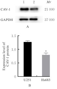
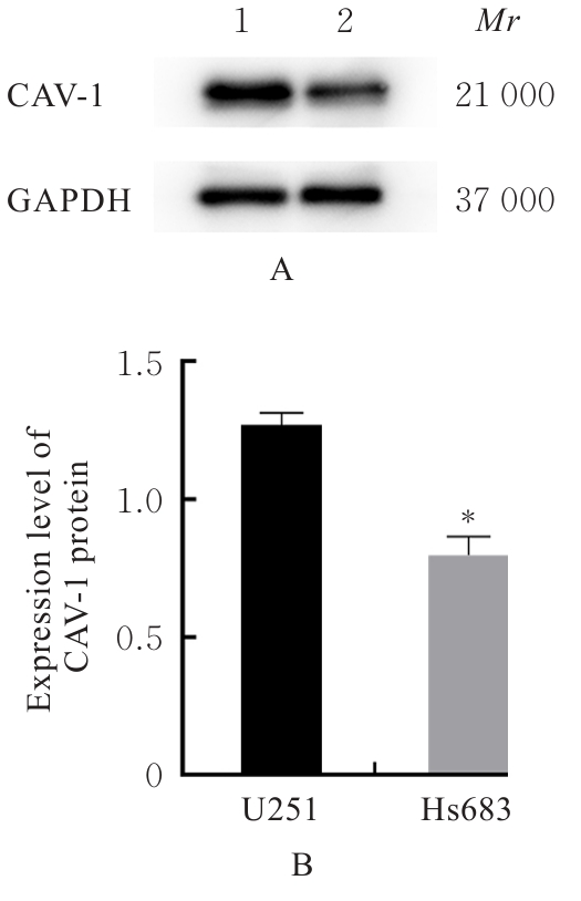
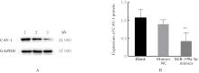
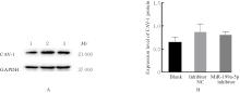
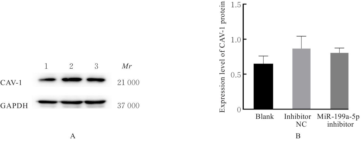
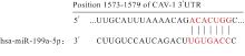
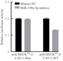
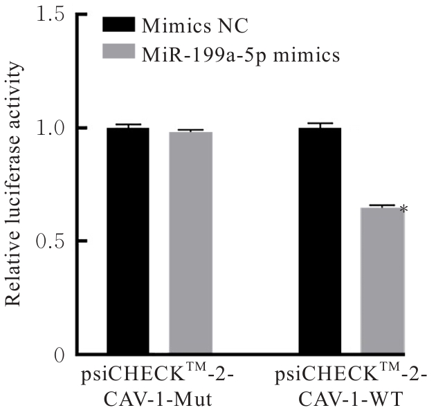
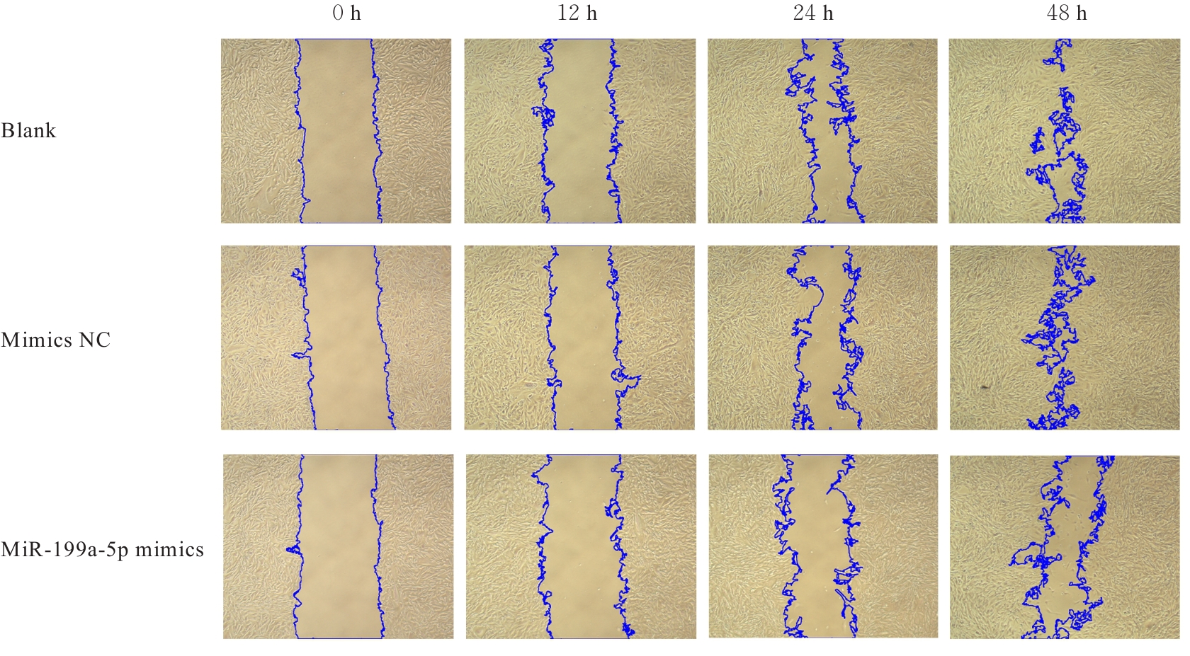
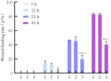
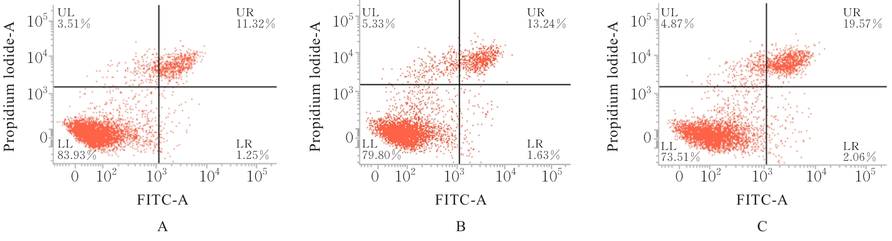
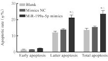
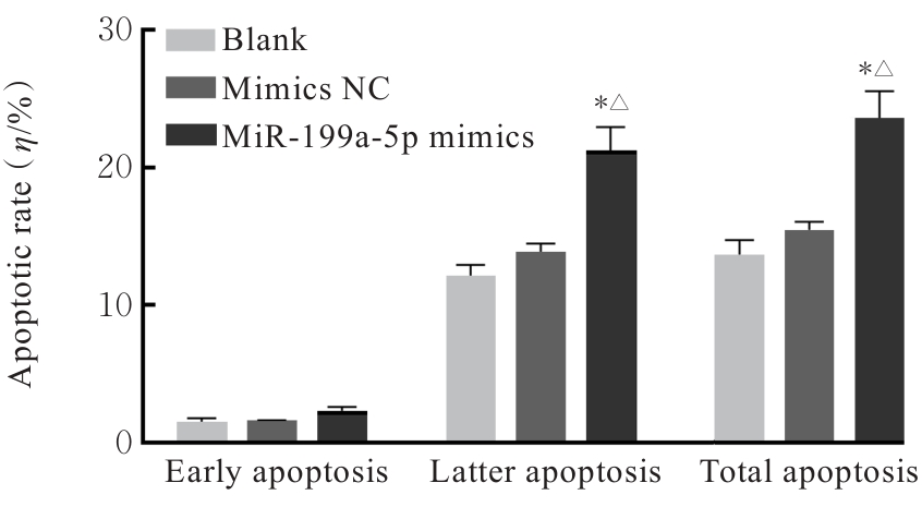
目的 探讨胶质母细胞瘤U251细胞过表达微小RNA(miR)-199a-5p后对细胞迁移和凋亡的影响,并阐明miR-199a-5p与小窝蛋白1(CAV-1)的靶向调控关系。 方法 体外培养胶质母细胞瘤U251细胞和少突胶质细胞瘤Hs683细胞,采用Western blotting法检测2种细胞中CAV-1蛋白表达水平,实时荧光定量PCR(RT-qPCR)法检测2种细胞中miR-199a-5p表达水平。U251细胞分为空白组(不进行转染)、mimics NC组(转染空载质粒)和miR-199a-5p mimics组(转染miR-199a-5p模拟物),Hs683细胞分为空白组(不进行转染)、inhibitor NC组(转染空载质粒)和miR-199a-5p inhibitor组(转染miR-199a-5p抑制物),采用RT-qPCR法检测各组细胞转染效率,Western blotting法检测各组细胞中CAV-1蛋白表达水平。TargetScan数据库预测miR-199a-5p与CAV-1在3'非翻译区(3'UTR)的结合位点,将psiCHECKTM-2-CAV-1-WT和psiCHECKTM-2-CAV-1-Mut分别与miR-199a-5p mimics和mimics NC共转染至U251细胞中,即psiCHECKTM-2-CAV-1-WT+mimics NC组、psiCHECKTM-2-CAV-1-WT+miR-199a-5p mimics组、psiCHECKTM-2-CAV-1-Mut+ mimics NC组和psiCHECKTM-2-CAV1-Mut+miR-199a-5p mimics组,双荧光素酶报告基因实验验证miR-199a-5p与CAV-1的靶向关系,细胞划痕实验检测各组U251细胞划痕愈合率,流式细胞术检测各组U251细胞凋亡率。 结果 Western blotting法和RT-qPCR法检测,与Hs683细胞比较,U251细胞中CAV-1蛋白表达水平明显降低(P<0.05);与U251细胞比较,Hs683细胞中miR-199a-5p表达水平明显升高(P<0.01)。与空白组和mimics NC组比较,miR-199a-5p mimics组U251细胞中miR-199a-5p表达水平明显升高(P<0.01),CAV-1蛋白表达水平明显降低(P<0.05)。与空白组比较,inhibitor NC组和miR-199a-5p inhibitor组Hs683细胞中miR-199a-5p表达水平均明显降低(P<0.01)。各组Hs683细胞中CAV-1蛋白表达水平比较差异均无统计学意义(P>0.05)。双荧光素酶报告基因实验验证,成功构建psiCHECKTM-2-CAV-1-野生型(WT)和psiCHECKTM-2-CAV-1-突变型(Mut)表达载体;与psiCHECKTM-2-CAV-1-WT-mimics NC组比较,psiCHECKTM-2-CAV-1-WT-miR-199a-5p mimics组U251细胞WT CAV-1相对荧光素酶活性明显降低(P<0.01)。细胞划痕实验,转染12、24和48 h时,与空白组比较,miR-199a-5p mimics组U251细胞划痕愈合率明显降低(P<0.05或P<0.01);转染48和72 h时,与mimics NC组比较,miR-199a-5p mimics组U251细胞划痕愈合率明显降低(P<0.01)。流式细胞术,与空白组和mimics NC组比较,miR-199a-5p mimics组U251细胞凋亡率明显升高(P<0.01)。 结论 通过向胶质母细胞瘤U251细胞转染miR-199a-5p成熟模拟物,可降低CAV-1蛋白表达水平,并抑制胶质瘤细胞的迁移,促进其凋亡,抑制肿瘤发生发展。miR-199a-5p与CAV-1之间的靶向关系可能为胶质瘤发生发展的潜在机制,并可能成为胶质瘤潜在的诊断和治疗靶点。

关键词: 脑胶质瘤细胞, 微小RNA-199a-5p, 小窝蛋白1, 细胞迁移, 细胞凋亡

Abstract:

Objective To discuss the effects of microRNA (miR)-199a-5p overexpression on cell migration and apoptosis in the glioblastoma U251 cells, and to clarify the targeting regulatory relationship between miR-199a-5p and caveolin-1 (CAV-1). Methods The glioblastoma U251 cells and oligodendroglioma Hs683 cells were cultured in vitro. Western blotting method was used to detect the expression levels of CAV-1 protein in 2 kinds of cells; real-time fluorescence quantitative PCR (RT-qPCR) method was used to detect the expression levels of miR-199a-5p in 2 kinds of cells. The U251 cells were divided into blank group (non-transfection), mimics NC group (transfected with empty vector), and miR-199a-5p mimics group (transfected with miR-199a-5p mimics). The Hs683 cells were divided into blank group (no transfection), inhibitor NC group (transfected with empty vector), and miR-199a-5p inhibitor group (transfected with miR-199a-5p inhibitor). RT-qPCR method was used to detect the transfection efficiency of the cells in various groups; Western blotting method was used to detect the expression levels of CAV-1 protein in the cells in various groups. TargetScan database was used to predict the binding sites between miR-199a-5p and CAV-1 in the 3' untrans lated region(3'UTR); psiCHECKTM-2-CAV-1-WT and psiCHECKTM-2-CAV-1-Mut were co-transfected with miR-199a-5p mimics and mimics NC into the U251 cells, respectively, forming psiCHECKTM-2-CAV-1-WT+mimics NC group, psiCHECKTM-2-CAV-1-WT+miR-199a-5p mimics group, psiCHECKTM-2-CAV-1-Mut+mimics NC group, and psiCHECKTM-2-CAV1-Mut+miR-199a-5p mimics group; dual-luciferase reporter gene assay was used to verify the targeting relationship between miR-199a-5p and CAV-1; cell scratch assay was used to detect the scratch healing rates of the U251 cells in various groups; flow cytometry was used to detect the apoptotic rates of the U251 cells in various groups. Results The Western blotting and RT-qPCR results showed that compared with Hs683 cells, the expression level of CAV-1 protein in the U251 cells was significantly decreased (P<0.05); compared with U251 cells, the expression level of miR-199a-5p in the Hs683 cells was significantly increased (P<0.01). Compared with blank group and mimics NC group, the expression level of miR-199a-5p in the U251 cells in miR-199a-5p mimics group was significantly increased (P<0.01), the expression level of CAV-1 protein in the U251 cells in miR-199a-5p mimics group was significantly decreased (P<0.05). Compared with blank group, the expression levels of miR-199a-5p in the Hs683 cells in inhibitor NC group and miR-199a-5p inhibitor group were significantly decreased (P<0.01). No significant differences were observed in the expression levels of CAV-1 protein in the Hs683 cells among various groups (P>0.05). The dual-luciferase reporter gene assay results showed that psiCHECKTM-2-CAV-1-wild type (WT) and psiCHECKTM-2-CAV-1-mutant (Mut) expression vectors were successfully constructed; compared with psiCHECKTM-2-CAV-1-WT-mimics NC group, the relative luciferase activity of WT CAV-1 in the U251 cells in psiCHECKTM-2-CAV-1-WT-miR-199a-5p mimics group was significantly decreased (P<0.01). The cell scratch assay results showed that at 12, 24, and 48 h after transfection, compared with blank group, the scratch healing rate of the U251 cells in miR-199a-5p mimics group was significantly decreased (P<0.05 or P<0.01). The flow cytometry results showed that compared with blank group and mimics NC group, the apoptotic rate of the U251 cells in miR-199a-5p mimics group was significantly increased (P<0.01). Conclusion Transfection of mature miR-199a-5p mimics into the glioblastoma U251 cells can reduce the expression of CAV-1 protein, inhibit glioma cell migration, promote apoptosis, and suppress tumorigenesis and development. The targeting relationship between miR-199a-5p and CAV-1 may represent a potential mechanism for glioma development and could serve as a potential diagnostic and therapeutic target for glioma.

Key words: Glioma cells, MicroRNA-199a-5p, Caveolin-1, Cell migration, Apoptosis

中图分类号: 

  • R739.41