吉林大学学报(医学版) ›› 2021, Vol. 47 ›› Issue (1): 82-88.doi: 10.13481/j.1671-587x.20210111

• 基础研究 • 上一篇    下一篇

3D打印钛合金种植体的制备及其骨结合性能

王蕊,李美华(),周万琳   

  1. 吉林大学第二医院口腔科,吉林 长春 130041
  • 收稿日期:2020-08-04 出版日期:2021-01-28 发布日期:2021-01-27
  • 通讯作者: 李美华 E-mail:meihl_699@163.com
  • 作者简介:王 蕊(1983-),女,黑龙江省大庆市人,主治医师,在读博士研究生,主要从事骨修复改建方面的研究。
  • 基金资助:
    吉林省科技厅自然科学基金项目(20180101111JC)

Preparation and bone-binding properties of 3D printed titanium alloy implants

Rui WANG,Meihua LI(),Wanlin ZHOU   

  1. Department of Stomatology,Second Hospital,Jilin University,Changchun 130041,China
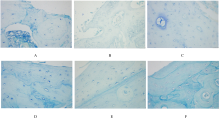
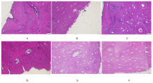
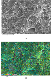
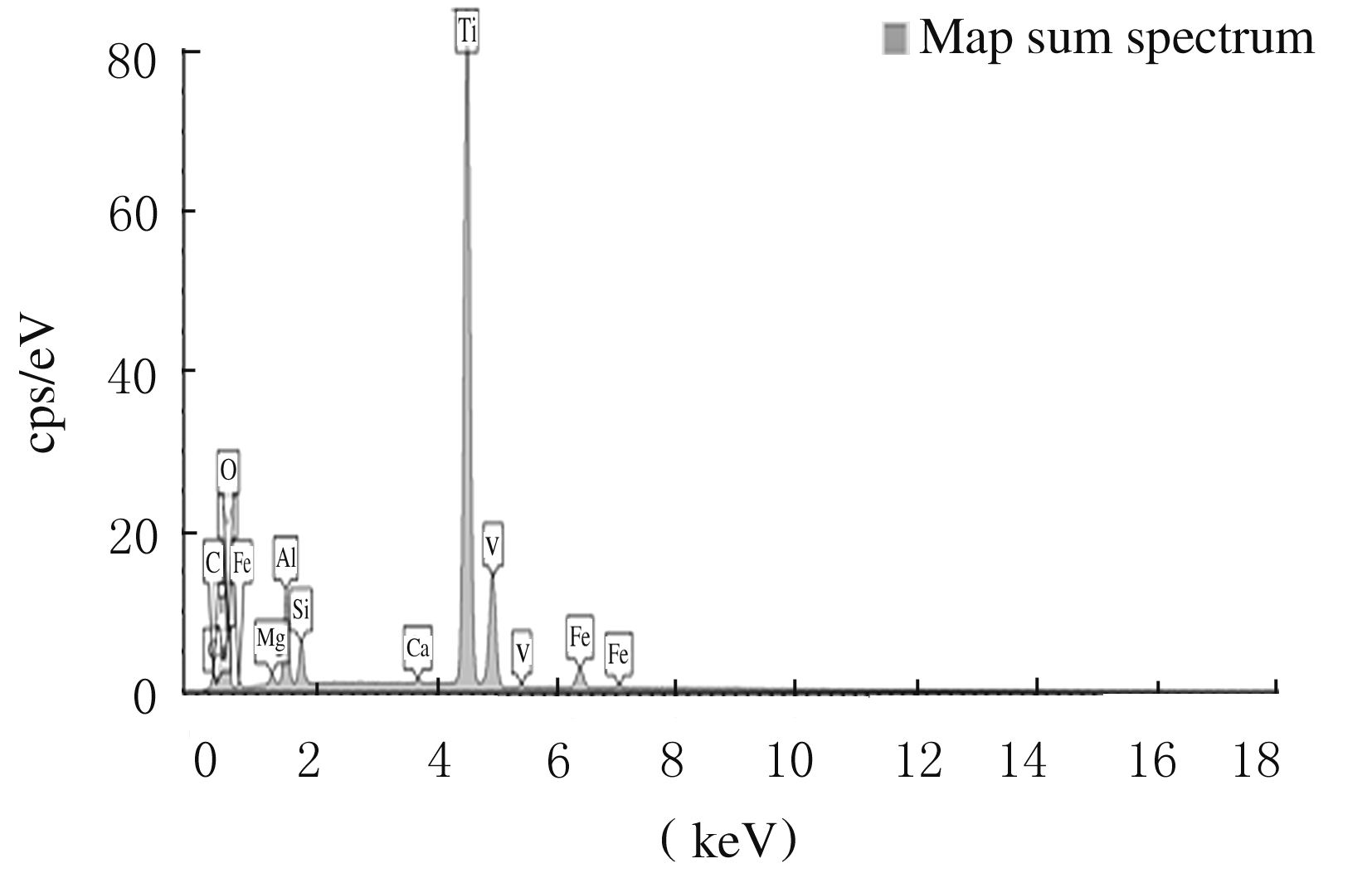
  • Received:2020-08-04 Online:2021-01-28 Published:2021-01-27
  • Contact: Meihua LI E-mail:meihl_699@163.com

摘要: 目的

观察3D打印种植体与经典Straumann种植体植入家兔体内后骨整合情况,为其临床应用提供理论依据。

方法

以3D打印的方法制作Ti-6Al-4V(TC4)种植体,以经典Straumann种植体作为对照,于6只家兔双侧股骨植入种植体建立家兔种植体模型,每侧各植入1颗TC4种植体和1颗Straumann种植体,共12颗TC4种植体(TC4种植体组)和12颗Straumann种植体(Straumann种植体组)。术后2、4和8周各处死2只家兔,取出种植体周围组织,甲苯胺蓝染色观察成骨细胞、新骨形成及骨组织修复情况,亚甲基蓝-酸性品红染色观察新骨形成及矿化情况,扫描电子显微镜(SEM)观察术后TC4种植体表面结构变化,采用能量色散X射线光谱仪(EDS)对术后TC4种植体进行元素构成分析。

结果

甲苯胺蓝染色,术后2周时,2组紧贴种植体的骨组织边缘有胞核深染、胞浆呈淡蓝色的成骨细胞线性排列;术后4周,2组种植体周围骨组织存在淡染的新生胶原纤维样结构,排列欠整齐;术后8周,2组家兔靠近种植体一侧骨组织可见淡蓝色、结构连续的新生骨形成,内含大量骨细胞,新生骨与原矿化骨界限清晰。亚甲基蓝-酸性品红染色,术后 2 周,2组种植体周围骨组织中有活跃的成骨细胞聚集;术后 4 周,2组种植体与原矿化骨组织间均可见新生的、红染的类骨质结构;术后 8 周,2组种植体表面均可见伴有一定程度钙化的新生骨形成,呈深红色,Straumann 种植体组钙化程度略优于 TC4 种植体组。SEM观察,完成体内实验的TC4种植体在表面结构上基本与术前TC4种植体保持一致,未见明显结构缺损形成。EDS扫描分析,完成体内实验的TC4种植体除含有与术前TC4种植体一致的元素外,尚存在常量元素钙(Ca)、锰(Mg)和微量元素硅(Si)等。

结论

3D打印TC4种植体具备良好的结构稳定性与生物相容性,在体内可以达到与Straumann种植体相似的骨结合。

关键词: 3D打印, 钛合金种植体, Straumann种植体, 骨结合

Abstract: Objective

To observe the osseointegration of 3D printed implants and classic Straumann implants after implanted in the rabbits,and to provide the theoretical basis for its clinical application.

Methods

The Ti-6A1-4V(TC4) implants were made by 3D printed method,and the classic Straumann implants were used as controls.The animal models were established by implanting bilateral femur implants in 6 rabbits; one TC4 implant and one Straumann implant were implanted on each side,and there were 12 T4C implants((TC4 implant group) and 12 Straumann implants (Straumann implant group) in all. At 2, 4 and 8 weeks after the operation, two rabbits in each group were killed respectively, and the tissue around the implants was taken out; toluidine blue staining was used to observe osteoblasts, new bone formation and bone tissue repair, and methylene blue acid fuchsin staining was used to observe the new bone repair and mineralization.The surface structure changes of TC4 implants after operation were observed by scanning electron microscope(SEM),and the element composition of TC4 implants after operation was analyzed by energy dispersive X-ray spectrometer(EDS).

Results

The toluidine blue staining results showed that at 2 weeks after the operation, the deep staining of nucleus and linear arrangement of light blue osteoblasts on the edge of bone tissue close to the implants of the rabbits in two groups were found; at 4 weeks after the operation, the existence of light stained new collagen fiber-like structure around the implants in two groups was seen, and the arrangement was not orderly; at 8 weeks after the operation,the light blue new bone formation with continuous structures was found in one side close to the implants of the rabbits in two groups, containing a large number of bone cells, and the boundary between new bone and original mineralized bone was clear. The methylene blue acid fuchsin staining results showed that at 2 weeks after the operation, the implants in two groups showed active osteoblast aggregation around the implants; at 4 weeks after the operation, new and red stained osteoid structures were found between the implants and the original mineralized bone in two groups; at 8 weeks after the operation, new bone formation with certain degree of calcification was seen on the surface of the implants in two groups, which was dark red,and the degree of calcification in Straumann group was slightly better than that in TC4 group. The SEM results showed that the TC4 implants completed in vivo experiment were basically consistent with the preoperative TC4 implants in surface structure, and no obvious structural defects were found.The results of EDS showed that the TC4 implant completed in vivo experiment contained not only the elements consistent with the preoperative TC4 implants, but also the major elements Ca, Mg and trace elements Si.

Conclusion

The 3D printed TC4 implants have good structural stability and biocompatibility, and can achieve the similar bone binding with Straumann implants invivo.

Key words: 3D printing, TC4 implants, Straumann implants, osseointegration

中图分类号: 

  • R783.1