吉林大学学报(医学版) ›› 2021, Vol. 47 ›› Issue (1): 89-95.doi: 10.13481/j.1671-587x.20210112

• 基础研究 • 上一篇    下一篇

MMP-3和CCN2在牙髓损伤模型大鼠牙髓组织中的表达及其意义

李梦洁1,2,谢金芳1,尹硕3,刘霞1()   

  1. 1.吉林大学口腔医院综合口腔治疗科,吉林 长春 130021
    2.河南省郑州市口腔医院牙体牙髓科,河南 郑州 450000
    3.吉林省长春市口腔医院修复科,吉林 长春 130041
  • 收稿日期:2020-07-12 出版日期:2021-01-28 发布日期:2021-01-27
  • 通讯作者: 刘霞 E-mail:liu_xia@jlu.edu.cn
  • 作者简介:李梦洁(1988-),女,山东省淄博市人,主治医师,医学硕士,主要从事牙体牙髓疾病临床及基础方面的研究。
  • 基金资助:
    吉林省教育厅科研基金项目(JJKH20190093KJ);吉林省财政厅科研基金项目(JCSZ2019378-9)

Expressions of MMP-3 and CCN2 in dental pulp tissue of rats with dental pulp injury and their significances

Mengjie LI1,2,Jinfang XIE1,Shuo YIN3,Xia LIU1()   

  1. 1.Department of Oral Comprehensive Treatment,Stomatology Hospital,Jilin University,Changchun 130021,China
    2.Department of Endodontics,Zhengzhou Dental Hospital,Henan Province,Zhengzhou 450000,China
    3.Department of Prosthodontics,Changchun Dental Hospital,Jilin Province,Changchun 130041,China
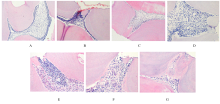
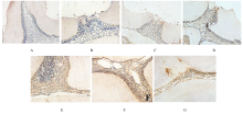
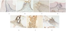
  • Received:2020-07-12 Online:2021-01-28 Published:2021-01-27
  • Contact: Xia LIU E-mail:liu_xia@jlu.edu.cn

摘要: 目的

建立大鼠牙髓损伤模型,初步探讨牙髓损伤后基质金属蛋白酶3(MMP-3)和结缔组织生长因子2 (CCN2)促进组织修复的作用。

方法

28只大鼠随机分为对照组和损伤后0 h、6 h、1 d、3 d、7 d及14 d组,每组4只,除对照组外,其余各组大鼠采用单纯开放法制备上颌磨牙牙髓损伤模型,损伤后不同时间点取各组大鼠牙髓组织,采用HE染色观察各组大鼠牙髓组织病理形态表现,免疫组织化学染色观察各组大鼠牙髓组织中MMP-3和CCN2的定位并检测其蛋白表达水平。

结果

HE染色,对照组大鼠牙髓组织中可见成牙本质细胞层、乏细胞层、多细胞层和固有牙髓分界清楚;损伤后0 h组大鼠牙髓组织可见血管轻度扩张;损伤后6 h组大鼠牙髓组织中血管扩张明显;损伤后3 d组大鼠牙髓组织中可见炎症细胞聚集;损伤后7 d组大鼠牙髓组织中可见新生血管,成纤维细胞增生活跃,并可见新生牙本质样细胞;损伤后14 d组大鼠牙髓组织中可见修复性牙本质形成。免疫组织化学染色,对照组大鼠牙髓组织中MMP-3和CCN2均呈阴性表达;损伤后0 和6 h组大鼠牙髓组织中可见MMP-3和CCN2弱阳性表达;损伤后1 d组大鼠开髓点下方牙髓组织成牙本质细胞和细胞基质中MMP-3和CCN2均呈阳性表达;损伤后3和7 d组大鼠牙髓组织成牙本质细胞、成纤维细胞和中性粒细胞中均可见MMP-3和CCN2强阳性表达;损伤后14 d组大鼠修复性牙本质中MMP-3和CCN2阳性表达减弱。与对照组比较,损伤后不同时间组大鼠牙髓组织中MMP-3和CCN2蛋白表达水平均明显升高(P<0.05);与损伤后0 h组比较,损伤后6 h组大鼠牙髓组织中MMP-3和CCN2蛋白表达水平差异无统计学意义(P>0.05),损伤后1 d、3 d、7 d和14 d组大鼠牙髓组织中MMP-3和CCN2蛋白表达水平均明显升高(P<0.05)。

结论

MMP-3和CCN2在大鼠牙髓损伤后的表达呈现先升高后降低的趋势,提示MMP-3和CCN2在促进牙髓损伤后的自身修复过程中起重要作用。

关键词: 牙髓损伤, 疾病模型,动物, 基质金属蛋白酶3, 结缔组织生长因子2

Abstract: Objective

To establish dental pulp injury rat models, and to preliminarily illustrate the role of matrix metalloproteinase-3(MMP-3) and connective tissue growth factor 2(CCN2) in promoting the repair of dental pulp tissue after injury.

Methods

A total of 28 rats were divided into control group and 0 h, 6 h, 1 d, 3 d, 7 d and 14 d after injury groups.Except control group,the rat models of pulp injury of maxillary molars were established by simple pulp exposure. The pulp tissue of rats in various groups were obtained at different time after injury. HE and immunohistochemistry staining methods were used to observe the pathomorphology of pulp tissue and the localization and the expression levels of MMP-3 and CCN2 proteins.

Results

The HE staining results showed that the four layers of odontoblasts, depleted cell layers, multicellular layers and pulp proper were clearly visible in the pulp tissue of the rats in control group;the blood vesels were slightly dilated in the pulp tissue of the rats in 0 h injury group; the blood vessels were significantly dilated in the pulp tissue of the rats in 6 h after injury group; the gathered inflammatory cells in the pulp tissue of the rats were found in 3 d after injury group; the new blood vessels, fibroblast proliferation, and new dentin-like cells in the pulp tissue of the rats were seen in 7 d after injury group; reparative dentin formation was seen in 14d after injury group. The immunochemistry results showed that no positive staining of MMP-3 and CCN2 was found in normal pulp of the rats in control group; in 0 h after injury group and 6 h after injury group, the weak expressions of MMP-3 and CCN2 were seen;the positive expressions of MMP-3 and CCN2 were found in odontoblasts and cellular matrix under the injury site in 1 d after injury group; in 3 d and 7 d after injury groups, strongly positive expressions of MMP-3 and CCN2 were found in the odontoblasts, fibroblasts and neutrophils (P<0.05); in 14 d after injury group, the positive expressions of MMP-3 and CCN2 in reparative dentin of the rats were significantly reduced.Compared with control group,the expression levels of MMP-3 and CCN2 proteins in pulp tissue of the rats in different time after injury groups were significantly increased(P<0.05);compared with 0 h after injury group, the expression levels of MMP-3 and CCN2 proteins in pulp tissue of the rats in 6 h after injury group had no significant difference(P>0.05),and the expression levels of MMP-3 and CCN2 proteins in pulp tissue of the rats in 1 d,3 d,7 d and 14 d after injury groups were significantly increased(P<0.05).

Conclusion

The expressions of MMP-3 and CCN2 in the rat injured dental pulp tissue first increase and then decrease, suggesting that MMP3 and CCN2 may promote self-repair after pulp injury.

Key words: dental pulp injury, disease models,animal, matrix metalloproteinase-3, connective tissue growth factor 2

中图分类号: 

  • R-332