吉林大学学报(医学版) ›› 2024, Vol. 50 ›› Issue (4): 1016-1025.doi: 10.13481/j.1671-587X.20240416

• 基础研究 • 上一篇    下一篇

PPC/PBS复合生物膜的屏障功能及其对兔胫骨骨缺损模型的促成骨作用

田野,石晓璐,翟少博,刘洋,杨征,吴毓川,储顺礼()   

  1. 吉林大学口腔医院种植科,吉林 长春 130021
  • 收稿日期:2023-09-28 出版日期:2024-07-28 发布日期:2024-08-01
  • 通讯作者: 储顺礼 E-mail:chusl@jlu.edu.cn
  • 作者简介:田 野(1998-),男,安徽省阜阳市人,在读硕士研究生,主要从事口腔种植区水平骨增量方面的研究。
  • 基金资助:
    吉林省科技厅省重点项目(20230203065SF);吉林省教育厅科研项目(JJKH20231291KJ)

Barrier function of PPC/PBS composite biofilm and its osteogenetic effect on tibial bone defect models of rabbits

Ye TIAN,Xiaolu SHI,Shaobo ZHAI,Yang LIU,Zheng YANG,Yuchuan WU,Shunli CHU()   

  1. Department of Prosthodontics,Stomatology Hospital,Jilin University,Changchun 130021,China
  • Received:2023-09-28 Online:2024-07-28 Published:2024-08-01
  • Contact: Shunli CHU E-mail:chusl@jlu.edu.cn

摘要:

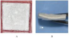
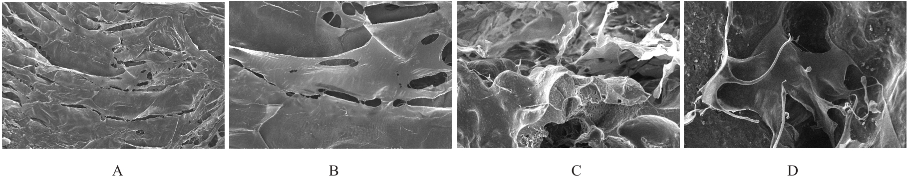
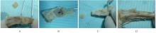
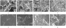
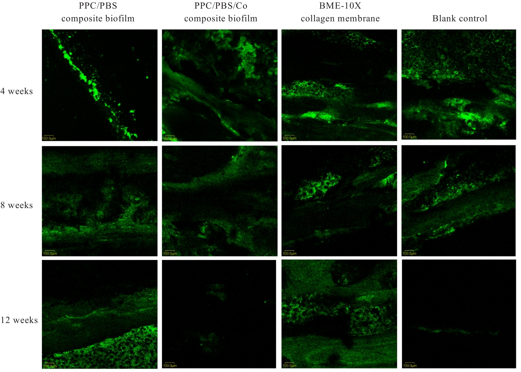
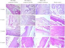
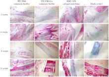
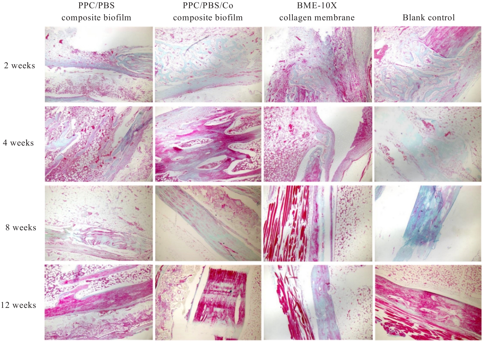
目的 探讨聚碳酸1,2-丙二酯(PPC)/聚丁二酸丁二醇酯(PBS)复合生物膜在兔胫骨骨缺损模型中的空间支撑能力及其对成骨效果的影响,阐明其屏障功能的可靠性和体内促成骨作用。 方法 制备PPC/PBS和PPC/PBS/Ⅰ型胶原(Col-Ⅰ)(PPC/PBS/Co)复合生物膜。选用18只日本大耳白兔,于兔每侧胫骨制备2处骨缺损,随机选择6只兔于骨缺损处放置PPC/PBS复合生物膜,术后4、8和12周各处死2只兔,扫描电子显微镜(SEM)观察兔骨缺损区PPC/PBS复合生物膜表面微观结构。 实验分为空白对照组、 PPC/PBS 复合生物膜组、 BME-10X 胶原膜组和PPC/PBS/Co复合生物膜组,分别行手术将上述生物膜放置于兔相应骨缺损处,空白对照组兔不放置复合生物膜,于术后2、4、8和12周时分别处死3只兔,采用软X线检测各组兔骨缺损区再生骨组织灰度值,荧光标记后采用激光共聚焦显微镜观察各组兔骨缺损区再生骨组织荧光强度,采用HE染色和改良Gomori三色染色法观察各组兔骨缺损区再生骨组织病理形态表现,免疫组织化学染色法检测各组兔骨缺损区再生骨组织中骨形态发生蛋白2(BMP-2)和骨桥蛋白(OPN)蛋白表达水平。 结果 大体观察, PPC/PBS 复合生物膜紧密覆盖于骨缺损区, 未见移位及塌陷。 SEM观察,PPC/PBS复合生物膜多孔面随时间延长表面出现微孔结构并数量增多,而光滑面基本未形成微孔样结构。软X线检测,各组兔骨缺损区再生骨组织灰度值均随时间延长而升高,12周时PPC/PBS/Co复合生物膜组兔骨缺损区再生骨组织灰度值明显高于其他各组(P<0.05)。共聚焦显微镜观察,4、8 和 12 周时 PPC/PBS/Co 复合生物膜组兔骨缺损区再生骨组织荧光强度与空白对照组相近;与PPC/PBS复合生物膜组和BME-10X胶原膜组比较,4周时PPC/PBS/Co复合生物膜组兔骨缺损区再生骨组织荧光强度升高(P<0.05),8和12 周时PPC/PBS/Co复合生物膜组兔骨缺损区再生骨组织荧光强度降低(P<0.05)。HE染色和改良Gomori染色,与PPC/PBS复合生物膜组和BME-10X胶原膜组比较,2和4周时PPC/PBS/Co复合生物膜组和空白对照组兔骨缺损区形成新骨的速度较快,在12周时骨缺损区形成的层板状骨矿化程度较高。免疫组织化学染色,2和4周时,与空白对照组、PPC/PBS复合生物膜组和BME-10X胶原膜组比较,PPC/PBS/Co复合生物膜组兔骨缺损区再生骨组织中BMP-2 和 OPN 蛋白表达水平升高(P<0.05 或 P<0.01); 与空白对照组和PPC/PBS复合生物膜组比较, BME-10X 胶原膜组兔骨缺损区再生骨组织中 BMP-2 和 OPN 蛋白表达水平均降低(P<0.05 或 P<0.01)。 结论 PPC/PBS 复合生物膜具有较好的空间支撑能力,物理屏障功能可靠,且PPC/PBS/Co复合生物膜具有良好的体内促成骨作用。

关键词: 引导骨再生, 复合生物膜, 聚碳酸1,2-丙二酯, 聚丁二酸丁二醇酯, 骨缺损

Abstract:

Objective To explore the spatial support capacity and its influence in osteogenic effect of composite biofilm based on poly(propylene carbonate) (PPC) /poly(butylene succinate) (PBS) in rabbit tibial bone defect models, and to clarify its barrier functional reliability and osteogenetic effect in vivo. Methods The composite biofilms of PPC/PBS and PPC/PBS/collegen type Ⅰ (Col-Ⅰ)(PPC/PBS/Co) were prepared.Eighteen Japanese big-ear rabbits were selected and two bone defects were prepared on each side of the tibia of the rabbits. Six rabbits were randomly selected to place PPC/PBS composite biofilm on the bone defects, 2 rabbits were executed at 2, 4, 8 and 12 weeks after operation, and the surface microstructures of PPC/PBS composite biofilm in the rabhit bone defect area were observed by scanning electron microscope (SEM). The experiment was divided into blank control group, PPC/PBS composite biofilm group, BME-10X collagen membrane group, and PPC/PBS/Co composite biofilm group. The above biofilms were placed on the corresponding bone defects of rabbits by operation, while no biofilm was placed in the rabbits in blank control group. Three rabbits were killed at 2, 4, 8 and 12 weeks after operation respectively, and the gray values of regenerated bone in the bone defect areas of the rabbits in varrous groups were detected by soft X-ray; the fluorescence intensities of regenerated bone tissue in the bone defect areas of the rabbits in various groups were observed by laser scanning confocal microscope after fluorescence labeling. The pathomorphology of regenerated bone tissue in the bone defect areas of the rabbits in various groups were observed by HE staining and modified Gomori staining, and the expression levels of bone morphogenetic protein 2 (BMP-2) and osteopontin (OPN) in the regenerated bone tissue in bone defect areas of the rabbits in various groups were detected by immunohistochemical staining. Results In general, the PPC/PBS composite biofilm was tightly covered in the bone defect area without displacement and collapse. The SEM results showed that the porous surface of PPC/PBS composite biofilm appeared micropore structure and the number of micropores was increased with the prolongation of time, while the smooth surface of biofilm basically did not form the micropore-like structure. The results of soft X-ray detection showed that the gray values of regenerated bone tissue in bone defect areas of the rabbits in various groups were increased with the prolongation of time, and the gray value of regenerated bone tissue in bone defect areas of the rabbits in PPC/PBS/Co composite biofilm group was significantly higher than those in other groups (P<0.05). The confocal micrscope results showed that the fluorescence intensity of regenerated bone tissue in bone defect areas of the rabbits in PPC/PBS/Co composite biofilm group was similar to those in blank control group at 4, 8, and 12 weeks; compared with PPC/PBS composite biofilm group and BME-10X collagen membrane group, the fluoresence intensity of regenerated bone tissue in bone defect areas of the rabbits in PPC/PB/Co composite biofilm group at 4 weeks was increased (P<0.05), and the fluoresence intensity of regenerated bone tissue in bone defect areas of the rabbits at 8 and 12 weeks were decreased (P<0.05). The results of HE staining and modified Gomori staining showed that compared with PPC/PBS composite biofilm group and BME-10X collagen membrane group, the new bone formed faster in PPC/PBS/Co composite biofilm group and blank control group at 2 and 4 weeks, and the lamellar bone mineralization was higher at 12 weeks. The immunohistochemical staining results showed that compared with blank control group, PPC/PBS composite biofilm group and BME-10X collagen membrane group, the expression levels of BMP-2 and OPN proteins in the regenerated bone tissue in bone defect areas of the rabbits in PPC/PBS/Co composite biofilm group at 2 and 4 weeks were increased (P<0.05 or P<0.01); compared with blank control group and PPC/PBS composite biofilm group, the expression levels of BMP-2 and OPN proteins in the regenerated bone tissue in bone defect areas of the rabbits in BME-10X collage membrane group were decreased (P<0.05 or P<0.01). Conclusion PPC/PBS composite biofilm has excellent spatial support capacity and reliable physical barrier function. The PPC/PBS/Co composite biofilm has a good effect in guiding bone regeneration in vivo.

Key words: Guided bone regeneration, Composite biofilm, Poly(propylene carbonate), Poly(butylene succinate), Bone defect

中图分类号: 

  • R783.1