,拔牙,骨性Ⅱ类高角,错??,上气道,舌骨位置," /> ,拔牙,骨性Ⅱ类高角,错??,上气道,舌骨位置,"/> 拔牙矫治对成年骨性Ⅱ类高角错𬌗患者上气道及周围组织的影响

吉林大学学报(医学版) ›› 2024, Vol. 50 ›› Issue (4): 1055-1061.doi: 10.13481/j.1671-587X.20240420

• 临床研究 • 上一篇    下一篇

拔牙矫治对成年骨性Ⅱ类高角错𬌗患者上气道及周围组织的影响

陈玉1,2,姜欢1,胡敏1()   

  1. 1.吉林大学口腔医院正畸科,吉林 长春 130021
    2.海军军医大学附属第一医院口腔科,上海 200433
  • 收稿日期:2023-10-01 出版日期:2024-07-28 发布日期:2024-08-01
  • 通讯作者: 胡敏 E-mail:humin@jlu.edu.cn
  • 作者简介:陈 玉(1993-),女,江苏省扬州市人,住院医师,医学硕士,主要从事口腔正畸临床方面的研究。
  • 基金资助:
    国家自然科学基金项目(81870795)

Effect of extraction orthodontic treatment on upper airway and surrounding tissue in adult patients with skeletal class Ⅱhigh angle malocclusion

Yu CHEN1,2,Huan JIANG1,Min HU1()   

  1. 1.Department of Orthodontics,Stomatology Hospital,Jilin University,Changchun 130021,China
    2.Department of Stomatology,First Affiliated Hospital,Naval Medical University,Shanghai 200433,China
  • Received:2023-10-01 Online:2024-07-28 Published:2024-08-01
  • Contact: Min HU E-mail:humin@jlu.edu.cn

摘要:

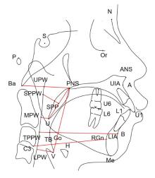
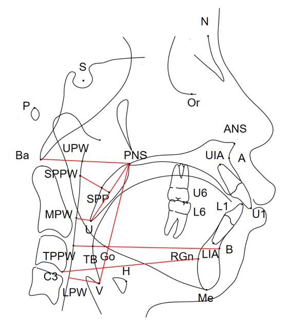
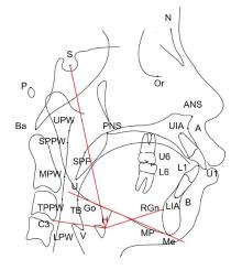
目的 通过对比骨性Ⅱ类高角错𬌗成年患者拔牙和不拔牙矫治前后上气道形态、舌骨位置和颅颌面结构的变化,分析拔牙矫治对该类患者上气道形态结构的影响,为其临床治疗方案的选择提供理论依据。 方法 收集就诊于吉林大学口腔医院正畸科已完成正畸治疗的骨性Ⅱ类高角错𬌗成年患者 60例,根据是否进行减数矫治分为拔牙组和非拔牙组,每组30例,对2组患者的临床资料进行回顾性分析。获取患者治疗前后的头颅定位侧位片,应用Dolphin软件对患者上气道形态、舌骨位置和颅颌面组织结构进行定点描绘和测量,采用SPSS 23.0统计软件对相关测量数据进行统计学分析。 结果 与矫治前比较,矫治后拔牙组患者悬雍垂尖与中咽壁点的距离(U-MPW)、会厌谷点与下咽壁点的距离(V-LPW)、上中切牙长轴与前颅底平面的下内交角(U1-SN)和下中切牙长轴与下颌平面的上内交角(L1-MP)明显减小(P<0.05),𬌗平面与前颅底平面的夹角(OP-SN)和上下中切牙长轴之间的夹角(U1-L1)明显增大,(P<0.05),其余测量指标差异无统计学意义(P>0.05);与矫治前比较,矫治后非拔牙组患者矫治后上牙槽座点-鼻根点-下牙槽座点角(ANB)明显减小(P<0.05),OP-SN和L1-MP明显增大(P<0.05),其余测量指标差异无统计学意义(P>0.05)。 结论 成年骨性Ⅱ类高角错𬌗患者在拔牙矫治后上气道矢状径变窄,主要发生在上气道口咽段和喉咽段,而舌骨位置未发生明显改变。

关键词: ')">, 拔牙, 骨性Ⅱ类高角, ??')">错??, 上气道, 舌骨位置

Abstract:

Objective To compare the changes of the upper airway morphology, hyoid bone position, and craniofacial structure before and after extraction treatment and non-extraction treatment in the adult patients with skeletal class Ⅱ high angle malocclusion,and to analyze the effect of extraction orthodontic treatment on the upper airway structure in these patients,and to provide the theoretical basis for the selection of clinical treatment plans. Methods A retrospective analysis on the clinical data of 60 adult patients with skeletal class Ⅱ high angle malocclusion who required orthodontic treatment was collected from the Orthodontics Department of Stomatology Hospital of Jilin University. The patients were divided into extraction group and non-extraction group according to whether treated with extraction orthodontic treatment, and there were 30 patients in each group.The lateral cephalometric radiographs of the patients before and after orthodontic treatment were obtained, and the upper airway, hyoid bone, and craniofacial tissues were delineated and detected by Dolphin software. SPSS 23.0 statistical software was used to perform the statistical analysis on the related measurement data. Results Compared with before orthodontic treatment, the distance between the uvula tip and midpharyngeal wall (U-MPW), aryngeal wall (V-LPW), the inner downward angle between the long axis of the upper central incisor and the anterior cranial base (U1-SN) as well as the inner upward angle between the lower central incisor long axis and mandibular plane (L1-MP)of the patients in extraction group with skeletal class Ⅱ high angle malocclusion after orthodontic treatment were significantly decreased(P<0.05),while the occlusal plane and the anterior cranial base (OP-SN) and the angle between the long axes of the upper and lower central incisors (U1-L1) were significantly increased (P<0.05).There were no significant differences in the other measurement indicators of the patients between two groups (P>0.05).Compared with before orthodontic treatment, the angle of posterior nasal spine (ANB) of the patients in non-extraction group with skeletal class Ⅱ high angle malocclusion after orthodontic treatment was significantly decreased (P<0.05), and the OP-SN and L1-MP significantly were increased (P<0.05). There were no significant differences in the other measurement indicators of the patients between two groups (P>0.05). Conclusion The sagittal diameter of upper airway of the adult patients with skeletal class Ⅱ high angle malocclusion tended to be orthodontic treatment after extraction orthodontic treatment,mainly in the oropharynx and laryngopharynx of the upper airway, while the hyoid bone position did not change significantly.

Key words: Extraction, Skeletal classⅡhigh angle, Malocclusion, Upper airway, Hyoid bone position

中图分类号: 

  • R783.5