吉林大学学报(医学版) ›› 2024, Vol. 50 ›› Issue (6): 1597-1605.doi: 10.13481/j.1671-587X.20240613

• 基础研究 • 上一篇    

罗伊氏乳杆菌对轮状病毒体内外复制的抑制作用及其对免疫因子表达的影响

李小凤1,程美慧2,刘洋1,刘长城1,贾雪娇1,刘梦琦1,赵微1()   

  1. 1.锦州医科大学基础医学院病原生物学实验室,辽宁 锦州 121000
    2.湖北省黄石市中心医院检验科,湖北 黄石 435000
  • 收稿日期:2023-12-21 出版日期:2024-11-28 发布日期:2024-12-10
  • 通讯作者: 赵微 E-mail:zhaowei-v@jzmu.edu.cn
  • 作者简介:李小凤(2000-),女,湖南省郴州市人,在读硕士研究生,主要从事病毒学基础方面的研究。
  • 基金资助:
    辽宁省教育厅基金项目(LJKMZ20221243);辽宁省科技厅科技计划联合计划基金项目(2023-MSLH-046)

Inhibitory effect of Lactobacillus reuteri on rotavirus replication in vivo and in vitro and its effect on expression of immune factors

Xiaofeng LI1,Meihui Cheng2,Yang LIU1,Changcheng LIU1,Xuejiao JIA1,Mengqi LIU1,Wei ZHAO1()   

  1. 1.Laboratory of Pathogenic Biology,School of Basic Medical Sciences,Jinzhou Medical University,Jinzhou 121000,China
    2.Department of Laboratory,Central Hospital,Huangshi City,Hubei Province,Huangshi 435000,China
  • Received:2023-12-21 Online:2024-11-28 Published:2024-12-10
  • Contact: Wei ZHAO E-mail:zhaowei-v@jzmu.edu.cn

摘要:

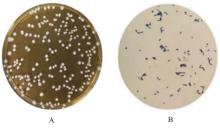
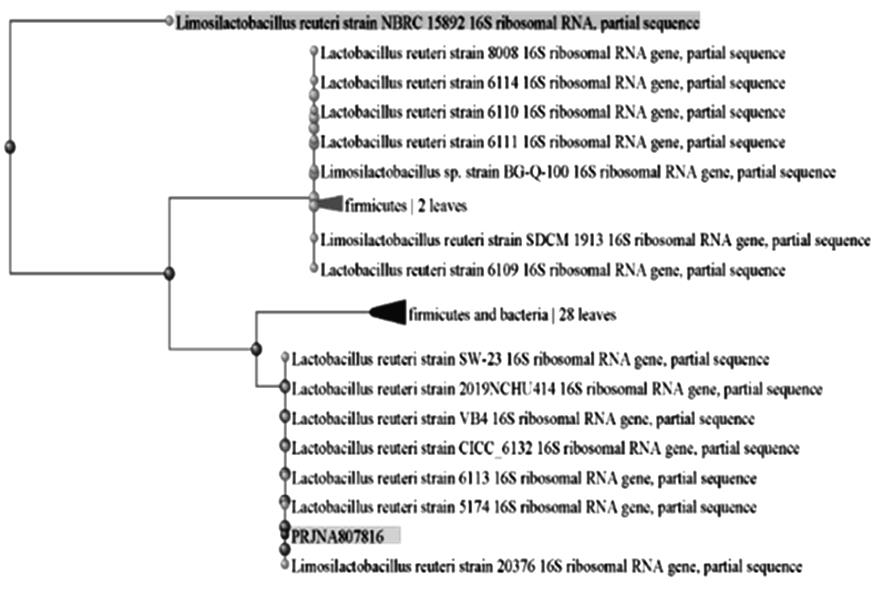
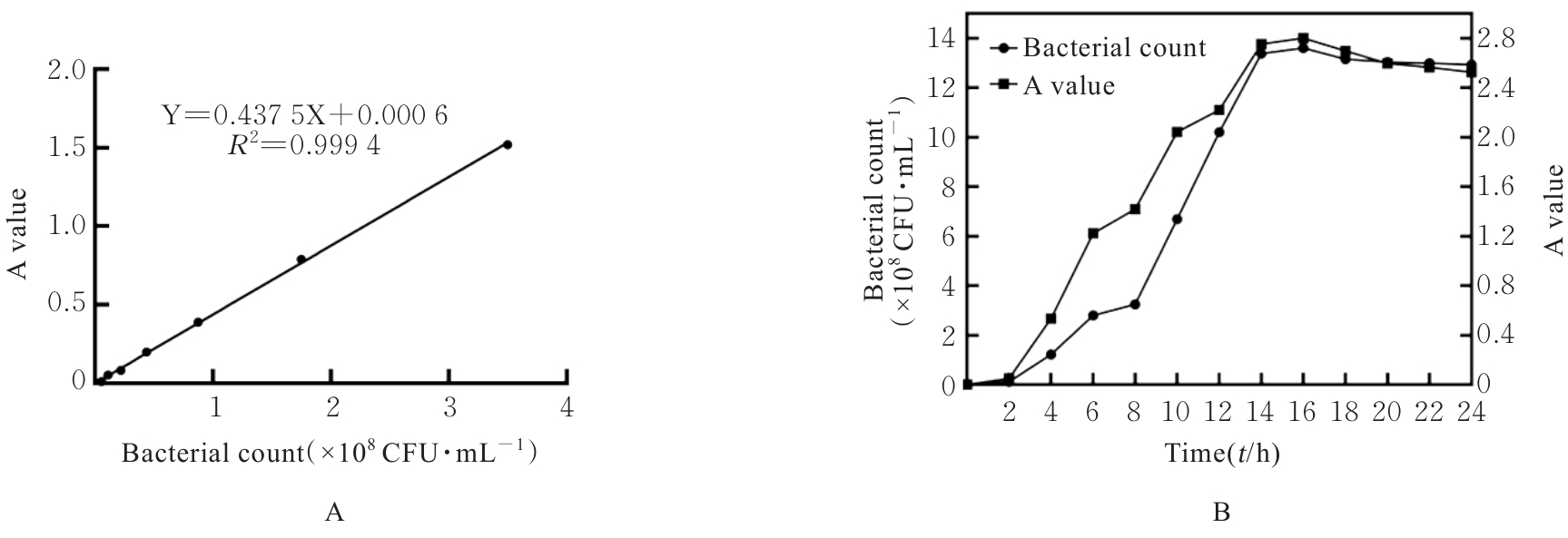
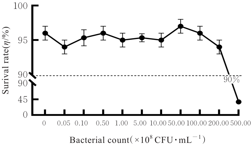
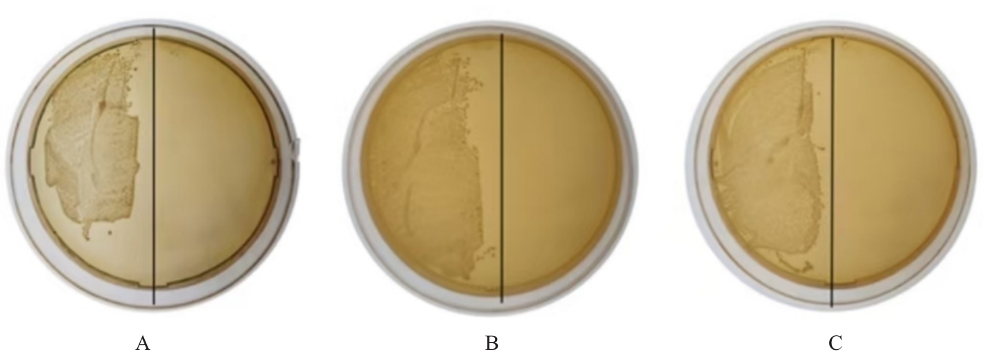
目的 探讨罗伊氏乳杆菌对轮状病毒(RV)SA11株体内外复制的抑制作用,并阐明其对相关免疫因子表达的影响。 方法 体外实验,培养并鉴定罗伊氏乳杆菌,绘制罗伊氏乳杆菌标准曲线和生长曲线,筛选罗伊氏乳杆菌培养最佳时间和最适浓度。采用5×10⁸、10×108、50×108、100×108、200×108和500×108 CFU·mL-1罗伊氏乳杆菌感染细胞,台盼蓝染色法检测Caco-2细胞存活率。将不同浓度罗伊氏乳杆菌与RV体外共孵育并作用于Caco-2细胞,Caco-2细胞分为阴性对照组(NC组)、阳性对照组(PC组)和107、108、109及1010 CFU·mL-1 罗伊氏乳杆菌组,免疫荧光灶法检测罗伊氏乳杆菌作用后Caco-2细胞中病毒滴度,实时荧光定量PCR(RT-qPCR)法检测不同浓度罗伊氏乳杆菌作用后Caco-2细胞中RV VP6基因拷贝数。体内实验,将25窝SPF级乳鼠分为对照组、RV组(感染SA11毒株)、Ab-NC组(抗生素处理耗竭肠道菌群)、Ab-RV组(耗竭肠道菌群后感染SA11毒株)和Ab-Lac-RV组(耗竭肠道菌群,并感染罗伊氏乳杆菌后感染SA11毒株)。收取各组乳鼠灌胃第2、4、6、8和10天的粪便样本和灌胃第4天结肠组织样本,RT-qPCR法检测各组乳鼠粪便中RV VP6基因拷贝数和结肠组织中白细胞介素(IL)-1β、IL-8、IL-10、γ干扰素(IFN-γ)和肿瘤坏死因子(TNF)-α mRNA表达水平。 结果 罗伊氏乳杆菌生长良好,形态圆润,呈圆形、光滑和乳白色的凸起样菌落,且边缘较整齐;革兰染色后菌体呈紫色、不规则和方形杆状;经16SrDNA测序后序列同源性为99%,提示罗伊氏乳杆菌活化成功;罗伊氏乳杆菌活菌数与吸光度(A)值呈线性关系,回归分析标准曲线为Y=0.437 5X+0.000 6,R2=0.999 4。培养0~2 h,细菌处于对数生长期,细菌生长迟缓;培养2~14 h,细菌快速生长,并于培养14~16 h时细菌生长趋于稳定,培养16 h时达到细菌生长速率顶峰,随后进入衰亡期。5×10⁸、10×108、50×108、100×108和200×108 CFU·mL-1罗氏乳杆菌感染后,Caco-2细胞存活率均>90%,因此选用上述浓度罗氏乳杆菌感染细胞。与PC组比较,5×108、10×108、50×108、100×108和200×108 CFU·mL-1罗氏乳杆菌组Caco-2细胞中RV VP6基因拷贝数均明显降低 (P<0.01)。与PC组比较,107、108、109和1010 CFU·mL-1罗伊氏乳杆菌组Caco-2细胞中病毒滴度均明显降低(P<0.01)。与对照组比较,Ab-NC组、Ab-RV组和Ab-Lac-RV组乳鼠肠道菌群菌落数量均明显减少,乳鼠肠道菌群耗竭成功。灌胃第2和4天,与RV组比较,Ab-RV组乳鼠粪便中RV VP6基因拷贝数明显降低(P<0.05);灌胃第4、6、8和10天,与Ab-RV组比较,Ab-Lac-RV组乳鼠粪便中RV VP6基因拷贝数明显降低(P<0.05)。与对照组比较,Ab-RV组和Ab-Lac-RV组乳鼠结肠组织中IL-1β、IL-10、IFN-γ及TNF-α mRNA表达水平均明显升高(P<0.05或P<0.01),IL-8 mRNA表达水平均明显降低(P<0.05),Ab-Lac-RV组乳鼠结肠组织中IL-10 mRNA表达水平明显升高(P<0.01)。 结论 罗伊氏乳杆菌可能通过上调IL-1β、IL-10、IFN-γ和TNF-α mRNA表达及下调IL-8 mRNA表达,抑制RV复制。

关键词: 罗伊氏乳杆菌, 轮状病毒, 乳鼠, 免疫因子, 标准曲线

Abstract:

Objectives To discuss the inhibitory effect of Lactobacillus reuteri on the replication of rotavirus (RV) strain SA11 in vivo and in vitro, and to clarify its effect on the expression of related immune factors. Methods For in vitro experiments, Lactobacillus reuteri was cultured and identified, and the standard curve and growth curve were plotted to screen the optimal time and concentration for Lactobacillus reuteri cultivation. The cells were infected with Lactobacillus reuteri at the concentrations of 5×10⁸, 10×108, 50×108, 100×108, 200×108, and 500×108 CFU·mL-1, and the surival rates of Caco-2 cells were detected by trypan blue staining method. Various concentrations of Lactobacillus reuteri were co-incubated with RV in vitro and applied to the Caco-2 cells. The cells were divided into negative control group (NC group), positive control group (PC group), and 107, 108, 109, and 1010 CFU·mL-1Lactobacillus reuteri groups. Immunofluorescence focus method was used to detect the viral titers in the Caco-2 cells after treated with Lactobacillus reuteri and real-time fluorescence quantitative PCR(RT-qPCR) method was used to detect the copy numbers of RV VP6 gene in the Caco-2 cells after treated with various concentrations of Lactobacillus reuteri. In in vivo experiments, 25 litters of SPF suckling mice were divided into control group, RV group (infected with SA11 strain), Ab-NC group (treated with antibiotic to deplete gut microbiota), Ab-RV group (depleting gut microbiota and then infected with SA11 strain), and Ab-Lac-RV group (depleting gut microbiota, treated with Lactobacillus reuteri, and then infected with SA11 strain). The fecal samples were collected on days 2, 4, 6, 8, and 10 gavage, colon tissue sample were collected on day 4 of and RT-qPCR method was used to detect the copy numbers of RV VP6 gene in feces and the mRNA expression levels of interleukin (IL)-1β, IL-8, IL-10, interferon-γ (IFN-γ), and tumor necrosis factor-α (TNF-α) in colon tissue of the suckling mice in vartious groups. Results The Lactobacillus reuteri grew well, with round, smooth, and milky white convex colonies and neat edges. After Gram staining, the bacteria appeared purple, irregular, and square-shaped rods. 16SrDNA sequencing showed 99% sequence homology, indicating successful activation of Lactobacillus reuteri. The number of live Lactobacillus reuteri was linearly related to the absorbance (A) value, and the standard curve for regression analysis was Y=0.437 5X+0.000 6, R2=0.999 4. During the 0-2 h cultivation period, the bacteria were at the logarithmic growth phase with slow growth; from 2-14 h, the bacteria grew rapidly and stabilized at 14-16 h, reaching the growth rate peak at 16 h, after which they entered the decline phase. Infection with Lactobacillus reuteri at concentrations of 5×10⁸, 10×108, 50×108, 100×108, and 200×108 CFU·mL-1 resulted in the survival rates of Caco-2 cells were all >90%, so these concentrations were selected for the further experiments. Compared with PC group, the copy numbers of RV VP6 gene in the Caco-2 cells in 5×10⁸, 10×108, 50×108, 100×108, and 200×108 CFU·mL-1Lactobacillus reuteri groups were significantly decreased (P<0.01). Compared with PC group, the viral titers in the Caco-2 cells in 107, 108, 109, and 1010 CFU·mL-1Lactobacillus reuteri groups were significantly decreased (P<0.01). Compared with control group, the numbers of gut microbiota colonies in Ab-NC, Ab-RV, and Ab-Lac-RV groups were significantly decreased, indicating successful depletion of gut microbiota in the suckling mice. On days 2 and 4 after gavage, the RV VP6 gene copy number in the feces in Ab-RV group was significantly lower than that in RV group (P<0.05). On days 4, 6, 8, and 10 after gavage, the RV VP6 gene copy number in the feces in Ab-Lac-RV group was significantly lower than that in Ab-RV group (P<0.05). Compared with control group, the expression levels of IL-1β, IL-10, IFN-γ, and TNF-α mRNA in colon tissue in Ab-RV and Ab-Lac-RV groups were significantly increased (P<0.05 or P<0.01), while the expression level of IL-8 mRNA was significantly decreased (P<0.05),and the expression level of IL-10 mRNA in colon tissue in Ab-LAC-RV group was significantly increased (P<0.01). Conclusion Lactobacillus reuteri may inhibit the RV replication by upregulating the expressions of IL-1β, IL-10, IFN-γ, and TNF-α mRNA and downregulating the expression of IL-8 mRNA.

Key words: Lactobacillus reuteri, Rotavirus, Suckling mice, Immune factor, Standard curve

中图分类号: 

  • R373.2