吉林大学学报(医学版) ›› 2024, Vol. 50 ›› Issue (6): 1587-1596.doi: 10.13481/j.1671-587X.20240612

• 基础研究 • 上一篇    

趋化因子CCL19诱导巨噬细胞M1极化对小鼠慢性胰腺炎的促进作用及其机制

崔连鸷1,张晓伟1,翟悦1,潘悦2,于秀艳1()   

  1. 1.吉林省肿瘤医院检验科,吉林 长春 130012
    2.大连理工大学医学部药学系,辽宁 大连 116024
  • 收稿日期:2024-02-22 出版日期:2024-11-28 发布日期:2024-12-10
  • 通讯作者: 于秀艳 E-mail:2360984054@qq.com
  • 作者简介:崔连鸷(1982-),女,吉林省长春市人,副主任技师,医学博士,主要从事临床免疫学和分子生物学方面的研究。
  • 基金资助:
    吉林省科技厅自然科学基金项目(YDZJ202401123ZYTS)

Promotion effect of chemokine CCL19-induced macrophage M1 polarization on chronic pancreatitis in mice and its mechanism

Lianzhi CUI1,Xiaowei ZHANG1,Hua ZHU1,Yue PAN2,Xiuyan YU1()   

  1. 1.Clinical Laboratory,Tumer Hospital,Jilin Province,Changchun 130012,China
    2.Department of Pharmacy,School of Medical Science,Dalian University of Technology,Dalian 116024,China
  • Received:2024-02-22 Online:2024-11-28 Published:2024-12-10
  • Contact: Xiuyan YU E-mail:2360984054@qq.com

摘要:

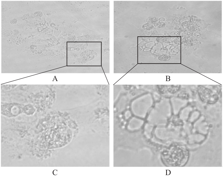
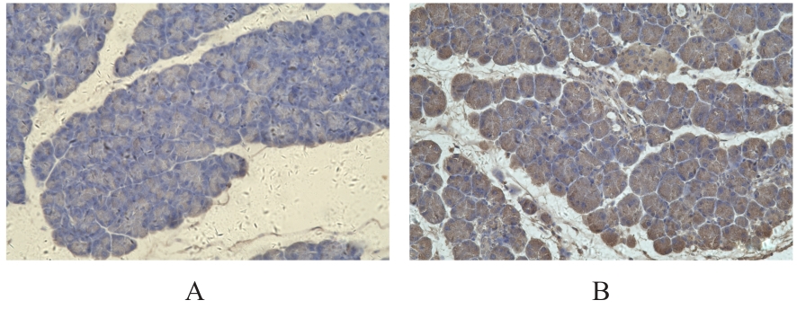
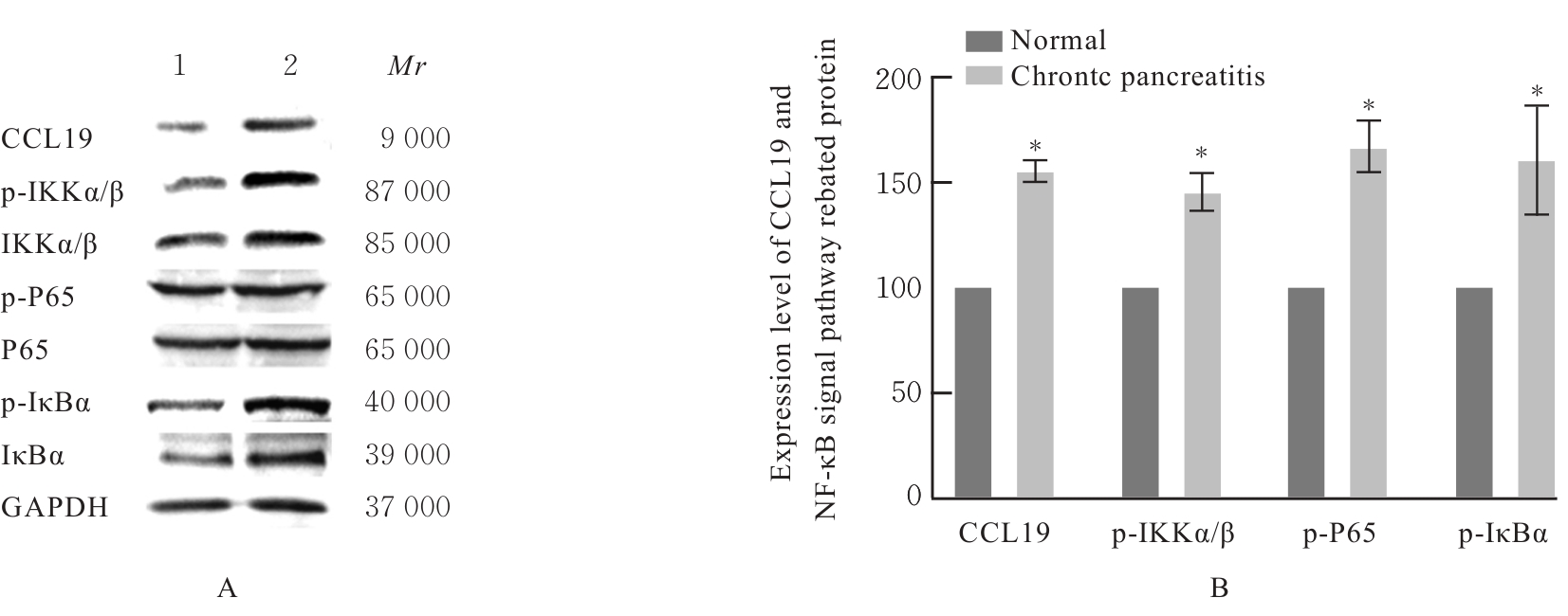
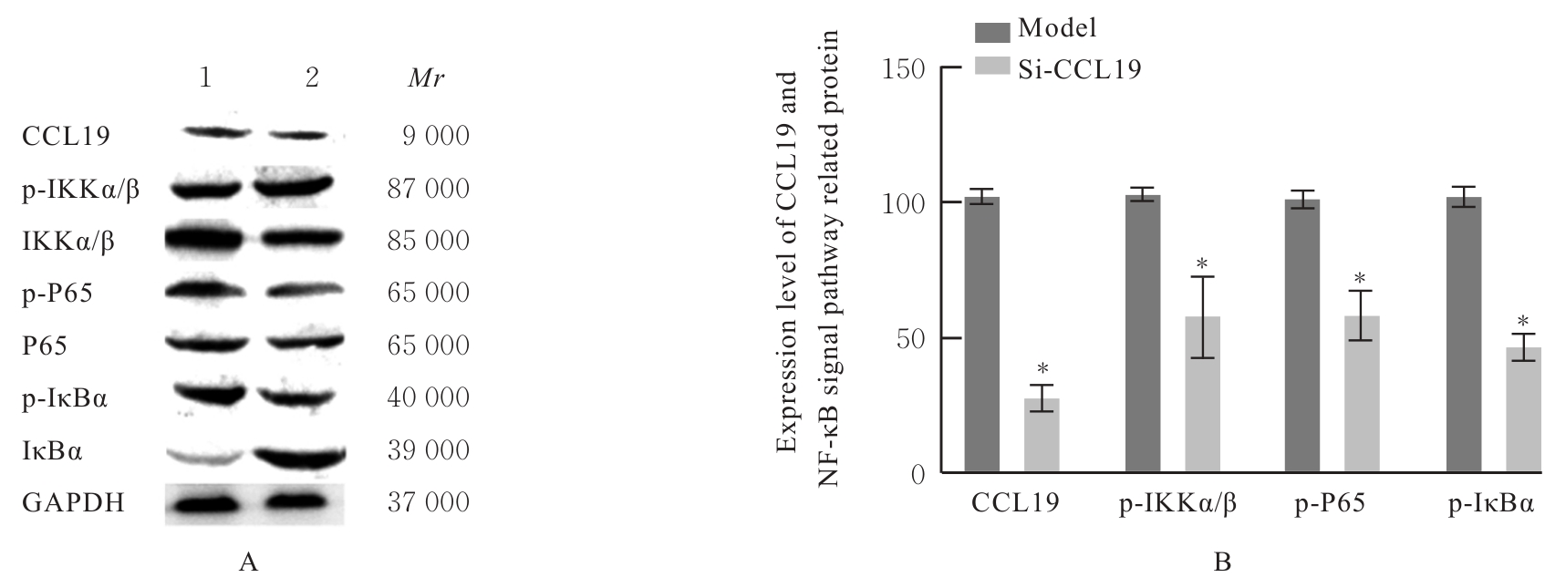
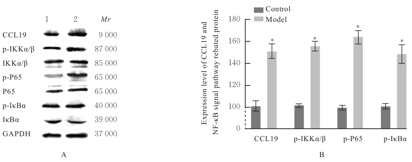
目的 探讨趋化因子C-C基序配体19(CCL19)诱导巨噬细胞M1极化对小鼠慢性胰腺炎的促进作用,并阐明其相关机制。 方法 选取10只雄性C57BL/6N小鼠,提取小鼠胰腺腺泡细胞和腹腔巨噬细胞,构建巨噬细胞-腺泡细胞共培养体系,共培养体系细胞分为对照组、模型组和小干扰RNA CCL19(si-CCL19)组,显微镜下观察各组腺泡细胞形态表现。随机选取40只小鼠,分为正常组和慢性胰腺炎组,每组20只。HE染色观察2组小鼠胰腺组织病理形态表现,免疫荧光染色法观察2组小鼠胰腺组织中角质蛋白19(CK19)、淀粉酶、M1型巨噬细胞相关标志物诱导型一氧化氮合酶(iNOS)和F4/80表达情况及各组共培养体系细胞中腺泡细胞形态表现及CK19和淀粉酶表达情况,酶联免疫吸附试验(ELISA)法检测2组小鼠血清和各组共培养体系细胞中肿瘤坏死因子α(TNF-α)、白细胞介素(IL)-6和IL-1β水平,免疫组织化学法观察2组小鼠胰腺组织中CCL19蛋白表达情况,Western blotting法检测2组小鼠胰腺组织和各组共培养体系细胞中CCL19蛋白和关键蛋白核因子κB(NF-κB)信号通路相关蛋白P65、磷酸化P65(p-P65)、κB抑制物激酶α/β(IKKα/β)、磷酸化IKKα/β(p-IKKα/β)、IκBα和磷酸化IκBα(p-IκBα)表达水平。 结果 HE染色,正常组小鼠胰腺组织腺泡细胞的紧密排列;与正常组比较,慢性胰腺炎组小鼠胰腺组织腺泡细胞产生了明显的空泡化,即腺泡细胞导管化,小鼠胰腺炎模型制备成功。免疫荧光染色法,与对照组比较,模型组腺泡细胞严重的空泡化明显,CK19表达明显增加,淀粉酶表达明显减少;与模型组比较,si-CCL19组中腺泡细胞导管化程度降低,CK19表达明显减少,淀粉酶表达明显增加;与正常组比较,慢性胰腺炎组小鼠胰腺组织中淀粉酶表达明显减少,CK19和M1型巨噬细胞标志物iNOS及F4/80表达均明显增加。ELISA法,与正常组比较,慢性胰腺炎组小鼠血清中TNF-α、IL-6和IL-1β水平均明显升高(P<0.05);与对照组比较,模型组细胞中TNF-α、IL-6和IL-1β水平均明显升高(P<0.05);与模型组比较,si-CCL19组细胞中TNF-α、IL-6和IL-1β水平均明显降低(P<0.05)。免疫组织化学法,与正常组比较,慢性胰腺炎组小鼠胰腺组织中CCL19蛋白表达明显增加。Western blotting法,与正常组比较,慢性胰腺炎组小鼠胰腺组织中CCL19蛋白表达水平和NF-κB信号通路相关蛋白p-IKKα/β、p-P65及p-IκBα蛋白表达水平均明显升高(P<0.05)。与对照组比较,模型组细胞中CCL19、p-IKKα/β、p-P65和p-IκBα蛋白表达水平均明显升高(P<0.05);与模型组比较,si-CCL19组细胞中CCL19、p-IKKα/β、p-P65和p-IκBα蛋白表达水平均明显降低(P<0.05)。 结论 CCL19通过NF-κB信号通路促进巨噬细胞M1型极化,诱导炎症微环境的产生,促进胰腺炎的发生发展。

关键词: 胰腺炎, C-C基序配体19, 巨噬细胞, M1型极化, 核因子κB

Abstract:

Objective To discuss the promotion effect of chemokine C-C motif ligand 19 (CCL19) induced macrophage M1 polarization on chronic pancreatitis of the mice, and to clarify its related mechanism. Methods Ten male C57BL/6N mice were selected, and the pancreatic acinar cells and peritoneal macrophages were extracted from these mice to construct the macrophage-acinar cell co-culture system. The co-culture system cells were divided into control group, model group, and small interfering RNA CCL19 (si-CCL19) group. The morphology of the acinar cells in various groups were observed under microscope. Forty mice were randomly selected and divided into normal group and chronic pancreatitis group, and there were 20 mice in each group. HE staining was used to observe the pathomorphology of pancreatic tissue of the mice in two groups; immunofluorescence staining was used to observe the expressions of cytokeratin 19 (CK19), amylase, M1 macrophage-related markers inducible nitric oxide synthase (iNOS), and F4/80 in pancreatic tissue of the mice in two groups and morphology of follicular cells and the expressions of CK19, amylase in the co-culture system cells in various groups; enzyme-linked immunosorbent assay (ELISA) was used to detect the levels of tumor necrosis factor-α (TNF-α), interleukin (IL)-6, and IL-1β in serum of the mice in two groups and in the co-culture system cells in various groups; immunohistochemistry was used to observe the expression of CCL19 protein in pancreatic tissue of the mice in two groups; Western blotting method was used to detect the expression levels of CCL19 protein and two nuclear factor-κB (NF-κB) signaling pathway-related proteins P65, phosphorylate P65 (p-P65), kappa B inhibitor of kinase α/β(IKKα/β), phosphorylated IKKα/β (p-IKKα/β), IkBα, phosphorylated IκBα(p-IκBα) in pancreatic tissue of the mice in two groups and in the co-culture system cells in various groups. Results The HE staining results showed that the acinar cells in pancreatic tissue of the mice in normal group were tightly arranged; compared with normal group, the acinar cells of the mice in chronic pancreatitis group showed obvious vacuolation and acinar cell ductal metaplasia, indicating successful preparation of the mouse pancreatitis model. The immunofluorescence staining results showed that compared with control group, the acinar cells in model group exhibited severe vacuolation, the CK19 expression was significantly increased, and the amylase expression was significantly decreased; compared with model group, the acinar cell ductal metaplasia in si-CCL19 group was decreased, the CK19 expression was significantly decreased, and the amylase expression was significantly increased; compared with normal group, the expression of amylase in pancreatic tissue of the mice in chronic pancreatitis group was significantly decreased, while the expressions of CK19 and M1 macrophage markers iNOS and F4/80 were significantly increased. The ELISA results showed that compared with normal group, the serum levels of TNF-α, IL-6, and IL-1β of the mice in chronic pancreatitis group were significantly increased (P<0.05); compared with control group, the levels of TNF-α, IL-6, and IL-1β in the cells in model group were significantly increased (P<0.05); compared with model group, the levels of TNF-α, IL-6, and IL-1β in the cells in si-CCL19 group were significantly decreased (P<0.05). The immunohistochemistry results showed that compared with normal group, the expression of CCL19 protein in pancreatic tissue of the mice in chronic pancreatitis group was significantly increased. The Western blotting results showed that compared with normal group, the expression levels of CCL19 protein and NF-κB signaling pathway-related proteins p-P65, p-IKKα/β, and p-IκBα in pancreatic tissue of the mice in chronic pancreatitis group were significantly increased(P<0.05); compared with control group, the expression levels of CCL19, p-IKKα/β, p-P65, and p-IκBα proteins in the cells in model group were significantly increased (P<0.05); compared with model group, the expression levels of CCL19, p-IKKα/β, p-P65, and p-IκBα proteins in the cells in si-CCL19 group were decreased (P<0.05). Conclusion CCL19 promotes the macrophage M1 polarization through the NF-κB signaling pathway, induces the formation of inflammatory microenvironment, and promotes the occurrence and development of pancreatitis.

Key words: Pancreatitis, C-C motif ligand 19, Macrophages, M1 polarization, Nuclear factor-κB

中图分类号: 

  • R364.5