吉林大学学报(医学版) ›› 2025, Vol. 51 ›› Issue (3): 716-726.doi: 10.13481/j.1671-587X.20250316

• 临床研究 • 上一篇    

缝隙连接蛋白β2对肺腺癌患者预后及肺腺癌A549细胞生物学行为的影响

王繁1,温馨2,3,王艺璇2,王远2,3()   

  1. 1.锦州医科大学基础医学院生物人类学研究所,辽宁 锦州 121001
    2.锦州医科大学基础医学院病理学教研室,辽宁 锦州 121001
    3.锦州医科大学附属第一医院病理科,辽宁 锦州 121001
  • 收稿日期:2024-07-18 接受日期:2024-09-29 出版日期:2025-05-28 发布日期:2025-07-18
  • 通讯作者: 王远 E-mail:wyxf880720@163.com
  • 作者简介:王 繁(1997-),女,陕西省榆林市人,助理实验师,医学硕士,主要从事肺癌及相关分子机制方面的研究。
  • 基金资助:
    国家自然科学基金项目(82103624)

Effect of gap junction β2 on prognosis of patients with lung adenocarcinoma and biological behavior of lung adenocarcinoma A549 cells

Fan WANG1,Xin WEN2,3,Yixuan WANG2,Yuan WANG2,3()   

  1. 1.Institute of Biological Anthropology,School of Basic Medical Sciences,Jinzhou Medical University,Jinzhou 121001,China
    2.Department of Pathology,School of Basic Medical Sciences,Jinzhou Medical University,Jinzhou 121001,China
    3.Department of Pathology,First Affiliated Hospital,Jinzhou Medical University,Jinzhou 121001,China
  • Received:2024-07-18 Accepted:2024-09-29 Online:2025-05-28 Published:2025-07-18
  • Contact: Yuan WANG E-mail:wyxf880720@163.com

摘要:

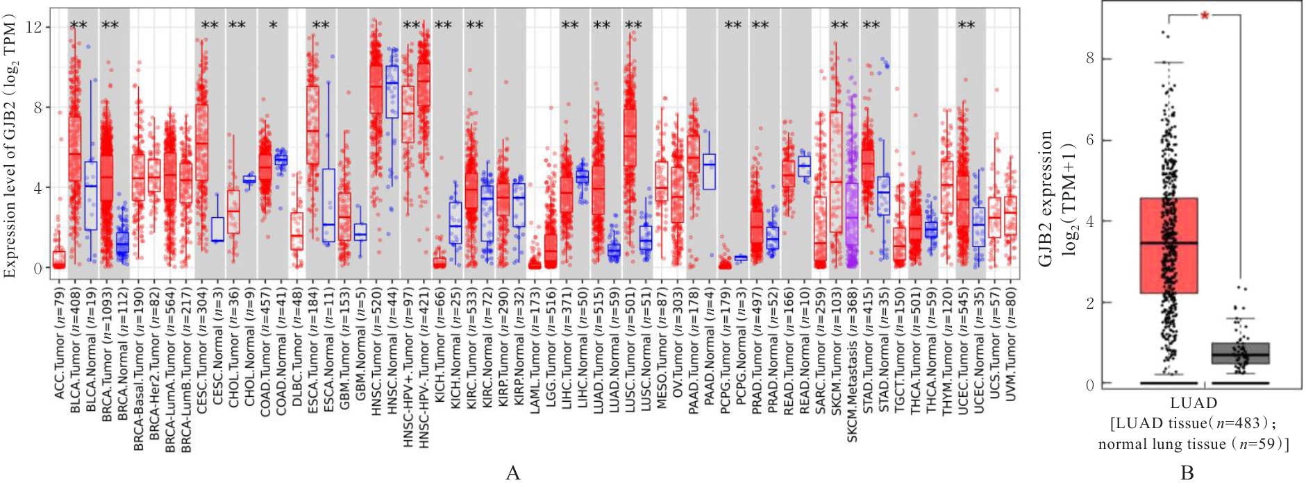
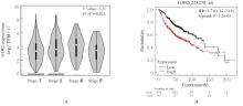
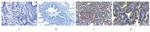
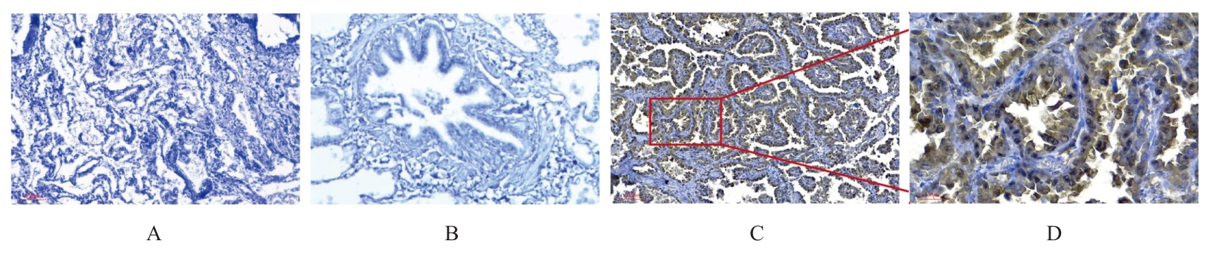
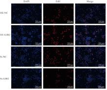
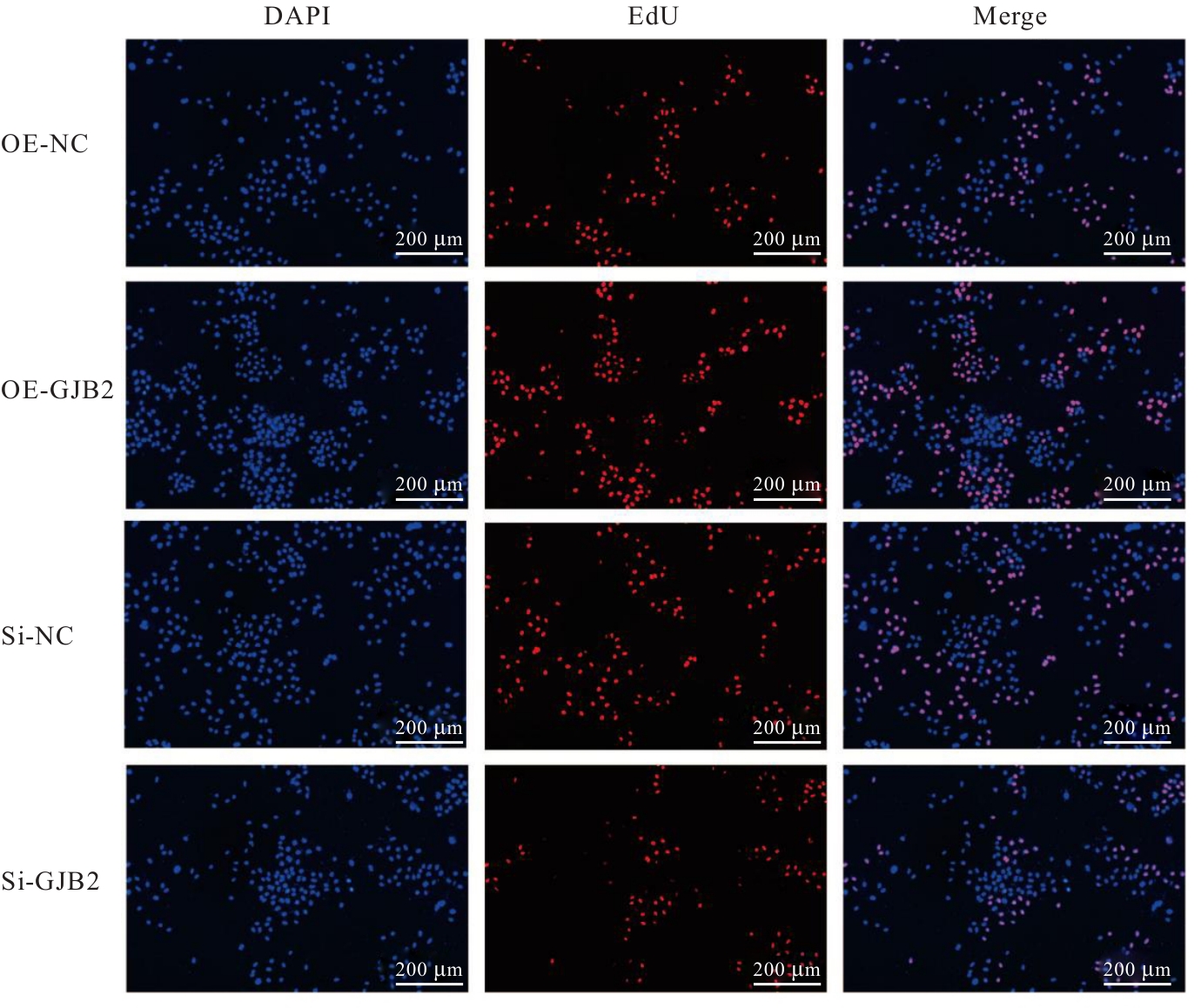
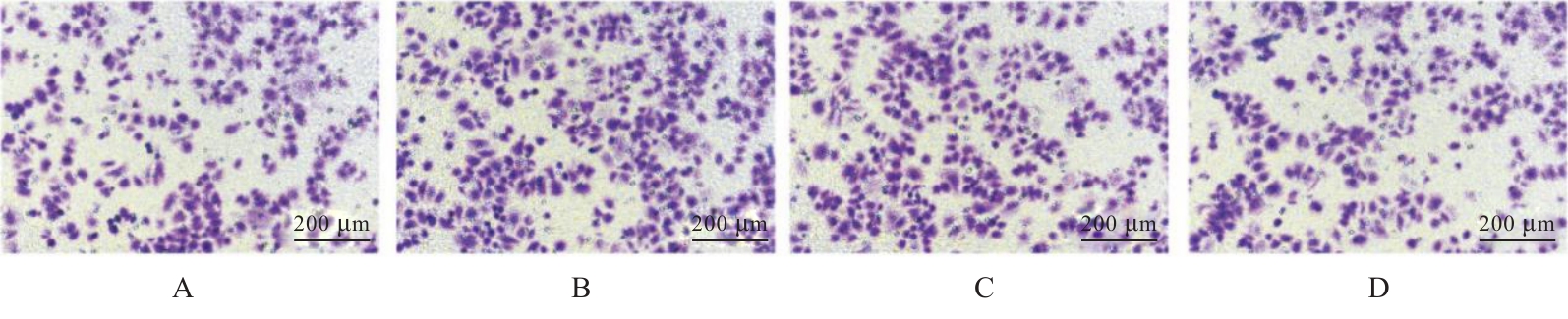
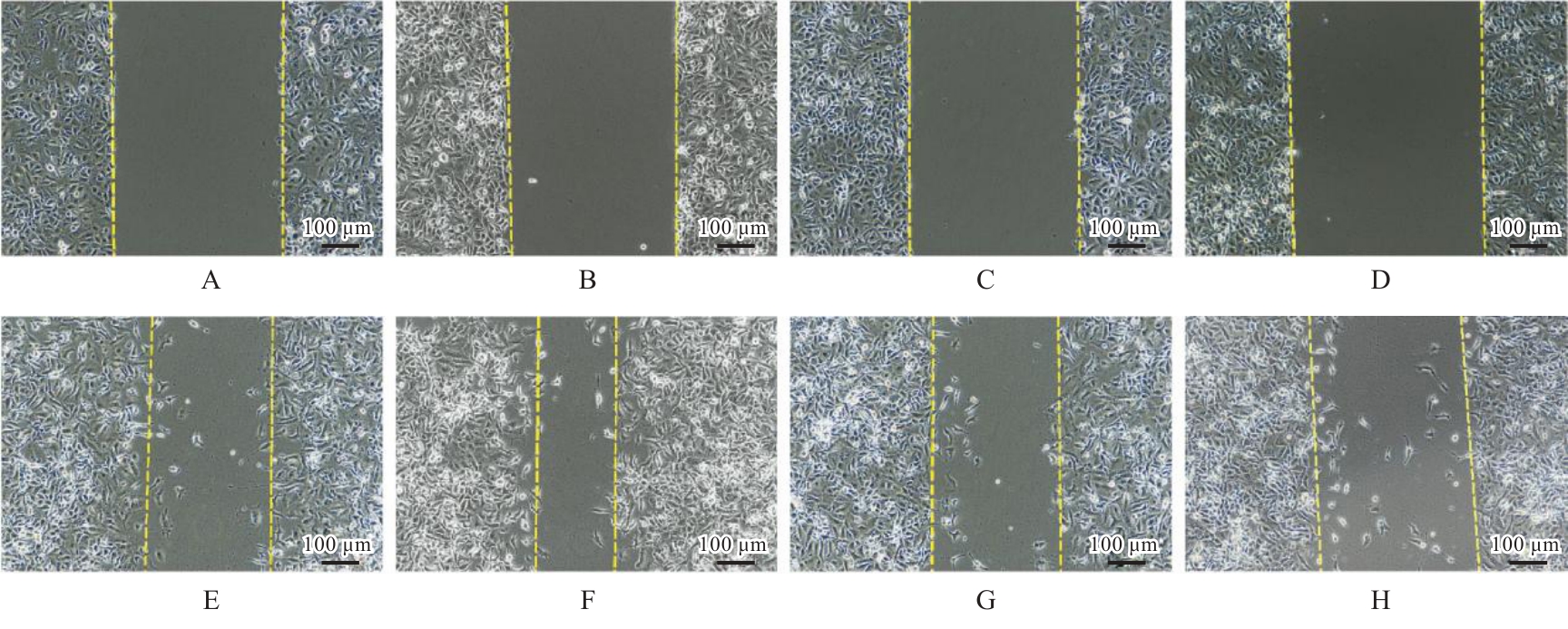
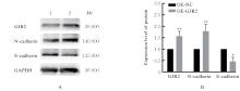
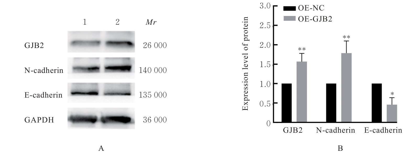
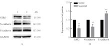
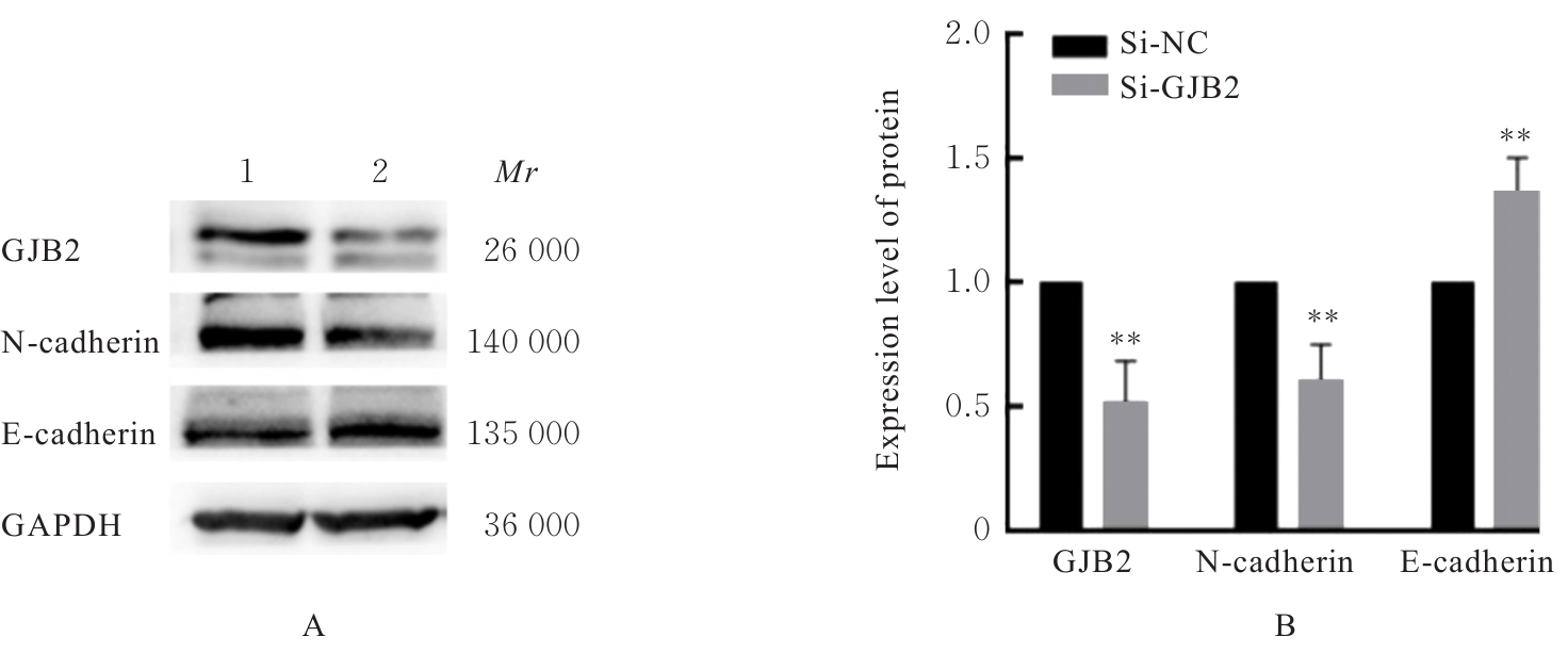
目的 探讨缝隙连接蛋白β2(GJB2)在肺腺癌(LUAD)A549细胞中的表达情况和生物学功能,为LUAD治疗提供参考。 方法 利用TIMER数据库分析多种肿瘤中GJB2基因表达差异,GEPIA数据库检测LUAD中GJB2 mRNA表达情况,分析GJB2基因与LUAD不同临床分期的相关性,采用Kaplan-Meier plotter数据库分析GJB2蛋白与LUAD患者预后的相关性。收集111例LUAD患者癌组织及癌旁正常肺组织样本,采用免疫组织化学染色法观察LUAD患者癌组织和癌旁正常肺组织中GJB2蛋白表达情况。体外培养人肺腺癌A549细胞,将A549细胞分为GJB2过表达组(OE-GJB2组,转染GJB2过表达质粒)及其阴性对照组(OE-NC组,转染GJB2空载质粒)、小干扰RNA(si-RNA)- GJB2组(si-GJB2组,转染GJB2-siRNA)及其阴性对照组(si-NC组,转染对照si-RNA),采用Western blotting法验证细胞转染效率。采用细胞计数试剂盒8(CCK-8)法检测各组A549细胞增殖活性,5-乙炔基-2'-脱氧尿苷(EdU)染色法检测各组A549细胞阳性表达率,克隆形成实验检测各组A549细胞克隆形成数,细胞划痕实验检测各组A549细胞迁移率,Transwell小室实验检测各组A549细胞中侵袭细胞数,Western blotting法检测各组A549细胞中GJB2蛋白和上皮-间质转化(EMT)相关蛋白表达水平。 结果 GJB2基因在多种肿瘤中均存在异常表达;与癌旁正常肺组织比较,LUAD患者癌组织中GJB2 mRNA表达水平明显升高(P<0.05),且GJB2的高表达与LUAD患者癌的临床病理分期具有关联性(P<0.05);与GJB2低表达LUAD患者比较,GJB2高表达LUAD患者的总生存期明显减少[风险比(HR)=1.71,95%CI:1.34~2.18,P<0.05];GJB2蛋白在癌旁正常肺泡和支气管上皮细胞中均阴性表达,与癌旁正常肺组织比较,LUAD患者癌组织中GJB2蛋白表达明显增强;LUAD患者癌组织中GJB2蛋白阳性表达与淋巴结转移具有明显关联性(P<0.05),但与患者性别(P=0.626)、年龄(P=0.639)和TNM分期(P=0.837)无关联性(P>0.05)。CCK-8法,与OE-NC组比较,OE-GJB2组A549细胞增殖活性明显升高(P<0.05或P<0.01);与si-NC组比较,si-GJB2组A549细胞增殖活性明显降低(P<0.05或P<0.01)。EdU染色法,与OE-NC组比较,OE-GJB2组A549细胞中EdU阳性表达率明显升高(P<0.01);与si-NC组比较,si-GJB2组A549细胞中EdU阳性表达率明显降低(P<0.01)。克隆形成实验,与OE-NC组比较,OE-GJB2组A549细胞克隆形成数明显增加(P<0.01);与si-NC组比较,si-GJB2组A549细胞克隆形成数明显减少(P<0.01)。Transwell小室实验,与OE-NC组比较,OE-GJB2组A549细胞侵袭细胞数明显增加(P<0.01);与si-NC组比较,si-GJB2组A549细胞侵袭细胞数明显减少(P<0.01)。细胞划痕实验,细胞划痕24 h后,与OE-NC组比较,OE-GJB2组A549细胞迁移率明显升高(P<0.01);与si-NC组比较,si-GJB2组A549细胞迁移率明显降低(P<0.01)。Western blotting法,与OE-NC组比较,OE-GJB2组A549细胞中GJB2和N钙黏蛋白(N-cadherin)蛋白表达水平均明显升高(P<0.01),E钙黏蛋白(E-cadherin)蛋白表达水平明显降低(P<0.05);与si-NC组比较,OE-GJB2组A549细胞中GJB2和N-cadherin蛋白表达水平均明显降低(P<0.01),E-cadherin蛋白表达水平明显升高(P<0.01)。 结论 GJB2在LUAD患者癌组织中高表达,并与LUAD患者的不良预后有关。GJB2可促进A549细胞的增殖、侵袭、迁移和EMT。

关键词: 缝隙连接蛋白β2, 肺腺癌, 细胞增殖, 细胞侵袭, 细胞迁移, 上皮-间质转化

Abstract:

Objective To discuss the expression and biological function of gap junction protein β2 (GJB2) in the lung adenocarcinoma (LUAD) A549 cells, and to procide the basis for the treatment of LUAD. Methods The TIMER database was used to analyze the differential expression of GJB2 gene in various tumors; the GEPIA database was used to detect the mRNA expression of GJB2 in LUAD and analyze its correlation with different clinical stages of LUAD; the Kaplan-Meier plotter database was used to analyze the correlation between GJB2 protein and the prognosis of the LUAD patients. A total of 111 pairs of cancer tissues and adjacent normal lung tissues of LUAD patients were collected, and immunohistochemical staining was used to observe the expression of GJB2 protein in LUAD tissues and adjacent normal lung tissues. The human LUAD A549 cells were cultured in vitro and divided into GJB2 overexpression group (OE-GJB2 group, transfected with GJB2 overexpression plasmid) and its negative control group (OE-NC group, transfected with GJB2 empty vector plasmid), small interfering RNA (siRNA)-GJB2 group (si-GJB2 group, transfected with GJB2-siRNA) and its negative control group (si-NC group, transfected with control siRNA). Western blotting method was used to verify the transfection efficiency of the cells; cell couting kit-8 (CCK-8) method was used to detect the proliferation activities of the A549 cells in various groups; 5-ethynyl-2'-deoxyuridine (EdU) staining was used to detect the positive expression rates of the A549 cells in various groups; colony formation assay was used to detect the number of colony formation of the A549 cells in various groups; cell scratch assay was used to detect the migration rate of the A549 cells in various groups; Transwell chamber assay was used to detect the number of the invasion A549 cells in various groups; Western blotting method was used to detect the expression levels of GJB2 protein and epithelial-mesenchymal transition (EMT)-related proteins in the A549 cells in various groups. Results The RT-PCR results showed that GJB2 gene was abnormally expressed in various tumors; compared with adjacent normal lung tissue, the mRNA expression level of GJB2 in cancer tissue of LUAD patients was significantly increased (P<0.05), and the high expression of GJB2 was correlated with the clinicopathological stage of LUAD (P<0.05). The Kaplan-Meier plotter results showed that compared with patients with low GJB2 expression, the overall survival of the patients with high GJB2 expression was significantly decreased [hazard ratio (HR)=1.71, 95% CI: 1.34-2.18, P<0.05]. GJB2 protein was negatively expressed in adjacent normal alveolar and bronchial epithelial cells; compared with adjacent normal lung tissue, the expression of GJB2 protein in cancer tissue of LUAD patients was significantly enhanced. In LUAD, the positive expression of GJB2 protein was significantly associated with lymphnode metastasis (P<0.05) but not with gender (P=0.626), age (P=0.639), or TNM stage (P=0.837) (P>0.05). The CCK-8 results showed that compared with OE-NC group, the proliferation activity of the A549 cells in OE-GJB2 group was significantly increased (P<0.05 or P<0.01); compared with si-NC group, the proliferation activity of the A549 cells in si-GJB2 group was significantly decreased (P<0.05 or P<0.01). The EdU staining results showed that compared with OE-NC group, the positive expression rate of EdU in the A549 cells in OE-GJB2 group was significantly increased (P<0.01); compared with si-NC group, the positive expression rate of EdU in the A549 cells in si-GJB2 group was significantly decreased (P<0.01). The colony formation assay results showed that compared with OE-NC group, the number of colony formation of the A549 cells in OE-GJB2 group was significantly increased (P<0.01); compared with si-NC group, the number of colony formation of the A549 cells in si-GJB2 group was significantly decreased (P<0.01). The Transwell chamber assay results showed that compared with OE-NC group, the number of invasion A549 cells in OE-GJB2 group was significantly increased (P<0.01); compared with si-NC group, the number of invasion A549 cells in si-GJB2 group was significantly decreased (P<0.01). The cell scratch assay results showed that 24 h after scratching, compared with OE-NC group, the migration rate of the A549 cells in OE-GJB2 group was significantly increased (P<0.01); compared with si-NC group, the migration rate of the A549 cells in si-GJB2 group was significantly decreased (P<0.01). The Western blotting results showed that compared with OE-NC group, the expression levels of GJB2 and N-cadherin proteins in the A549 cells in OE-GJB2 group were significantly increased (P<0.01), while the expression level of E-cadherin protein was significantly decreased (P<0.05); compared with si-NC group, the expression levels of GJB2 and N-cadherin proteins in the A549 cells in si-GJB2 group were significantly decreased (P<0.01), while the expression level of E-cadherin protein was significantly increased (P<0.01). Conclusion GJB2 is highly expressed in cancer tissue of LUAD patients and is associated with the poor prognosis in LUAD patients. GJB2 promotes the proliferation, invasion, migration, and EMT of the A549 cells.

Key words: Gap junction protein β2, Lung adenocarcinoma, Cell proliferation, Cell invasion, Cell migration, Epithelial-mesenchymal transition

中图分类号: 

  • R730.5