吉林大学学报(医学版) ›› 2026, Vol. 52 ›› Issue (1): 1-9.doi: 10.13481/j.1671-587X.20260101

• 基础研究 •    下一篇

沉默GPR139基因对乳腺癌细胞增殖、凋亡和自噬的影响及其机制

姚晓含1,姚明辰2,王志清2,李赫阳2,闫燕1,雷宁静3()   

  1. 1.郑州大学第一附属医院医学研究中心,河南 郑州 450052
    2.郑州大学第一临床医学院,河南 郑州 450052
    3.郑州大学基础医学院微生物与免疫学系,河南 郑州 450001
  • 收稿日期:2025-02-13 接受日期:2025-03-24 出版日期:2026-01-28 发布日期:2026-02-25
  • 通讯作者: 雷宁静 E-mail:lnj717@zzu.edu.cn
  • 作者简介:姚晓含(1990-),女,河南省许昌市人,实验师,生物学硕士,主要从事肿瘤分子生物学方面的研究。
  • 基金资助:
    国家自然科学基金青年科学基金项目(82303343)

Effect of silencing GPR139 gene on proliferation, apoptosis and autophagy of breast cancer cells and its mechanism

Xiaohan YAO1,Mingchen YAO2,Zhiqing WANG2,Heyang LI2,Yan YAN1,Ningjing LEI3()   

  1. 1.Medical Research Center,First Affiliated Hospital,Zhengzhou University,Zhengzhou 450052,China
    2.First Clinical Academy,Zhengzhou University,Zhengzhou 450052,China
    3.Department of Microbiology and Immunology,School of Basic Medical Sciences,Zhengzhou University,Zhengzhou 450001,China
  • Received:2025-02-13 Accepted:2025-03-24 Online:2026-01-28 Published:2026-02-25
  • Contact: Ningjing LEI E-mail:lnj717@zzu.edu.cn

摘要:

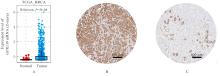
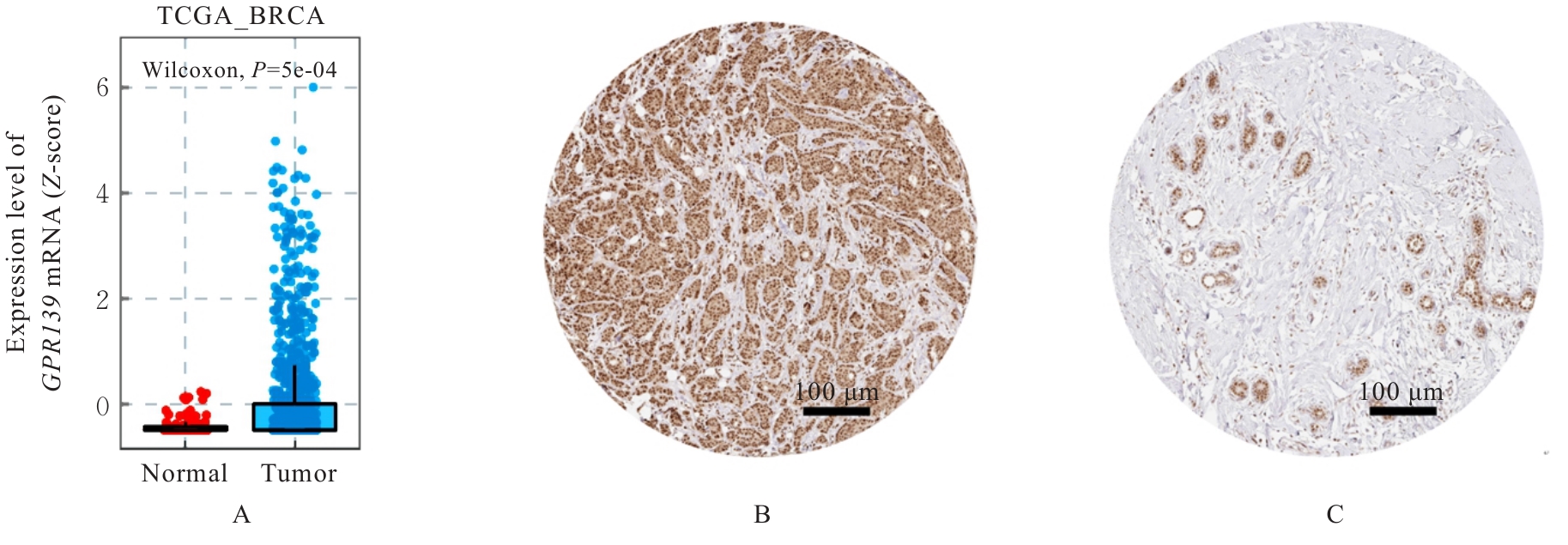
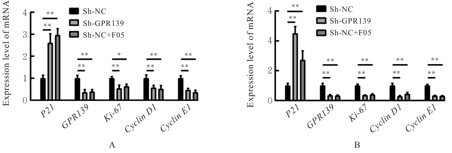
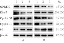
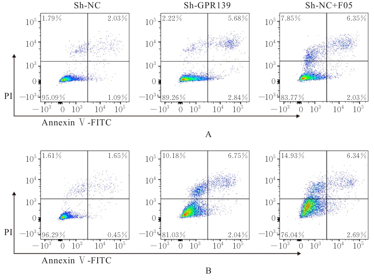
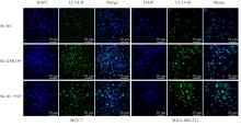
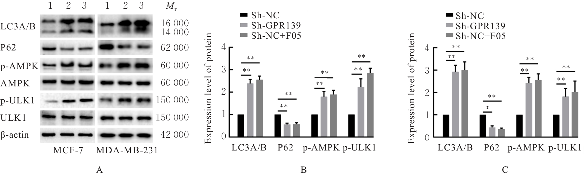
目的 探讨沉默G蛋白偶联受体139(GPR139)基因对乳腺癌细胞增殖、凋亡和自噬的影响,并阐明其可能的作用机制。 方法 通过癌症基因组图谱(TCGA)和人类蛋白质图谱(HPA)数据库下载GPR139 mRNA和蛋白在乳腺癌组织与正常组织中的表达情况。利用慢病毒干扰技术将GPR139短发夹RNA(shRNA)转染至人乳腺癌MCF-7和MDA-MB-231细胞系中,分为sh-NC组(感染阴性对照慢病毒)、sh-GPR139(感染GPR139 shRNA干扰慢病毒)和sh-NC+F05组(感染阴性对照慢病毒后,加入GPR139拮抗剂NCRW0005-F05)。采用噻唑蓝(MTT)法检测各组乳腺癌细胞增殖活性,实时荧光定量PCR(RT-qPCR)法和Western blotting法检测各组乳腺癌细胞中细胞增殖标志物Ki-67、细胞周期素(CyclinD1Cyclin E1P21 mRNA及蛋白表达水平,流式细胞术检测各组乳腺癌细胞凋亡率,Western blotting法检测各组乳腺癌细胞中腺苷酸活化蛋白激酶(AMPK)、磷酸化AMPK、Unc-51样自噬激活激酶1(ULK1)、磷酸化ULK1、微管相关蛋白1轻链3A/B(LC3A/B)和P62蛋白表达水平,免疫荧光法检测各组乳腺癌细胞中LC3A/B蛋白表达水平。 结果 TCGA和HPA数据库,在人乳腺癌组织中的GPR139 mRNA表达水平明显高于正常组织(P<0.001),GPR139蛋白表达量明显高于正常组织。MTT法,与sh-NC组比较,sh-GPR139组和sh-NC+F05组乳腺癌细胞增殖活性明显降低(P<0.01)。Western blotting法,与sh-NC组比较,sh-GPR139组和sh-NC+F05组乳腺癌细胞中GPR139、Ki67、Cyclin D1和Cyclin E1蛋白表达水平降低(P<0.05或P<0.01),P21蛋白表达水平升高(P<0.01)。RT-qPCR法,与sh-NC组比较,sh-GPR139组和sh-NC+F05组乳腺癌细胞中GPR139Ki67Cyclin D1Cyclin E1 mRNA表达水平降低(P<0.05或P<0.01),P21 mRNA表达水平升高(P<0.01)。流式细胞术,与sh-NC组比较,sh-GPR139组和sh-NC+F05组乳腺癌细胞凋亡率明显增加(P<0.01)。免疫荧光法,在MCF-7细胞和MDA-MB-231细胞中,与sh-NC组比较,sh-GPR139组和sh-NC+F05组细胞中LC3A/B蛋白表达水平明显升高(P<0.01)。Western blotting法,与sh-NC组比较,sh-GPR139组和sh-NC+F05组LC3A/B、p-AMPK和p-ULK1蛋白表达水平明显升高(P<0.01),P62蛋白表达水平明显降低(P<0.05或P<0.01)。 结论 沉默GPR139基因可诱导乳腺癌细胞自噬和凋亡,抑制细胞增殖,其机制可能与升高细胞中AMPK和ULK1磷酸化水平有关。

关键词: 乳腺癌, G蛋白偶联受体139, 细胞增殖, 细胞凋亡, 自噬, 腺苷酸活化蛋白激酶

Abstract:

Objective To discuss the effect of silencing G protein-coupled receptor 139 (GPR139) gene on the proliferation, apoptosis and autophagy of breast cancer cells, and to clarify its possible mechanism. Methods The expression of GPR139 mRNA and protein in breast cancer tissue and normal tissue was downloaded from The Cancer Genome Atlas (TCGA) and Human Protein Atlas (HPA) databases; the GPR139 short hairpin RNA (shRNA) was transfected into the human breast cancer MCF-7 and MDA-MB-231 cell lines by lentiviral interference technology, and the cells were divided into sh-NC group (infected with negative control lentivirus), sh-GPR139 group (infected with GPR139 shRNA interference lentivirus), and sh-NC+F05 group (after infected with negative control lentivirus, GPR139 antagonist NCRW0005-F05 was added); methylthiazolyldiphenyl-tetrazolium bromide(MTT) method was used to detect the proliferation activities of the cells in various groups; Western blotting and real-time fluorescence quantitative PCR (RT-qPCR) methods were used to detect the expression levels of proliferation markers Ki-67Cyclin D1Cyclin E1, and P21 mRNA and proteins in the cells in various groups; flow cytometry was used to detect the apoptotic rates of the breast cancer cells in various groups; Western blotting method was used to detect the expression levels of adenosine monophosphate-activated protein kinase (AMPK), phosphorylated AMPK, Unc-51 like autophagy activating kinase 1 (ULK1), phosphorylated ULK1, microtubule-associated protein 1 light chain 3A/B (LC3A/B), and P62 proteins in the cells in various groups; immunofluorescence method was used to detected the expression level of LC3A/B protein in the breast cancer cells in various groups. Results The TCGA and HPA database results showed that the expression level of GPR139 mRNA in human breast cancer tissue was significantly higher than that in normal tissue (P<0.001), and the expression level of GPR139 protein was significantly higher than that in normal tissue. The MTT assay results showed that compared with sh-NC group, the proliferation activities of the breast cancer cells in sh-GPR139 group and sh-NC+F05 group were significantly decreased (P<0.01). The Western blotting results showed that compared with sh-NC group, the expression levels of GPR139, Ki67, Cyclin D1 and Cyclin E1 proteins in the breast cancer cells in sh-GPR139 group and sh-NC+F05 group were decreased (P<0.05 or P<0.01), and the expression level of P21 protein was increased (P<0.01). The RT-qPCR results showed that compared with sh-NC group, the expression levels of GPR139Ki67Cyclin D1 and Cyclin E1 mRNA in the breast cancer cells in sh-GPR139 group and sh-NC+F05 group were decreased (P<0.05 or P<0.01), and the expression level of P21 mRNA was increased (P<0.01). The flow cytometry results showed that compared with sh-NC group, the apoptotic rates of the breast cancer cells in sh-GPR139 group and sh-NC+F05 group were significantly increased (P<0.01). The immunofluorescence results showed that in MCF-7 cells and MDA-MB-231 cells, compared with sh-NC group, the expression levels of LC3A/B protein in the cells in sh-GPR139 group and sh-NC+F05 group were significantly increased (P<0.01). The Western blotting results showed that compared with sh-NC group, the expression levels of LC3A/B, p-AMPK and p-ULK1 proteins in sh-GPR139 group and sh-NC+F05 group were significantly increased (P<0.01), and the expression level of P62 protein was significantly decreased (P<0.05 or P<0.01). Conclusion Silencing GPR139 gene can induce autophagy and apoptosis of breast cancer cells and inhibit cell proliferation, and its mechanism may be related to the increased phosphorylation levels of AMPK and ULK1 in the cells.

Key words: Breast cancer, G protein-coupled receptor 139, Cell proliferation, Apoptosis, Autophagy, Adenosine monophosphate-activated protein kinase

中图分类号: 

  • R737.9