吉林大学学报(医学版) ›› 2026, Vol. 52 ›› Issue (1): 162-170.doi: 10.13481/j.1671-587X.20260117

• 基础研究 • 上一篇    下一篇

凉膈散对LPS诱导的急性肺损伤的保护作用及其机制

于鑫1,董铮2,张引1,蔡相龙1,刘亮亮1,孙娜1,牛文博3,杨磊4,杨文杰5()   

  1. 1.武警特色医学中心综合重症医学科,天津 300162
    2.武警天津总队医院内二科,天津 300162
    3.锦州海关综合技术服务中心,辽宁 锦州 121013
    4.天津市南开医院急腹症研究所,天津 300100
    5.天津市第一中心医院感染科,天津 300190
  • 收稿日期:2025-01-21 接受日期:2025-03-31 出版日期:2026-01-28 发布日期:2026-02-24
  • 通讯作者: 杨文杰 E-mail:yangm8006@sina.com
  • 作者简介:于 鑫(1983-),男,黑龙江省哈尔滨市人,主治医师,医学硕士,主要从事呼吸与危重症方面的研究。
  • 基金资助:
    天津市卫健委医学重点学科建设资助项目(TJYXZDXK-3-001D)

Protective effect of Liangge powder on LPS-induced acute lung injury and its mechanism

Xin YU1,Zheng DONG2,Yin ZHANG1,Xianglong CAI1,Liangliang LIU1,Na SUN1,Wenbo NIU3,Lei YANG4,Wenjie YANG5()   

  1. 1.Department of Respiratory and Critical Care Medicine,Characteristic Medical Center of Chinese People’s Armed Police Forces,Tianjin 300162,China
    2.Department of Internal Medicine,Tianjin Armed Police Corps Hospital,Tianjin 300162,China
    3.Comprehensive Technical Service Center of Jinzhou Customs,Jinzhou 121013,China
    4.Tianjin Key Laboratory of Acute Abdomen Disease Associated Organ Injury and ITCWM Repair,Tianjin Nankai Hospital,Tianjin,300100,China
    5.Department of Infection,First Central Hospital,Tianjin City,Tianjin 300190,China
  • Received:2025-01-21 Accepted:2025-03-31 Online:2026-01-28 Published:2026-02-24
  • Contact: Wenjie YANG E-mail:yangm8006@sina.com

摘要:

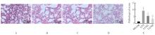
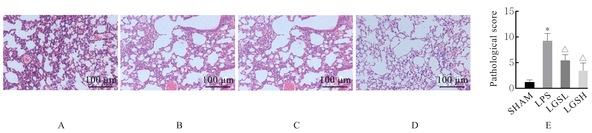
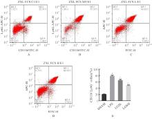
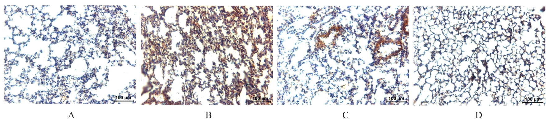
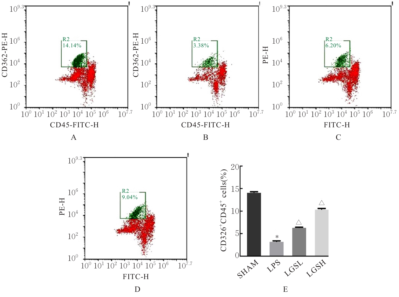
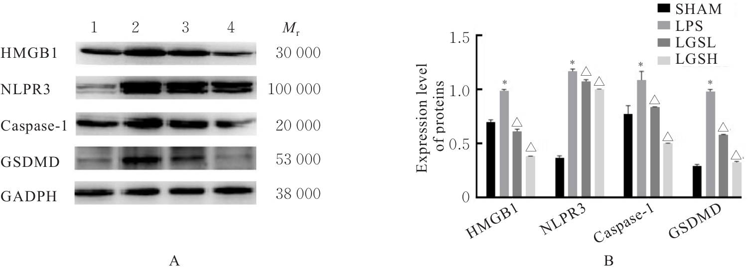
目的 探讨凉膈散(LGS)对脂多糖(LPS)诱导的急性肺损伤(ALI)的保护作用,并阐明其作用机制。 方法 将40只雄性的C57BL/6小鼠随机分为假手术组(SHMA组)、模型组(LPS组)、低剂量LGS组(LGSL组,8.58 g·kg-1 LGS)和高剂量LGS组(LGSH组,17.16 g·kg-1 LGS),每组10只。LPS组、LGSL组和LSGH组小鼠采用气管滴注LPS诱导建立ALI模型,造模24 h后取材。采用HE染色法观察各组小鼠肺组织病理形态表现并进行病理评分,流式细胞术检测各组小鼠肺组织中中性粒细胞和肺泡上皮细胞百分率,免疫组织化学染色法检测各组小鼠肺组织中髓过氧化物酶(MPO)水平,酶联免疫吸附试验(ELISA)法检测各组小鼠肺泡灌洗液中相关炎性介质白细胞介素18(IL-18)、白细胞介素1β(IL-1β)和白蛋白水平,Western blotting法检测各组小鼠肺组织中高迁移率族蛋白B1(HMGB1)、NLR家族Pyrin域蛋白3(NLRP3)、含半胱氨酸的天冬氨酸蛋白水解酶1(Caspase-1)和消皮素D(GSDMD)蛋白表达水平。 结果 HE染色,与SHMA组比较,LPS组小鼠肺组织病理损伤加重,病理评分明显升高(P<0.05);与LPS组比较,LGSL组和LGSH组小鼠肺组织病理学评分明显降低(P<0.05)。流式细胞术,与SHMA组比较,LPS组小鼠肺组织中中性粒细胞百分率明显升高(P<0.05),肺泡上皮细胞百分率明显降低(P<0.05);与LPS组比较,LGSL组和LGSH组小鼠肺组织中中性粒细胞百分率明显降低(P<0.05),肺泡上皮细胞百分率明显升高(P<0.05)。免疫组织化学染色法,与SHMA组比较,LPS组小鼠肺组织中MPO水平明显升高(P<0.05);与LPS组比较,LGSL组和LGSH组小鼠肺组织中MPO水平明显降低(P<0.05)。ELISA法,与SHMA组比较,LPS组小鼠肺泡灌洗液中IL-18、IL-1β和白蛋白水平明显升高(P<0.05);与LPS组比较,LGSL组和LGSH组小鼠肺泡灌洗液中IL-18、IL-1β和白蛋白水平明显降低(P<0.05)。Western blotting法,与SHMA组比较,LPS组小鼠肺组织中HMGB1、NLRP3、Caspase-1和GSDMD蛋白表达水平均明显升高(P<0.05);与LPS组比较,LGSL组和LGSH组小鼠肺组织中HMGB1、NLRP3、Caspase-1和GSDMD蛋白表达水平明显降低(P<0.05)。 结论 LGS可通过降低中性粒细胞浸润和上皮细胞焦亡以减轻LPS诱导的病理肺损伤和炎症反应,其机制可能与其通过HMGB1信号通路下调NLRP3、Caspase-1和GSDMD蛋白表达有关。

关键词: 凉膈散, 急性肺损伤, 细胞焦亡, NOD样受体蛋白3, 含半胱氨酸的天冬氨酸蛋白水解酶1, 消皮素D

Abstract:

Objective To discuss the protective effect of Liangge Powder (LGS) on lipopolysaccharide (LPS)-induced acute lung injury (ALI), and to clarify its mechanism. Methods Forty male C57BL/6 mice were randomly divided into sham operation group (SHMA group), model group (LPS group), low dose of LGS group (LGSL group, 8.58 g·kg?1 LGS), and high dose of LGS group (LGSH group, 17.16 g·kg?1 LGS), with 10 mice in each group. The mice in LPS group, LGSL group, and LGSH group were intratracheally instilled with LPS to induce the ALI model, and samples were taken 24 h after modeling. HE staining was used to observe the pathomorphology of lung tissue of the mice in various groups and to perform the pathological scores; flow cytometry was used to detect the percentages of neutrophils and epithelial cells in lung tissue of the mice in various groups; immunohistochemistry staining assay was used to detect the level of myeloperoxidase (MPO) in lung tissue of the mice in various groups; enzyme-linked immunosorbent assay (ELISA) was used to detect the levels of related inflammatory mediators interleukin-18 (IL-18), interleukin-1β (IL-1β), and albumin in bronchoalveolar lavage fluid of the mice in various groups; Western blotting method was used to detect the expression levels of high mobility group box 1 (HMGB1), NLR family pyrin domain containing 3 (NLRP3), cysteinyl aspartate specific proteinase-1 (Caspase-1), and gasdermin D (GSDMD) proteins in lung tissue of the mice in various groups. Results The HE staining results showed that compared with SHMA group, the pathological injury of lung tissue of the mice in LPS group was aggravated, and the pathological score was significantly increased (P<0.05); compared with LPS group, the pathological scores of lung tissue of the mice in LGSL group and LGSH group were significantly decreased (P<0.05). The flow cytometry results showed that compared with SHMA group, the percentage of neutrophils in lung tissue of the mice in LPS group was increased (P<0.05), and the percentage of epithelial cells was decreased (P<0.05); compared with LPS group, the percentages of neutrophils in lung tissue of the mice in LGSL group and LGSH group were decreased (P<0.05), and the percentages of epithelial cells were increased (P<0.05). The immunohistochemistry staining results showed that compared with SHMA group, the MPO level in lung tissue of the mice in LPS group was significantly increased (P<0.05); compared with LPS group, the MPO levels in lung tissue of the mice in LGSL group and LGSH group were significantly decreased (P<0.05). The ELISA results showed that compared with SHMA group, the levels of IL-1β, IL-18, and albumin in bronchoalveolar lavage fluid of the mice in LPS group were significantly increased (P<0.05); compared with LPS group, the levels of IL-1β, IL-18, and albumin in bronchoalveolar lavage fluid of the mice in LGSL group and LGSH group were significantly decreased (P<0.05). The Western blotting results showed that compared with SHMA group, the expression levels of HMGB1, NLRP3, Caspase-1, and GSDMD proteins in lung tissue of the mice in LPS group were significantly increased (P<0.01); compared with LPS group, the expression levels of HMGB1, NLRP3, Caspase-1, and GSDMD proteins in lung tissue of the mice in LGSL group and LGSH group were significantly decreased (P<0.05). Conclusion LGS can alleviate LPS-induced pathological lung injury and inflammatory response by reducing neutrophil infiltration and epithelial cell pyroptosis, and its mechanism may be related to the down-regulation of NLRP3, Caspase-1, and GSDMD protein expressions through the HMGB1 signaling pathway.

Key words: Liangge San, Acute lung injury, Pyroptosis, NOD-like receptor protein 3, Cysteinyl aspartate specific proteinase-1, Gasdermin D

中图分类号: 

  • R285.5