| 1 |
TENORIO R, FERNÁNDEZ DE CASTRO I, KNOWLTON J J, et al. Reovirus σNS and μNS proteins remodel the endoplasmic reticulum to build replication neo-organelles[J]. mBio,2018,9(4):e01253.
|
| 2 |
FERNÁNDEZ DE CASTRO I, TENORIO R, RISCO C. Virus assembly factories in a lipid world[J]. Curr Opin Virol, 2016, 18: 20-26.
|
| 3 |
POWERS R E, WANG S Y, LIU T Y, et al. Reconstitution of the tubular endoplasmic reticulum network with purified components[J]. Nature, 2017, 543(7644): 257-260.
|
| 4 |
CHUA K B, VOON K, YU M, et al. Investigation of a potential zoonotic transmission of orthoreovirus associated with acute influenza-like illness in an adult patient[J]. PLoS One, 2011, 6(10): e25434.
|
| 5 |
WONG A H, CHENG P K, LAI M Y, et al. Virulence potential of fusogenic orthoreoviruses[J]. Emerg Infect Dis, 2012, 18(6): 944-948.
|
| 6 |
VOON K, TAN Y F, LEONG P P, et al. Pteropine orthoreovirus infection among out-patients with acute upper respiratory tract infection in Malaysia[J]. J Med Virol, 2015, 87(12): 2149-2153.
|
| 7 |
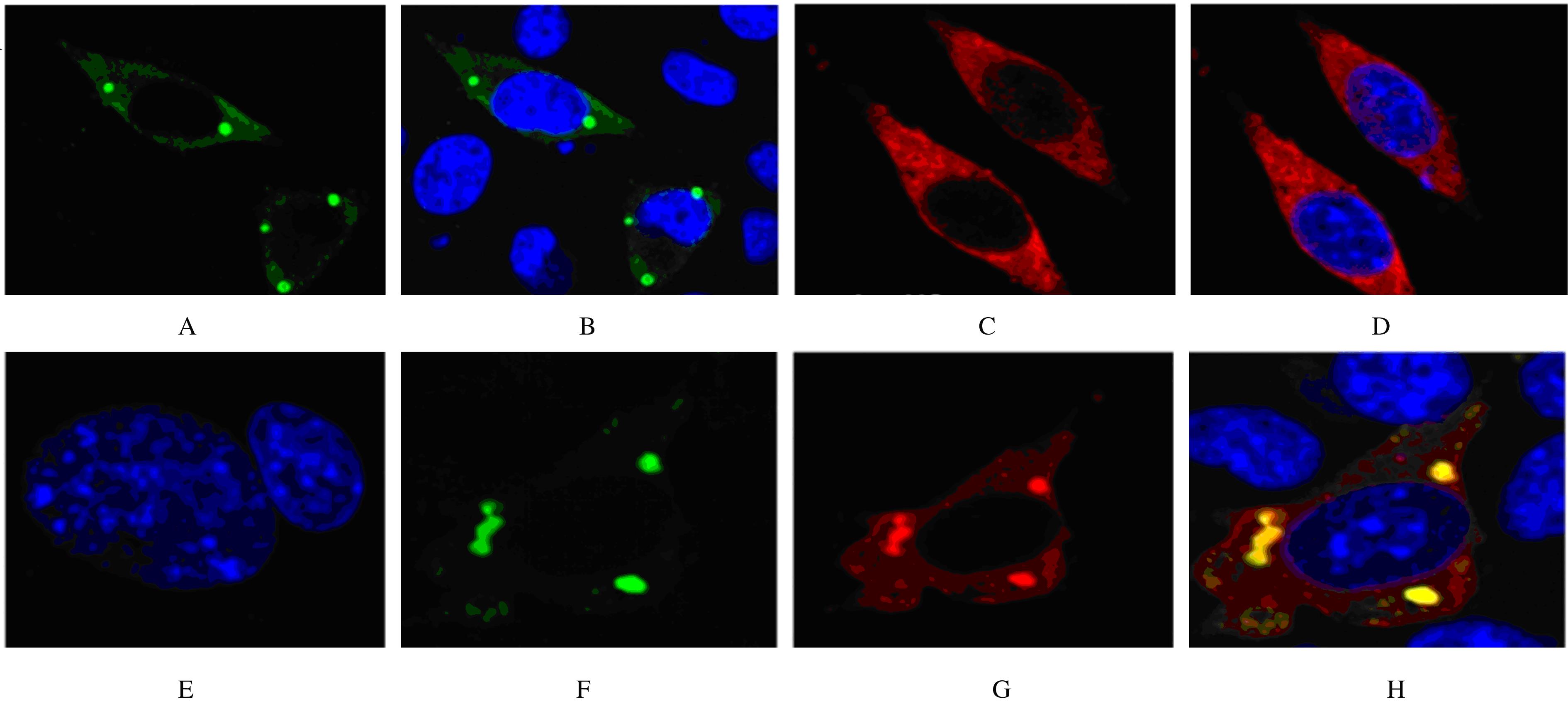
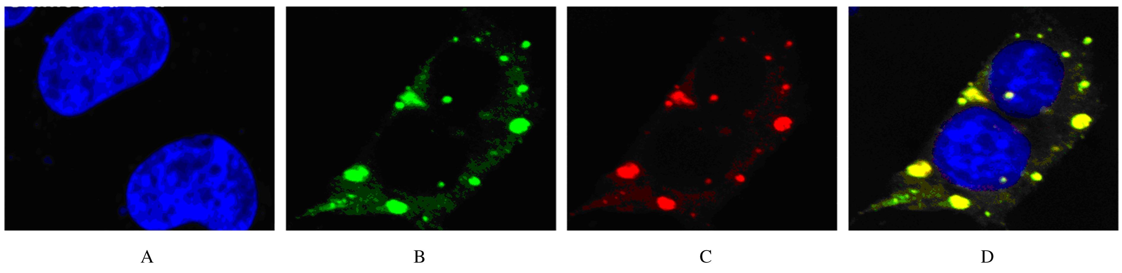
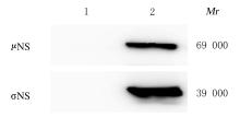
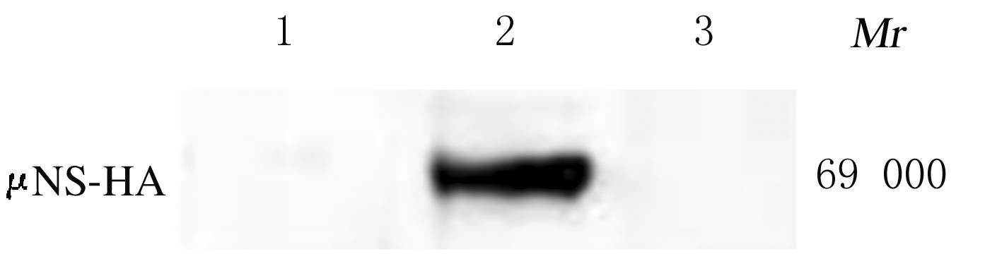
BECKER M M, PETERS T R, DERMODY T S. Reovirus sigma NS and mu NS proteins form cytoplasmic inclusion structures in the absence of viral infection[J]. J Virol, 2003, 77(10): 5948-5963.
|
| 8 |
BROERING T J, PARKER J S L, JOYCE P L, et al. Mammalian reovirus nonstructural protein microNS forms large inclusions and colocalizes with reovirus microtubule-associated protein micro2 in transfected cells[J]. J Virol, 2002, 76(16): 8285-8297.
|
| 9 |
MILLER C L, BROERING T J, PARKER J S, et al. Reovirus sigma NS protein localizes to inclusions through an association requiring the mu NS amino terminus[J]. J Virol, 2003, 77(8): 4566-4576.
|
| 10 |
TOURIS-OTERO F, MARTı́NEZ-COSTAS J, VAKHARIA V N, et al. Avian reovirus nonstructural protein μNS forms viroplasm-like inclusions and recruits protein σNS to these structures[J]. Virology, 2004, 319(1): 94-106.
|
| 11 |
PARKER J S, BROERING T J, KIM J, et al. Reovirus core protein mu2 determines the filamentous morphology of viral inclusion bodies by interacting with and stabilizing microtubules[J]. J Virol, 2002, 76(9): 4483-4496.
|
| 12 |
BROERING T J, MCCUTCHEON A M, CENTONZE V E, et al. Reovirus nonstructural protein muNS binds to core particles but does not inhibit their transcription and capping activities[J]. J Virol, 2000, 74(12): 5516-5524.
|
| 13 |
ARNOLD M M, MURRAY K E, NIBERT M L. Formation of the factory matrix is an important, though not a sufficient function of nonstructural protein mu NS during reovirus infection[J]. Virology, 2008, 375(2): 412-423.
|
| 14 |
KOBAYASHI T, CHAPPELL J D, DANTHI P,et al. Gene-specific inhibition of reovirus replication by RNA interference[J]. J Virol, 2006, 80(18): 9053-9063.
|
| 15 |
ZAMORA P F, HU L Y, KNOWLTON J J, et al. Reovirus nonstructural protein σNS Acts as an RNA stability factor promoting viral genome replication[J]. J Virol, 2018. DOI:10.1128/jvi.00563-18 .
doi: 10.1128/jvi.00563-18
|
| 16 |
KNOWLTON J J, DE CASTRO I F, ASHBROOK A W,et al. The TRiC chaperonin controls reovirus replication through outer-capsid folding[J].Nat Microbiol, 2018,3(4):481-493.
|
| 17 |
BECKER M M, GORAL M I, HAZELTON P R,et al. Reovirus sigmaNS protein is required for nucleation of viral assembly complexes and formation of viral inclusions[J]. J Virol, 2001, 75(3): 1459-1475.
|
| 18 |
BORODAVKA A, AULT J, STOCKLEY P G, et al. Evidence that avian reovirus σNS is an RNA chaperone: implications for genome segment assortment[J]. Nucleic Acids Res, 2015, 43(14): 7044-7057.
|
| 19 |
TENORIO R, FERNÁNDEZ DE CASTRO I, KNOWLTON J J, et al. Function, architecture, and biogenesis of reovirus replication neoorganelles[J]. Viruses, 2019, 11(3): 288.
|
| 20 |
ZAMORA P F, HU L Y, KNOWLTON J J, et al. Reovirus nonstructural protein σNS Acts as an RNA stability factor promoting viral genome replication[J]. J Virol, 2018, 92(15). DOI:10.1128/jvi.00563-18 .
doi: 10.1128/jvi.00563-18
|
| 21 |
GILLIAN A L, SCHMECHEL S C, LIVNY J, et al. Reovirus protein sigmaNS binds in multiple copies to single-stranded RNA and shares properties with single-stranded DNA binding proteins[J].J Virol,2000,74(13):5939-5948.
|
| 22 |
BORODAVKA A, DYKEMAN E C, SCHRIMPF W,et al. Protein-mediated RNA folding governs sequence-specific interactions between rotavirus genome segments[J]. Elife,2017,6:e27453.
|
| 23 |
YANG J, XIA H J, QIAN Q, et al. RNA chaperones encoded by RNA viruses[J]. Virol Sin, 2015,30(6): 401-409.
|
| 24 |
TOURÍS-OTERO F, MARTÍNEZ-COSTAS J, VAKHARIA V N, et al. Characterization of the nucleic acid-binding activity of the avian reovirus non-structural protein sigma NS[J].J Gen Virol,2005, 86(pt4):1159-1169.
|
 )
)