吉林大学学报(医学版) ›› 2021, Vol. 47 ›› Issue (4): 926-933.doi: 10.13481/j.1671-587X.20210415

• 基础研究 • 上一篇    下一篇

氯喹通过影响胰腺癌细胞自噬和线粒体功能对吉西他滨耐药细胞的作用及其机制

陆路,黎东明,王学国,宋丹,王太成,赵红岩,吴晓勇()   

  1. 海南医学院第二附属医院肝胆胰外科,海南 海口 570100
  • 收稿日期:2020-11-05 出版日期:2021-07-28 发布日期:2021-07-22
  • 通讯作者: 吴晓勇 E-mail:lugegewu@163.com
  • 作者简介:陆 路(1985-),男,湖北省随州市人,主治医师,医学硕士,主要从事肝胆胰腺及脾脏疾病诊断和治疗方面的研究。
  • 基金资助:
    海南省卫健委卫生健康行业科研项目(19A200007)

Effect of chloroquine on gemcitabine-resistant cells by affecting autophagy and mitochondrial function of pancreatic cancer cells and its mechanism

Lu LU,Dongming LI,Xueguo WANG,Dan SONG,Taicheng WANG,Hongyan ZHAO,Xiaoyong WU()   

  1. Department of Hepatobiliary and Pancreatic Surgery,Second Affiliated Hospital,Hainan Medical College,Haikou 570100,China
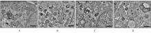
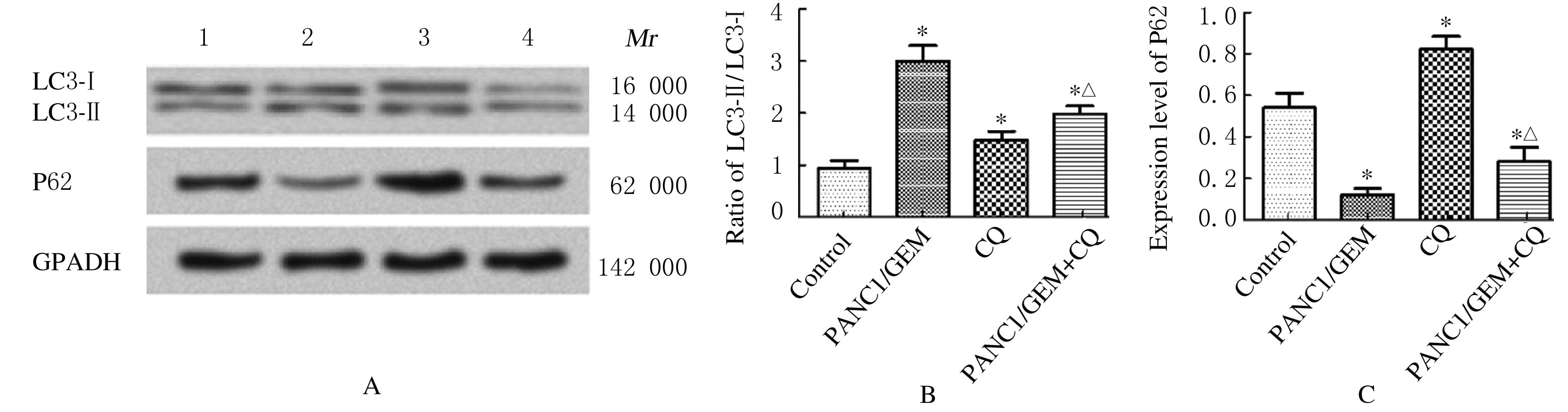
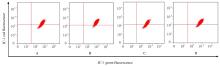
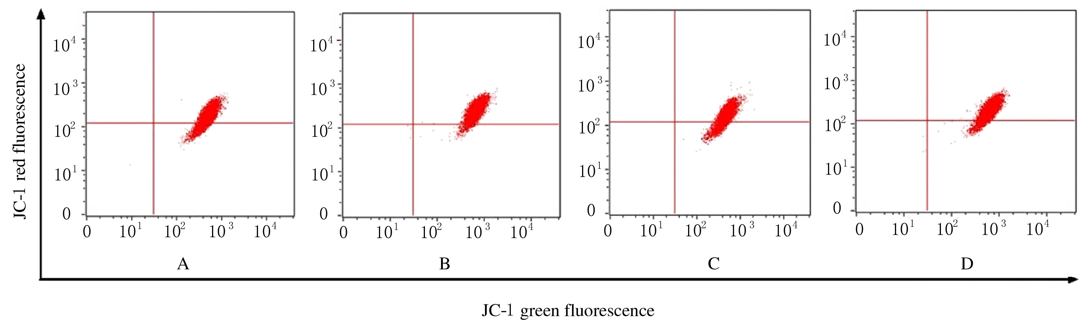
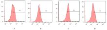
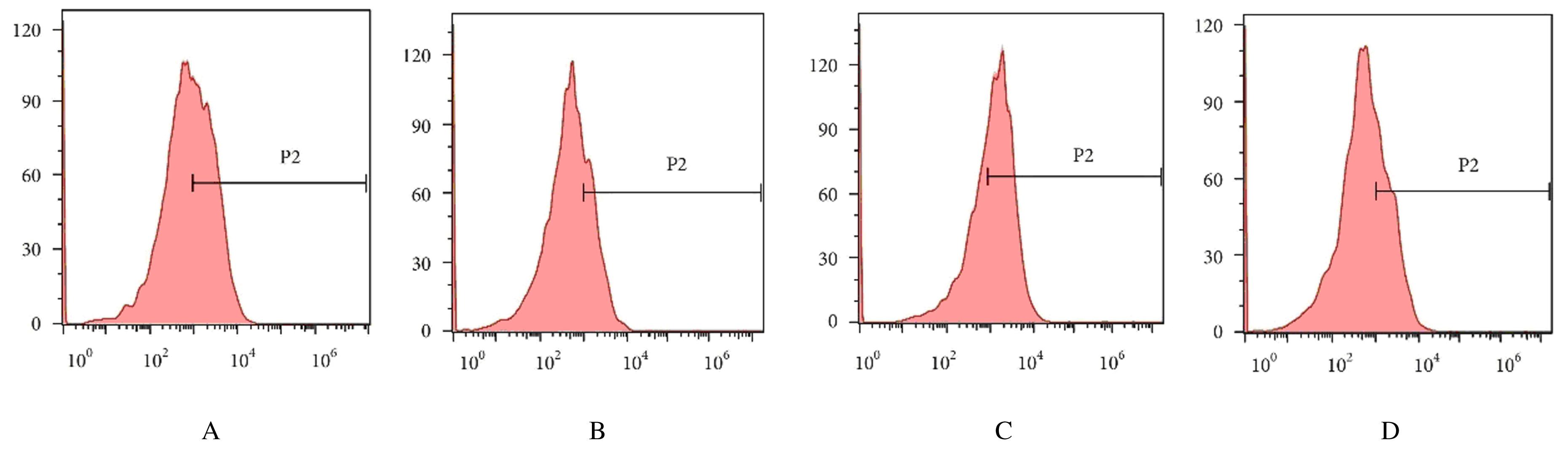
  • Received:2020-11-05 Online:2021-07-28 Published:2021-07-22
  • Contact: Xiaoyong WU E-mail:lugegewu@163.com

摘要: 目的

探讨氯喹(CQ)对吉西他滨(GEM)耐药胰腺癌(PC)细胞的影响,阐明其相关作用机制。

方法

利用低浓度依次递增法建立GEM耐药PC PANC1细胞株(PANC1/GEM),分为GEM耐药(PANC1/GEM)组、CQ组和GEM耐药组+CQ(PANC1/GEM+CQ)组,并以PANC1细胞为对照组。CCK-8法验证PANC1/GEM细胞株是否成功建立;透射电子显微镜下观察各组细胞中自噬小体数量,Western blotting法检测自噬标志蛋白胞浆型微管相关蛋白1轻链3(LC3-Ⅰ)、膜型微管相关蛋白1轻链3(LC3-Ⅱ)和P62蛋白表达水平,分别应用JC-1法、DCFH-DA法和Annexin Ⅴ-FITC/PI双染法采用流式细胞仪检测各组细胞中线粒体膜电位、活性氧(ROS)水平和细胞凋亡率,采用Western blotting法检测各组细胞中B细胞淋巴瘤2(Bcl-2)、Bcl-2相关X蛋白(Bax)和裂解的半胱氨酸天冬氨酸蛋白酶3(Cleaved caspase-3)蛋白表达水平。

结果

成功建立PANC1/GEM细胞株, 与PANC1细胞比较,GEM对PANC1/GEM细胞的半数抑制浓度(IC50)明显增加(P<0.01)。与对照组比较,PANC1/GEM组、CQ组和PANC1/GEM+CQ组细胞中自噬小体数量明显增加且LC3-Ⅱ/LC3-Ⅰ比值均明显升高(P<0.05),PANC1/GEM组和PANC1/GEM+CQ组细胞中P62蛋白表达水平明显降低(P<0.05),CQ组细胞中P62蛋白表达水平明显升高(P<0.05);与PANC1/GEM组比较, PANC1/GEM+CQ组细胞中LC3-Ⅱ/LC3-Ⅰ比值明显降低(P<0.05),P62蛋白表达水平明显升高(P<0.05)。与对照组比较,PANC1/GEM组和PANC1/GEM+CQ组细胞线粒体膜电位、ROS水平和细胞凋亡率明显降低(P<0.05),CQ组细胞线粒体膜电位、ROS水平和细胞凋亡率明显升高(P<0.05);与PANC1/GEM组比较,PANC1/GEM+CQ组细胞线粒体膜电位、ROS水平和细胞凋亡率明显升高(P<0.05)。与对照组比较,PANC1/GEM组和PANC1/GEM+CQ组细胞中Bcl-2蛋白表达水平明显升高(P<0.05),Bax和Cleaved caspase-3蛋白表达水平明显降低(P<0.05),CQ组细胞中Bcl-2蛋白表达水平明显降低(P<0.05),Bax和Cleaved caspase-3蛋白表达水平明显升高(P<0.05);与PANC1/GEM组比较,PANC1/GEM+CQ组细胞中Bcl-2蛋白表达水平明显降低(P<0.05),Bax和Cleaved caspase-3蛋白表达水平明显升高(P<0.05)。

结论

CQ能够明显促进耐药PC细胞对GEM的敏感性,其机制可能与抑制PC细胞自噬,降低细胞线粒体膜电位,促进细胞中ROS的产生,从而上调GEM所诱导的细胞凋亡有关。

关键词: 氯喹, 胰腺肿瘤, 吉西他滨, 耐药细胞, 细胞自噬, 线粒体

Abstract: Objective

To investigate the effect of chloroquine (CQ) on the gemcitabine(GEM) resistant pancreatic cancer(PC) cells, and to clarify its related mechanism;

Methods

The GEM resistant PANC1 cell line (PANC1/GEM) was established by low concentration sequential increasing method, and the cells were divided into GEM resistant group (PANC1/GEM group), CQ group and GEM resistant+CQ group (PANC1/GEM+CQ group), and the PANC1 cells were used as control group at the same time. CCK-8 method was used to verify whether the cell line was successfully established.The number of autophagy bodies was observed by transmission electron microscope, and the expression levels of autophagy marker proteins cytoplas microtubule-associated protein 1 light chain 3 (LC3-Ⅰ),membrane microtubule-associated protein 1 light chain 3 (LC3-Ⅱ) and P62 proteins were detected by Western blotting method; JC-1 method, DCFH-DA, Annexin Ⅴ-FITC/PI double staining and flow cytometry were used to detect the mitochondrial membrane potential,the reactive oxygen species (ROS) levels and the apoptotic rates of the cells in various groups. The expression levels of apoptosis related proteins B cell lymphoma-2(Bcl-2), Bcl-2 associated X protein(Bax) and Cleaved caspase-3 proteins in the cells in various groups were detected by Western blotting method.

Results

The PANC1/GEM cell line was successfully established, and the IC50 value of GEM to the PANC1/GEM cells was significantly increased compared with PANC1 cells(P<0.01). Compared with control group, the number of autophagy bodies and the LC3-Ⅱ/LC3-Ⅰ ratios in PANC1/GEM group, CQ group and PANC1/GEM+CQ group were significantly increased (P<0.05), and the levels of P62 protein in PANC1/GEM group and PANC1/GEM+CQ group were significantly decreased (P < 0.05), and the expression level of P62 protein in CQ group was significantly increased(P<0.05). Compared with PANC1/GEM group, the LC3-Ⅱ/LC3-Ⅰ ratio in PANC1/GEM+CQ group was significantly decreased(P<0.05), and the expression level of P62 protein was significantly increased (P<0.05).Compared with control group, the mitochondrial membrane potentials of the cells in PANC1/GEM group and PANC1/GEM+CQ group, the ROS levels and the apoptotic rates were significantly decreased (P<0.05),and the mitochondrial membrane potential,the ROS levels and the apoptotic rate in CQ group were significantly increased (P<0.05);compared with PANC1/GEM group,the mitochondrial membrane potential,the ROS level and the apoptotic rate in PANC1/GEM+CQ group were significantly increased(P<0.05).Compared with control group, the expression levels of Bcl-2 protein in PANC1/GEM and PANC1/GEM+CQ groups were increased (P<0.05), while the expression levels of Bax and Cleaved caspase-3 proteins were significantly decreased (P<0.05);the expression level of Bcl-2 protein in CQ group was significantly decreased (P<0.05),and the expression levels of Bax and Cleaved caspase-3 proteins were significantly increased (P<0.05). Compared with PANC1/GEM group, the expression level of Bcl-2 protein in PANC1/GEM+CQ group was significantly decreased (P<0.05), while the expression levels of Bax and Cleaved caspase-3 proteins were significantly increased (P<0.05).

Conclusion

CQ can significantly promote the sensitivity of drug-resistant PC cells to GEM, the mechanism of which may be related to the inhibition of autophagy of pancreatic cancer cells, the reduction of mitochondrial membrane potential, the promotion of ROS production in the cells, and the up-regulation of GEM-induced apoptosis.

Key words: chloroquine, pancreatic neoplasms, gemcitabine, resistant cells, autophagy, mitochondria

中图分类号: 

  • R735.9