吉林大学学报(医学版) ›› 2023, Vol. 49 ›› Issue (1): 84-93.doi: 10.13481/j.1671-587X.20230111

• 基础研究 • 上一篇    下一篇

经微弧氧化/碱处理并加载RGD肽涂层多孔钛合金支架的制备及其生物学特性

陈丽燕,林景广()   

  1. 锦州医科大学附属第二医院口腔预防科,辽宁 锦州 121000
  • 收稿日期:2022-04-12 出版日期:2023-01-28 发布日期:2023-02-03
  • 通讯作者: 林景广 E-mail:379311389@qq.com
  • 作者简介:陈丽燕(1996-),女,山东省泰安市人,在读硕士研究生,主要从事口腔种植材料方面的研究。
  • 基金资助:
    辽宁省科技厅自然科学基金项目(20170540332)

Preparation and biological properties of porous titanium alloy scaffolds treated by micro-arc oxidation/alkali and loaded with RGD peptide coating

Liyan CHEN,Jingguang LIN()   

  1. Department of Oral Prevention,Second Affiliated Hospital,Jinzhou Medical University,Jinzhou 121000,China
  • Received:2022-04-12 Online:2023-01-28 Published:2023-02-03
  • Contact: Jingguang LIN E-mail:379311389@qq.com

摘要:

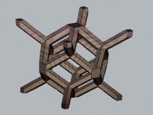
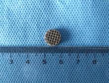
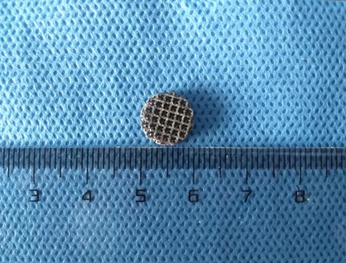
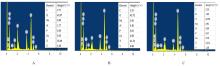
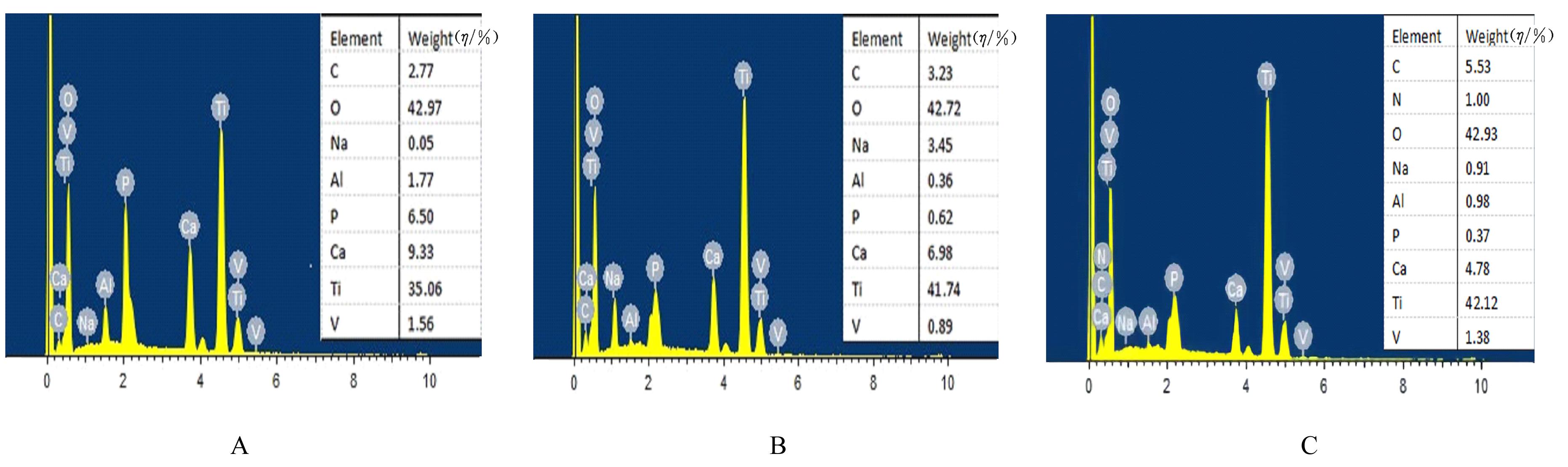
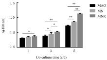
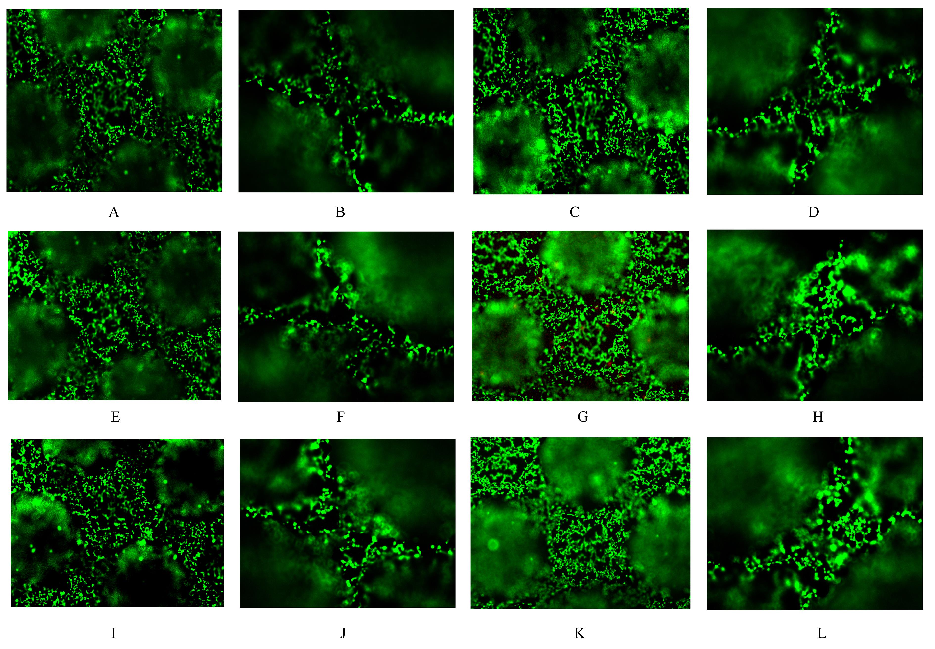
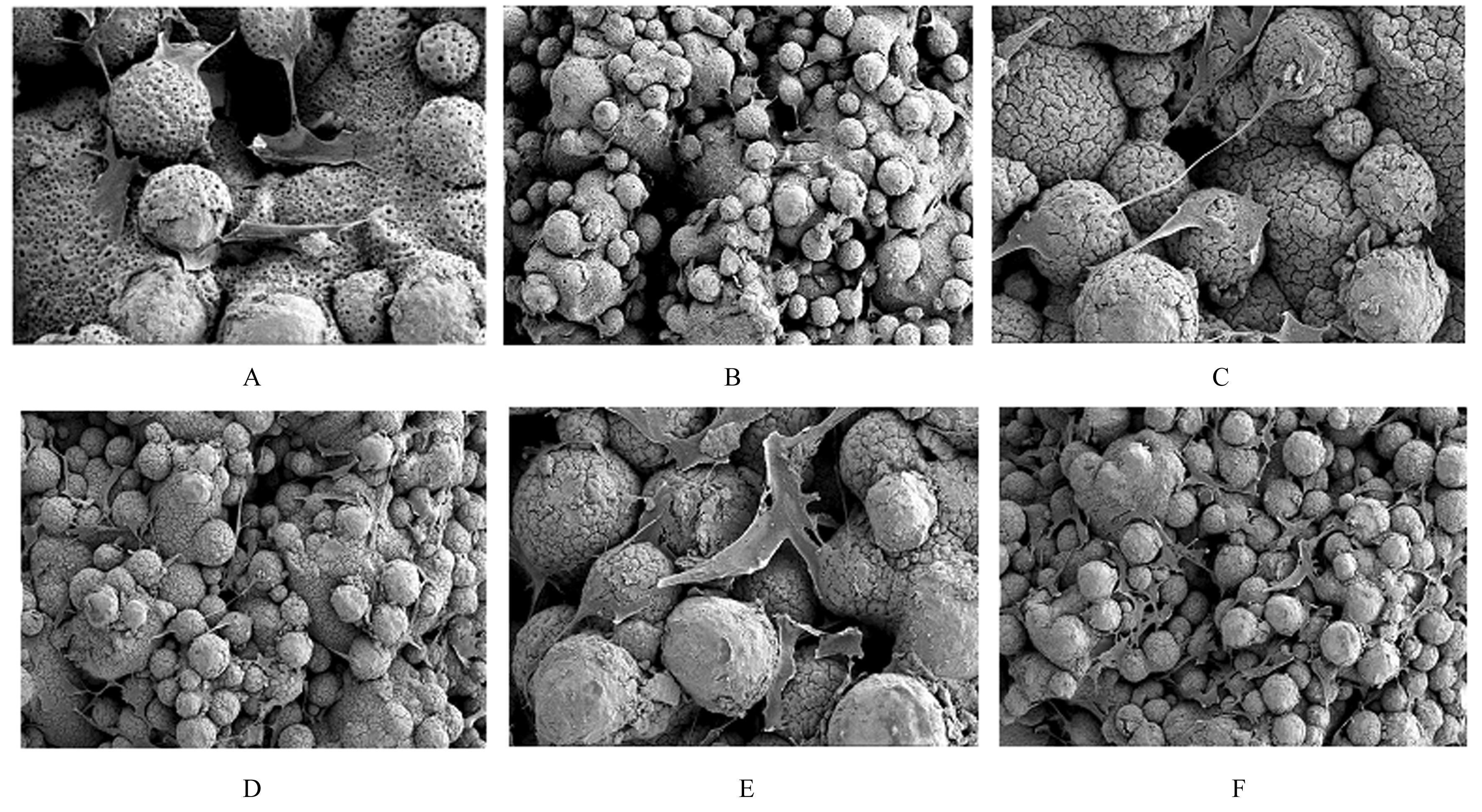
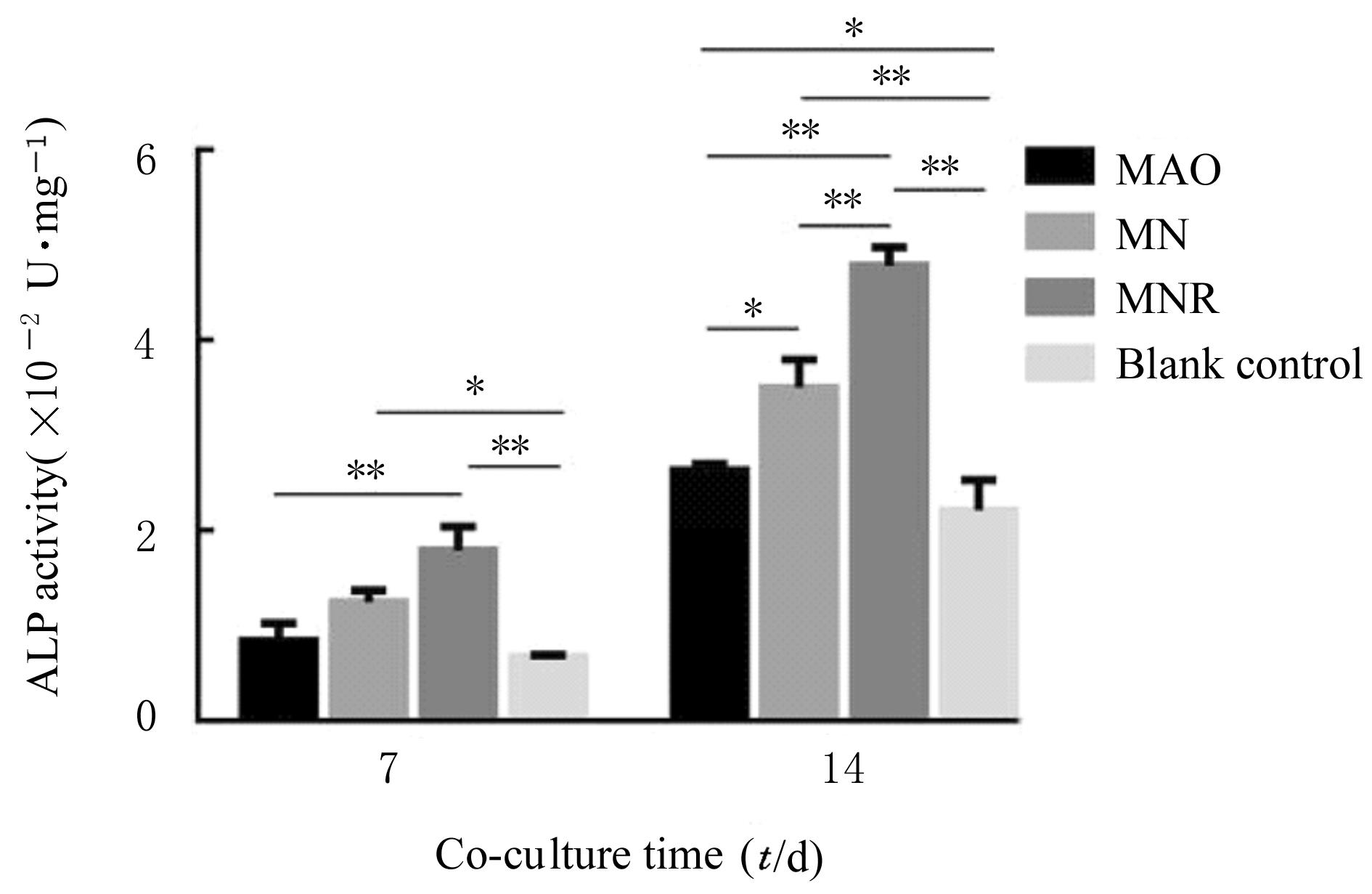
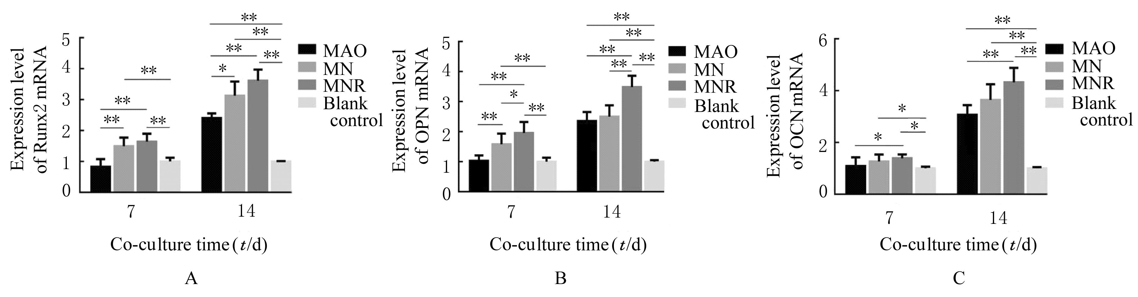
目的 通过3D打印技术打印多孔钛合金支架,研究其表面微弧氧化(MAO)/碱处理并加载精氨酸-甘氨酸-天冬氨酸(RGD)肽涂层对前成骨细胞生物学行为的影响。 方法 设计并打印3D多孔钛合金支架,对其进行不同表面处理后分为MAO组、MAO/碱处理(MN)组、MAO/碱处理加载RGD肽(MNR)组,另设空白对照组。检测各组多孔钛合金支架的弹性模量,扫描电子显微镜(SEM)观察各组多孔钛合金支架表面微观结构,能谱分析仪(EDS)检测各组多孔钛合金支架表面元素构成,接触角测量仪检测各组多孔钛合金支架表面水滴接触角大小。将小鼠胚胎成骨细胞前体细胞(MC3T3-E1细胞)与各组支架共培养,CCK-8法检测各组多孔钛合金支架表面细胞增殖活性,Live/Dead细胞染色法检测各组多孔钛合金支架的生物相容性,细胞黏附实验观察各组细胞在多孔钛合金支架表面黏附情况,采用碱性磷酸酶(ALP)试剂盒检测各组细胞ALP活性,实时荧光定量PCR(RT-qPCR)法检测各组细胞中Runt相关转录因子2(Runx2)、骨桥素(OPN)和骨钙素(OCN)mRNA表达水平。 结果 3D打印多孔钛合金支架的弹性模量为(1.17±0.62) GPa。SEM观察,MAO处理后的多孔钛合金支架表面呈现火山口样形貌,碱处理后出现细小裂纹并呈现纳米级鱼鳞结构,加载RGD肽涂层的多孔钛合金支架表面观察到散在的RGD颗粒。EDS检测,MNR组支架表面RGD涂层成功加载。接触角测量仪检测,多孔钛合金支架表面接触角MAO组>MN组>MNR组。CCK-8法,培养第1、3和5 天时3组细胞增殖活性均呈增长趋势,培养第3和5 天时各组细胞增殖活性组间比较差异均有统计学意义(P<0.05或P<0.01)。Live/Dead细胞染色,3组支架均具备良好的体外相容性。细胞黏附实验,共培养48 h后,MNR组细胞数量和形态伸展均优于MAO组和MN组。培养第7天时,与MAO组比较,MNR组细胞ALP活性明显升高(P<0.01),培养第14天时3组细胞ALP活性组间两两比较差异均有统计学意义(P<0.05或P<0.01)。RT-qPCR法检测,培养第7天时,与空白对照组和MAO组比较,MN组和MNR组细胞中Runx2和OPN mRNA表达水平均明显升高(P<0.01),MNR组细胞中OCN mRNA表达水平明显升高(P<0.05);培养第14天时,MAO组、MN组和MNR组细胞中Runx2和OPN mRNA表达水平组间两两比较差异均有统计学意义(P<0.05或P<0.01),与空白对照组比较,MAO组、MN组和MNR组细胞中OCN mRNA表达水平均明显升高(P<0.01)。 结论 3D打印多孔钛合金支架具有与人体骨组织匹配的弹性模量,支架表面MAO/碱处理并加载RGD肽涂层对MC3T3-E1细胞无毒性且对其成骨分化有促进作用。

关键词: 3D打印, 钛合金, 微弧氧化, 碱处理, 精氨酸-甘氨酸-天冬氨酸肽

Abstract:

Objective To print the porous titanium alloy scaffolds by 3D printing technology,and to study the effect of micro arc oxidation (MAO)/alkali treatment and arginine-glycine-aspartic acid(RGD) coating on the biological behavior of osteoblasts. Methods The 3D porous titanium alloy scaffolds were designed and printed, and they were divided into MAO group, MAO/alkali treatment (MN) group, MAO/alkali treatment loaded with RGD peptide coating (MNR) group after different surface treatments,another blank control group was set up.The elastic modulus of the scaffolds in various groups was detected.The microstructures of the scaffold surface in various groups were observed by scanning electron microscope (SEM), the elemental compositions of the scaffold surface were detected by energy dispersive spectroscopy (EDS), and the contact angles of water droplets on the scaffold surface were measured by contact angle measuring instrument.The mouse embryonic osteoblast precursor cells (MC3T3-E1 cells) were co-cultured with the scaffolds in various groups.CCK-8 assay was used to detect the cell proliferation activities,Live/Dead cell staining was used to detect the biocompatibility of the scaffolds in various groups, cell adhesion test was used to observe the adhesion of cells on the material surface, and alkaline phosphatase (ALP) kit was used to detect the ALP activities.Real-time fluorescence quantitative PCR (RT-qPCR) method was used to detect the expression levels of Runt-related transcription factor 2(Runx2), osteopontin(OPN) and osteocalcin(OCN) mRNA in the cells in various groups. Results The elastic modulus of 3D printed scaffolds was (1.17±0.62) GPa. The SEM observation results showed that the surface of the scaffolds after MAO treatment showed crater-like morphology. After alkali treatment, small cracks and nano scale structure appeared.The scattered RGD particles were found on the surface of the scaffolds loaded with RGD peptide coating.The ERS detection showed that the RGD peptide coating was successfully loaded on the surface of scaffolds. The contact angle measuring instrument detection results showed that the surface contact angles were MAO group>MN group>MNR group. The cell proliferation activities in three groups showed an increasing trend on the 1st, 3rd and 5th days detected by CCK-8 assay, and there were statistically significant differences in the cell proliferation activities between various groups on the 3rd and 5th days of culture(P<0.05 or P<0.01). The results of Live/Dead cell staining showed that the scaffolds in three groups had good in vitro compatibility. In cell adhesion test, after 48 h of co-culture, the number and morphological extension of cells in MNR group were better than those in MAO group and MN group. On the 7th day of culture, compared with MAO group, the activity of ALP in the cells in MNR group was significantly increased (P<0.01); on the 14th day of culture, there were significant differences in the ALP activities between three groups (P<0.05 or P<0.01).The results of RT-qPCR method showed that the expression levels of Runx2 and OPN mRNA in MN group and MNR group were higher than those in blank control group and MAO group on the 7th day of culture (P<0.01), and the expression level of OCN mRNA in MNR group was higher than those in blank control group and MAO group (P<0.05); on the 14th day of culture,there were significant differences in the expression levels of Runx2 and OPN mRNA between three groups(P<0.05 or P<0.01). Compared with blank control group, the expression levels of OCN mRNA in MAO group,MN group,and MNR group were significantly increased (P<0.01). Conclusion The 3D printed porous titanium alloy scaffolds hve the elastic modulus matching with the human bone tissue. The surface MAO/alkali treatment and loading with RGD peptide coating are non-toxic to the MC3T3-E1 cells and can promote their osteogenic differentiation.

Key words: 3D printing, Titanium alloy, Micro-arc oxidation, Alkali treatment, Arginine-glycine-aspartic acid peptide

中图分类号: 

  • R318.08