吉林大学学报(医学版) ›› 2025, Vol. 51 ›› Issue (5): 1384-1389.doi: 10.13481/j.1671-587X.20250527

• 临床医学 • 上一篇    

左下肢非典型脂肪瘤样肿瘤1例报告及文献复习

陈明1(),罗清华1,金红光2,韩亮3   

  1. 1.内蒙古自治区呼伦贝尔市人民医院 内蒙古民族大学呼伦贝尔临床医学院骨科,内蒙古 呼伦贝尔 021000
    2.内蒙古民族大学附属医院骨科,内蒙古 通辽 028000
    3.吉林大学中日联谊医院病理科,吉林 长春 130033
  • 收稿日期:2025-02-10 接受日期:2025-04-25 出版日期:2025-09-28 发布日期:2025-11-05
  • 通讯作者: 陈明 E-mail:1057744806@qq.com
  • 作者简介:陈 明(1990-),男,内蒙古自治区呼伦贝尔市人,主治医生,医学硕士,主要从事骨科临床疾病方面的研究。
  • 基金资助:
    内蒙古自治区科技厅自然科学基金项目(2024LHMS08036)

Atypical lipomatous tumor of left lower limb: A case report and literature review

Ming CHEN1(),Qinghua LUO1,Hongguang JIN2,Liang HAN3   

  1. 1.Department of Orthopedics,People’s Hospital,Hulunbuir City,Inner Mongolia Autonomous Region,College of Hulunbuir Clinical Medical Sciences,Inner Mongolia Minzu University,Hulunbuir 021000,China
    2.Department of Orthopedics,Affiliated Hospital,Inner Mongolia Minzu University,Tongliao 028000,China
    3.Department of Pathology,China-Japan Union Hospital,Jilin University,Changchun 130033,China
  • Received:2025-02-10 Accepted:2025-04-25 Online:2025-09-28 Published:2025-11-05
  • Contact: Ming CHEN E-mail:1057744806@qq.com

摘要:

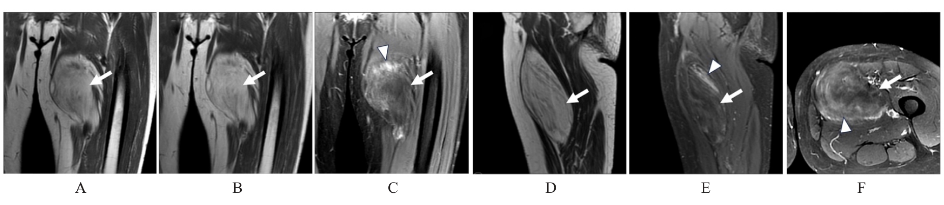
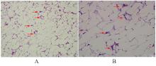
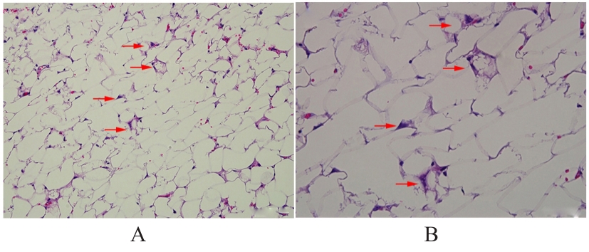
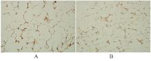
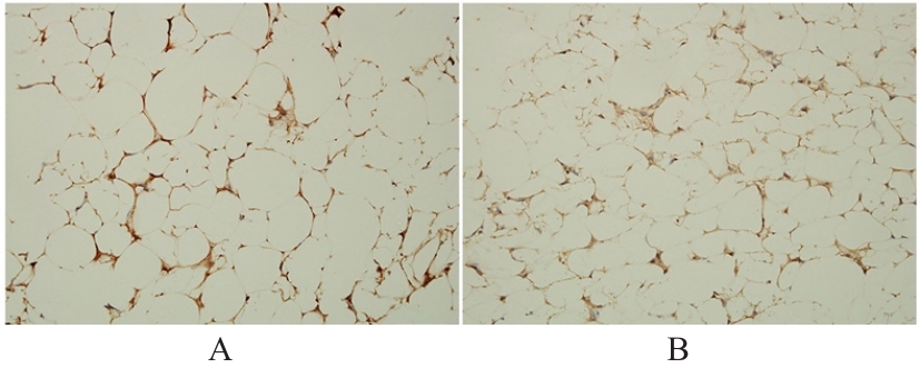
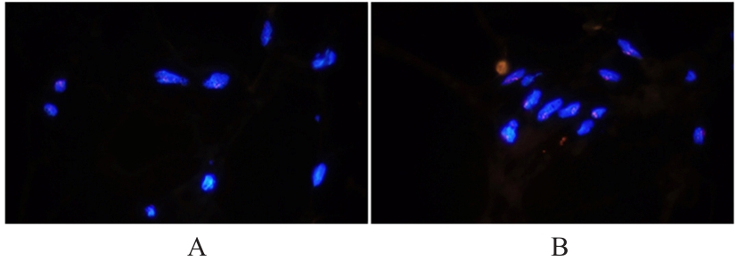
非典型脂肪瘤样肿瘤(ALT)是一种起源于脂肪细胞组织且少见的软组织肉瘤。现报道1例下肢ALT患者的临床表现、影像学资料及病理检查资料,为该疾病的临床诊治提供参考。患者,男性,64岁,因左大腿肿物伴胀痛5年入院。查体于左大腿内侧触及皮下肿物,质软,活动性良好,压痛阳性。磁共振成像(MRI)平扫检查结果显示肿物边界清楚,包膜完整;T1WI图像呈高信号,其信号强度似皮下脂肪信号;T2加权成像(T2WI)-Ideal序列同相位图像呈高信号,水像显示肿块大部分呈低信号,其内可见多发高信号;T2WI脂肪抑制序列可见低信号肿块,内有多发斑片状高信号。患者入院3 d后行病损切除术。术中见肿物位于筋膜内、股四头内侧肌肉间,呈脂肪样,有完整包膜,与周围组织局部粘连较轻,肌肉部分有侵蚀,完整切除肿物及侵蚀肌肉。术后病理检查见灰黄色结节1枚,体积为16.0 cm×10.0 cm×4.0 cm,表面光滑,包膜完整,实质呈灰黄色,脂肪样,质软;另见灰黄色组织数块,总体积为5.0 cm×4.0 cm×1.2 cm,实质呈灰褐色,质地中等。显微镜下见病变组织主要由相对成熟的脂肪组织构成,可见细胞核增大、浓染,散在分布单核或多核的不典型间质细胞,纤维分隔区内存在数量不等的单泡状或多泡状脂肪母细胞。免疫组织化学染色显示细胞周期蛋白依赖性激酶4(CDK4)和鼠双微体2(MDM2)阳性,荧光原位杂交(FISH)基因检测显示肿瘤细胞内MDM2基因扩增。病理诊断为左下肢ALT。术后6个月随访,未见肿瘤复发。术前MRI检查可为ALT诊断提供有效依据,术后病理学检查可验证ALT诊断,有助于判定患者预后。

关键词: 下肢, 非典型脂肪瘤样肿瘤, 脂肪细胞, 预后, 鼠双微体2

Abstract:

Atypical lipomatous tumor (ALT) is a rare soft tissue sarcoma originating from adipocytic tissue. The clinical manifestations, imaging findings, and pathological examinations of one patient with ALT in the lower extremity were reported to provide reference for the clinical diagnosis and treatment of this disease. The patient was a 64-year-old male who was admitted due to a mass in the left thigh with swelling and pain for 5 years. The physical examination results showed a subcutaneous mass was palpable on the medial side of the left thigh, soft in texture, demonstrating good mobility and positive tenderness.The magnetic resonance imaging (MRI) non-contrast scan showed a well-defined lesion with an intact capsule. The lesion presented high signal intensity on T1-weighted image(T1WI), similar to subcutaneous fat signal; on T2-weighted image (T2WI)-Ideal in-phase images, the lesion showed high signal, while water images showed low signal, with multiple high-signal foci inside. On T2WI fat-suppression sequence, a low-signal mass was observed with multiple patchy high signals inside.Three days after admission, the patient underwent lesion resection. Intraoperatively, the tumor was located within the fascia, between the medial quadriceps muscles, presenting as lipomatous tissue with an intact capsule, mildly adherent to surrounding tissue, and with partial muscle invasion. The mass and invaded muscle were completely excised. The postoperative pathological examination results revealed a gray-yellow nodule measuring 16.0 cm×10.0 cm×4.0 cm, with a smooth surface and intact capsule. The cut surface was gray-yellow, fatty-like, and soft in texture. Additional gray-yellow tissue fragments were found, with a total volume of 5.0 cm×4.0 cm×1.2 cm, exhibiting a gray-brown cut surface with moderate texture. Under microscope, the lesion consisted mainly of relatively mature adipose tissue. Enlarged and hyperchromatic nuclei were observed, with scattered mononuclear or multinuclear atypical stromal cells. Fibrous septa contained variable numbers of univacuolated or multivacuolated lipoblasts. The immunohistochemistry results showed positivity for cyclin-dependent kinase 4 (CDK4) and murine double minute 2(MDM2). The fluorescence in situ hybridization (FISH) results demonstrated MDM2 gene amplification in tumor cells. The pathological diagnosis was ALT of the left lower extremity. At 6-month follow-up after operation, no tumor recurrence was observed. The preoperative MRI detection may provide effective evidence for the diagnosis of ALT, while postoperative pathological examination can confirm the diagnosis and help evaluate the prognosis of the patients.

Key words: Lower limb, Atypical lipomatous tumor, Adipocyte, Prognosis, Murine double minute 2

中图分类号: 

  • R737.33