吉林大学学报(医学版) ›› 2026, Vol. 52 ›› Issue (2): 440-450.doi: 10.13481/j.1671-587X.20260216

• 基础研究 • 上一篇    

基于卡介苗感染过程中MAPK-Mcl-1信号通路和巨噬细胞极化调控机制的生物信息学分析及其实验验证

葛睿涵1,2,李晨1,2,王生鹏1,3,4,卢洋1,2,谭彩霞1,2,崔皓天1,3,4,王新敏3,4(),章乐1,2,4()   

  1. 1.石河子大学医学院病理生理学教研室,新疆 石河子 832008
    2.新疆地方与民族高发病教育部 重点实验室,新疆 石河子 832008
    3.石河子大学第一附属医院泌尿外科,新疆 石河子 832008
    4.国家卫健委中亚高发病防治重点实验室,新疆 石河子 832008
  • 收稿日期:2025-05-05 接受日期:2025-06-16 出版日期:2026-03-28 发布日期:2026-04-15
  • 通讯作者: 王新敏,章乐 E-mail:1977602697@qq.com;1257067540@qq.com
  • 作者简介:葛睿涵(1998-),女,山东省潍坊市人,在读硕士研究生,主要从事感染性疾病病理生理学方面的研究。
  • 基金资助:
    新疆生产建设兵团科技局指导性科技计划项目(2022ZD045);新疆生产建设兵团科技局指导性科技计划项目(2022ZD073);新疆生产建设兵团教育局研究生创新项目(BTYJXM-2024-K63);新疆生产建设兵团第八师指导性计划项目(2025ZDSF01)

Bioinformatic analysis on regulatory mechanism of MAPK-Mcl-1 signaling pathway and macrophage polarization during Bacillus Calmette-Guérin infection and its experimental validation

Ruihan GE1,2,Chen LI1,2,Shengpeng WANG1,3,4,Yang LU1,2,Caixia TAN1,2,Haotian CUI1,3,4,Xinmin WANG3,4(),Le ZHANG1,2,4()   

  1. 1.Department of Pathophysiology,School of Medicine,Shihezi University,Shihezi 832008,China
    2.Xinjiang Provincial and Ethnic High Incidence Key Laboratory,Ministry of Education,Shihezi 832008,China
    3.Department of Urology,First Affiliated Hospital,Shihezi University,Shihezi 832008,China
    4.Key Laboratory for Prevention and Treatment of High-Incidence Diseases in Central Asia,National Health Commission,Shihezi 832008,China
  • Received:2025-05-05 Accepted:2025-06-16 Online:2026-03-28 Published:2026-04-15
  • Contact: Xinmin WANG,Le ZHANG E-mail:1977602697@qq.com;1257067540@qq.com

摘要:

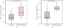
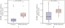
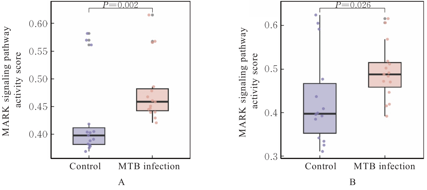
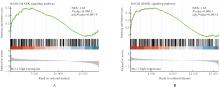
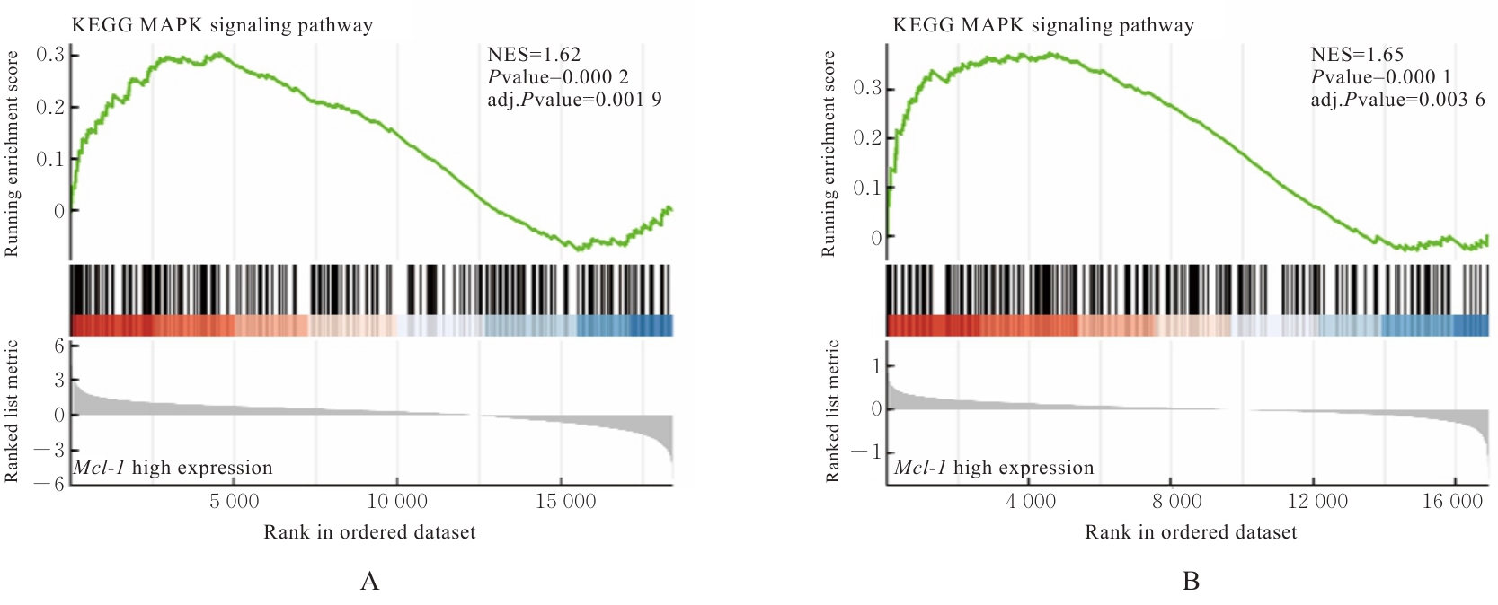
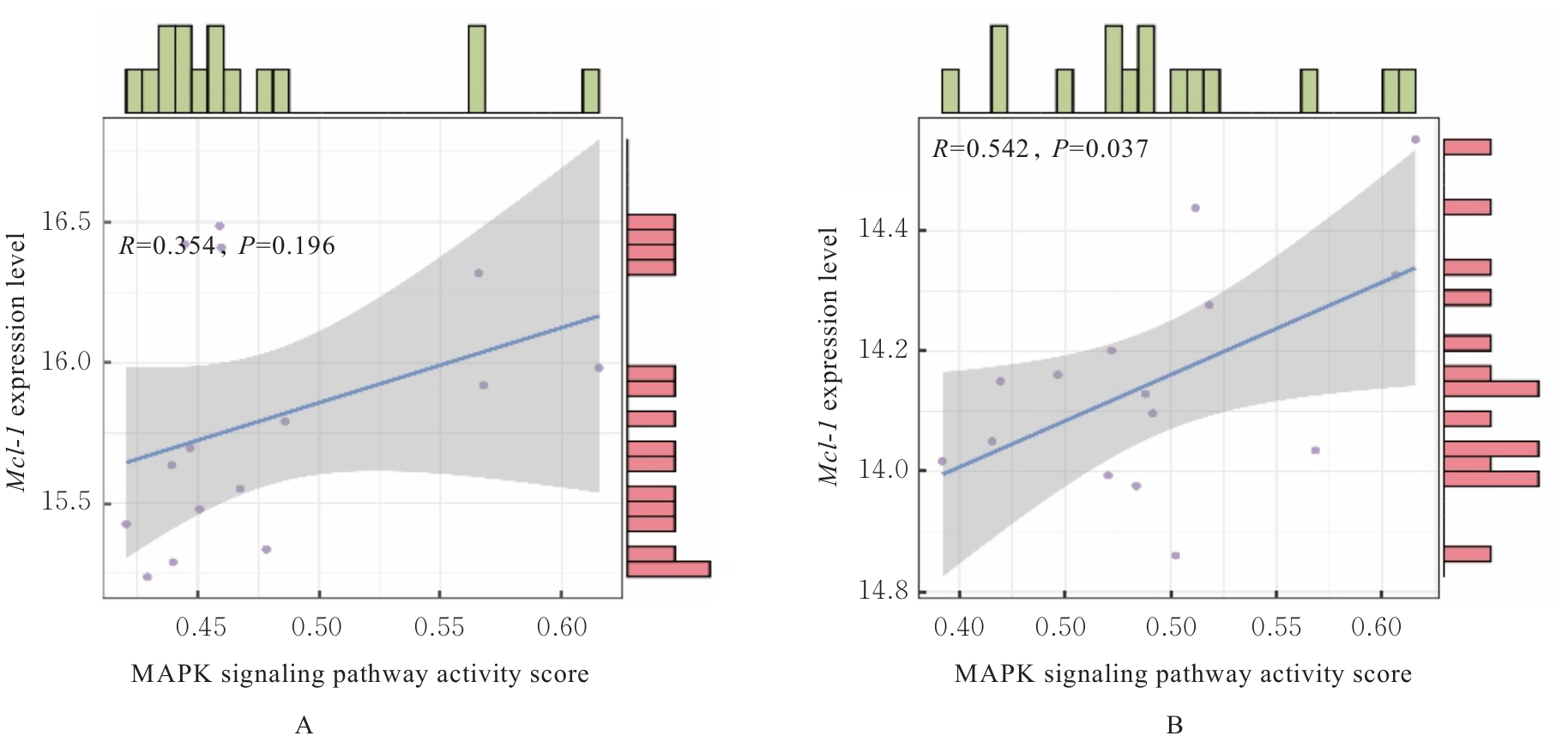
目的 探讨丝裂原活化蛋白激酶(MAPK)-髓系细胞白血病序列1(Mcl-1)信号轴在卡介苗(BCG)感染巨噬细胞极化进程中的作用,并阐明其可能的分子机制。 方法 从高通量基因表达(GEO)数据库下载GSE89391和GSE51029转录组测序数据,从分子特征数据库(MSigDB)下载MAPK信号通路相关基因集。采用基因集变异分析(GSVA)包对GEO数据库中巨噬细胞数据进行单样本基因集富集分析(ssGSEA),计算MAPK信号通路活性评分。根据Mcl-1表达水平中位数将感染样本分为Mcl-1高表达组和Mcl-1低表达组,并进行基因集富集分析(GSEA)。采用Spearman相关分析法评估GEO数据库中巨噬细胞中Mcl-1表达水平与MAPK通路活性的相关性。配制细胞外信号调节激酶(ERK)通路阻断剂PD98059(50 μmol·L-1)、c-Jun氨基末端激酶(JNK)通路阻断剂SP600125(30 μmol·L-1)和p38通路阻断剂SB203580(60 μmol·L-1)。将Raw264.7细胞分为对照组、各阻断剂处理组、BCG组及BCG感染后各阻断剂处理组,制备BCG菌液并感染相应组细胞,各组加入相应阻断剂。0、12和24 h后收集各组细胞上清及细胞样本。采用酶联免疫吸附试验(ELISA)法检测各组巨噬细胞上清中Mcl-1、白细胞介素6(IL-6)、肿瘤坏死因子α(TNF-α)、白细胞介素10(IL-10)及转化生长因子β(TGF-β)水平,实时荧光定量PCR(RT-qPCR)法检测各组巨噬细胞中M1型标志物诱导型一氧化氮合酶(iNOS)mRNA和M2型标志物炎症区域分子1(Fizz1)mRNA表达水平。 结果 GEO数据库,与未被结核分枝杆菌(MTB)感染的对照巨噬细胞比较,MTB感染的巨噬细胞中Mcl-1基因表达水平明显升高(P<0.05)。ssGSEA分析,与未被MTB感染的对照巨噬细胞比较,MTB感染的巨噬细胞中MAPK信号通路活性评分明显升高(P<0.05)。GSEA分析,Mcl-1高表达组差异表达基因显著富集于MAPK通路(P<0.05)。Spearman相关性分析,Mcl-1基因表达水平与MAPK信号通路活性评分呈正相关关系(P<0.05)。干预12和24 h后,与BCG组比较,阻断剂处理的各组巨噬细胞上清液中Mcl-1水平明显降低(P<0.05)。干预12 h后,与对照组比较,BCG组巨噬细胞上清液中IL-6和TNF-α水平均明显升高(P<0.05);与BCG组比较,BCG+SP组和BCG+PD+SP组巨噬细胞上清液中IL-6及TNF-α水平均明显降低(P<0.05)。干预24 h后,与对照组比较,BCG组巨噬细胞上清液中TNF-α水平明显升高(P<0.05);与BCG组比较,BCG+PD组、BCG+SP组、BCG+SB组和BCG+PD+SP组巨噬细胞上清液中IL-6水平均明显降低(P<0.05),加入阻断剂的各组巨噬细胞上清液中TNF-α水平均明显降低(P<0.05)。干预12 h后,与对照组比较,BCG组巨噬细胞上清液中TGF-β水平明显升高(P<0.05);与BCG组比较,BCG+SP组、BCG+PD+SP组、BCG+PD+SB组、BCG+SP+SB组和BCG+PD+SP+SB组巨噬细胞上清液中TGF-β水平均明显升高(P<0.05)。干预24 h后,与对照组比较,BCG组巨噬细胞上清液中IL-10水平明显升高(P<0.05);与BCG组比较,BCG+PD组、BCG+SP组、BCG+PD+SP组、BCG+PD+SB组和BCG+SP+SB组巨噬细胞上清液中IL-10及TGF-β水平均明显降低(P<0.05)。感染后0 h,与对照组比较,BCG组巨噬细胞中iNOSFizz1 mRNA表达水平均明显升高(P<0.05)。干预12 h后,与对照组比较,BCG组巨噬细胞中iNOS mRNA表达水平明显降低(P<0.01),Fizz1 mRNA表达水平明显升高(P<0.01);与BCG组比较,加入阻断剂后各组巨噬细胞中iNOS mRNA表达水平均明显升高(P<0.01),BCG+PD组、BCG+PD+SP组、BCG+PD+SB组、BCG+SP+SB组和BCG+PD+SP+SB组巨噬细胞中Fizz1 mRNA表达水平明显升高(P<0.01)。干预24 h后,与对照组比较,BCG组巨噬细胞中iNOSFizz1 mRNA表达水平均明显升高(P<0.05);与BCG组比较,加入阻断剂的各组巨噬细胞中iNOS mRNA表达水平均无明显变化,差异无统计学意义(P>0.05);BCG+PD+SP组巨噬细胞中Fizz1 mRNA表达水平明显降低(P<0.01)。 结论 MAPK信号通路可能通过调控Mcl-1活性介导BCG感染巨噬细胞的极化进程,其中JNK通路发挥核心调控作用,p38与ERK通路协同参与调控。

关键词: 生物信息学, 卡介苗, 巨噬细胞极化, 髓系细胞白血病序列1, 丝裂原活化蛋白激酶信号通路, c-Jun氨基末端激酶

Abstract:

Objective To discuss the role of mitogen-activated protein kinase (MAPK)-myeloid cell leukemia sequence 1 (Mcl-1) signaling axis in the polarization process of macrophages infected with Bacillus Calmette-Guérin (BCG), and to clarify its possible molecular mechanism. Methods The transcriptome sequencing data of GSE89391 and GSE51029 were downloaded from Gene Expression Omnibus (GEO) database, and the MAPK signaling pathway-related gene sets were downloaded from Molecular Signatures Database (MSigDB). The gene set variation analysis (GSVA) package was used to perform single-sample gene set enrichment analysis (ssGSEA) on the macrophage data in GEO database, and the activity scores of MAPK signaling pathway were calculated. The infected samples were divided into Mcl-1 high expression group and Mcl-1 low expression group according to the median of Mcl-1 expression level, and gene set enrichment analysis (GSEA) was performed. Spearman correlation analysis was used to assess the correlation between Mcl-1 expression level and MAPK pathway activity in macrophages in GEO database. The extracellular signal-regulated kinase (ERK) pathway blocker PD98059 (50 μmol·L?1), c-Jun N-terminal kinase (JNK) pathway blocker SP600125 (30 μmol·L?1), and p38 pathway blocker SB203580 (60 μmol·L?1) were prepared.The Raw264.7 cells were divided into control group, various blocker-treated groups, BCG group, and various blocker-treated groups after BCG infection. BCG bacterial solution was prepared and used to infect the cells in corresponding groups, and the corresponding blockers were added to the cells in various groups. The cell supernatants and cell samples in various groups were collected at 0, 12, and 24 h after treatment. Enzyme-linked immunosorbent assay (ELISA) method was used to detect the levels of Mcl-1, interleukin-6 (IL-6), tumor necrosis factor-α (TNF-α), interleukin-10 (IL-10), and transforming growth factor-β (TGF-β) in supernatant of the macrophages in various groups; real-time fluorescence quantitative PCR (RT-qPCR) method was used to detect the expression levels of M1 marker inducible nitric oxide synthase (iNOS) mRNA and M2 marker found in inflammatory zone 1 (Fizz1) mRNA in the macrophages in various groups. Results Based on GEO databases, compared with uninfected control macrophages, the level of Mcl-1 gene in the macrophages infected with Mycobacterium tuberculosis(MTB) was significantly increased (P<0.05). The ssGSEA analysis results showed that compared with uninfected control macrophages, the MAPK signaling pathway activity score in MTB-infected macrophages was significantly increased (P<0.05). The GSEA analysis results revealed that differentially expressed genes in Mcl-1 high expression groups were significantly enriched in the MAPK pathway (P<0.05). The Spearman correlation analysis results indicated there was a positive correlation between the expression level of Mcl-1 gene and MAPK signaling pathway activity score (P<0.05). At 12 and 24 h after treatment, compared with BCG group, the levels of Mcl-1 in supernatant of the macrophages in various blocker-treated groups were significantly decreased (P<0.05). At 12 h after treatment, compared with control group, the levels of IL-6 and TNF-α in supernatant of the macrophages in BCG group were significantly increased (P<0.05). Compared with BCG group, the levels of IL-6 and TNF-α in supernatants of the macrophages in BCG+SP and BCG+PD+SP groups were significantly decreased (P<0.05). At 24 h after treatment, compared with control group, the level of TNF-α in supernatant of the macrophages in BCG group was significantly increased (P<0.05). Compared with BCG group, the levels of IL-6 in supernatants of the macrophages in BCG+PD, BCG+SP, BCG+SB, and BCG+PD+SP groups were significantly decreased (P<0.05), and the levels of TNF-α in supernatants of the macrophages in all blocker-treated groups were significantly decreased (P<0.05). At 12 h after treatment, compared with control group, the level of TGF-β in supernatant of the macrophages in BCG group was significantly increased (P<0.05). Compared with BCG group, the levels of TGF-β in supernatants of the macrophages in BCG+SP, BCG+PD+SP, BCG+PD+SB, BCG+SP+SB, and BCG+PD+SP+SB groups were significantly increased (P<0.05). At 24 h after treatment, compared with control group, the level of IL-10 in supernatant of the macrophages in BCG group was significantly increased (P<0.05). Compared with BCG group, the levels of IL-10 and TGF-β in supernatants of the macrophages in BCG+PD, BCG+SP, BCG+PD+SP, BCG+PD+SB, and BCG+SP+SB groups were significantly decreased (P<0.05). At 0 h after infection, compared with control group, the expression levels of iNOS and Fizz1 mRNA in the macrophages in BCG group were significantly increased (P<0.05). At 12 h after treatment, compared with control group, the expression level of iNOS mRNA in the macrophages in BCG group was significantly decreased (P<0.01), while the expression level of Fizz1 mRNA was significantly increased (P<0.01). Compared with BCG group, the expression levels of iNOS mRNA in the macrophages in all blocker-treated groups were significantly increased (P<0.01), and the expression levels of Fizz1 mRNA in the macrophages in BCG+PD, BCG+PD+SP, BCG+PD+SB, BCG+SP+SB, and BCG+PD+SP+SB groups were significantly increased (P<0.01). At 24 h after treatment, compared with control group, the expression levels of iNOS and Fizz1 mRNA in the macrophages in BCG group were significantly increased (P<0.05). The expression levels of iNOS mRNA in the macrophages between BCG group and all blocker-treated groups showed no statistically significant difference (P>0.05). Compared with BCG group, the expression level of Fizz1 mRNA in the macrophages in BCG+PD+SP group was significantly decreased (P<0.01). Conclusion MAPK signaling pathway may mediate the polarization process of macrophages infected with BCG by regulating Mcl-1 activity, in which the JNK pathway plays a core regulatory role, and the p38 and ERK pathways are synergistically involved in the regulation.

Key words: Bioinformatics, Bacillus Calmette-Guérin, Macrophage polarization, Myeloid cell leukemia sequence 1, Mitogen-activated protein kinase signaling pathway, c-Jun N-terminal kinase

中图分类号: 

  • R363.2