吉林大学学报(医学版) ›› 2026, Vol. 52 ›› Issue (2): 398-409.doi: 10.13481/j.1671-587X.20260212

• 基础研究 • 上一篇    

柴芪益肝方调控巨噬细胞极化对肝癌HepG2细胞恶性生物学行为的抑制作用

张希倩1,党志博2(),杜雨楠1,吴培1,谢航3,谭高峰1   

  1. 1.河南省中医院 河南中医药大学第二附属医院老年病科,河南 郑州 450053
    2.河南省中医院 河南中医药大学第二附属医院肝胆脾胃病科,河南 郑州 450053
    3.河南省中医院 河南中医药大学第二附属医院重症医学科,河南 郑州 450053
  • 收稿日期:2025-05-31 接受日期:2025-07-06 出版日期:2026-03-28 发布日期:2026-04-15
  • 通讯作者: 党志博 E-mail:1622097861@qq.com
  • 作者简介:张希倩(1988-),女,河南省郑州市人,医学硕士,主要从事中医内科老年病方面的研究。
  • 基金资助:
    河南省卫健委中医药科学研究专项课题项目(2023ZY2062)

Inhibitory effect of Chaiqi Yigan formula on malignant biological behaviors of liver cancer HepG2 cells by regulating macrophage polarization

Xiqian ZHANG1,Zhibo DANG2(),Yunan DU1,Pei WU1,Hang XIE3,Gaofeng TAN1   

  1. 1.Department of Geriatrics,Henan Provincial Hospital of Traditional Chinese Medicine,Second Affiliated Hospital,Henan University of Chinese Medicine,Zhengzhou 450053,China
    2.Department of Hepatobiliary,Spleen and Stomach Diseases,Henan Provincial Hospital of Traditional Chinese Medicine,Second Affiliated Hospital,Henan University of Chinese Medicine,Zhengzhou 450053,China
    3.Department of Critical Care Medicine,Henan Provincial Hospital of Traditional Chinese Medicine,Second Affiliated Hospital,Henan University of Chinese Medicine,Zhengzhou 450053,China
  • Received:2025-05-31 Accepted:2025-07-06 Online:2026-03-28 Published:2026-04-15
  • Contact: Zhibo DANG E-mail:1622097861@qq.com

摘要:

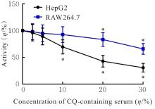
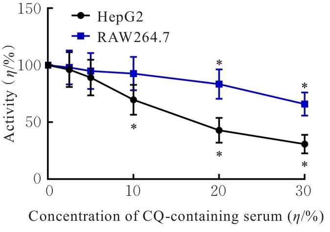
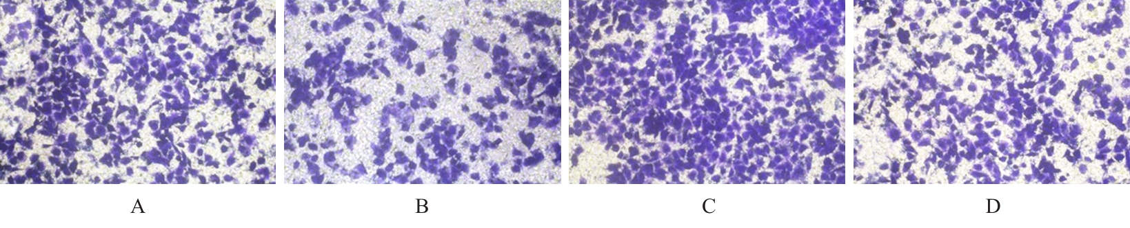
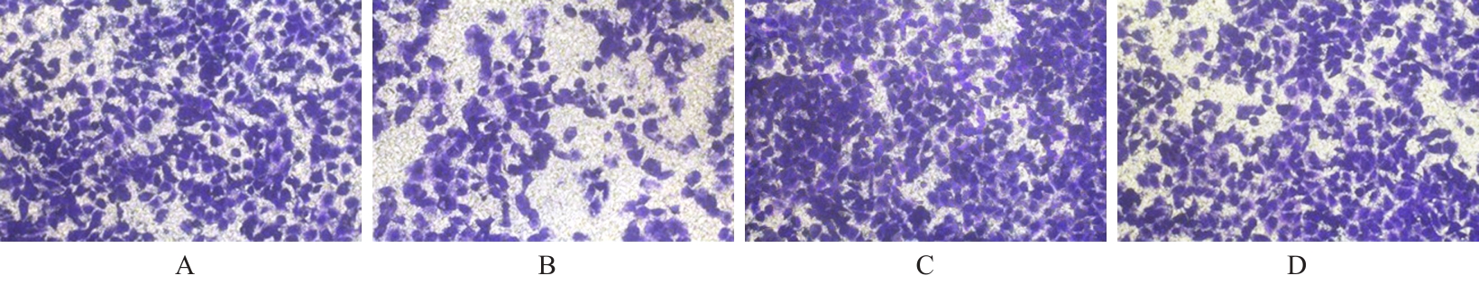
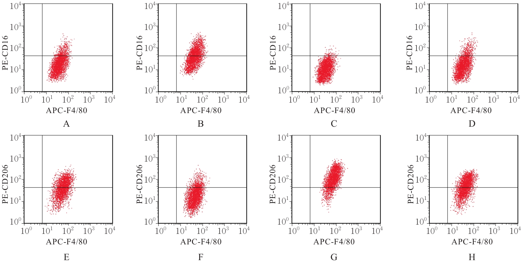
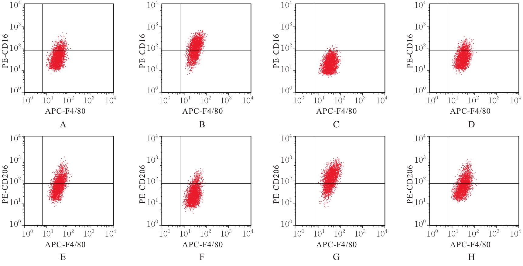
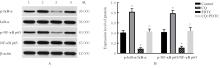
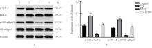
目的 探讨柴芪益肝方(CQ)通过激活核因子κB(NF-κB)信号通路调控巨噬细胞极化对肝癌HepG2细胞恶性生物学行为的抑制作用,为CQ应用于肝癌的治疗提供理论依据。 方法 制备含CQ血清,稀释后浓度为2.5%、5.0%、10.0%、20.0%和30.0%。采用细胞计数试剂盒8(CCK-8)法检测含CQ血清作用后各组RAW264.7细胞和HepG2细胞活力,酶联免疫吸附测定(ELISA)试剂盒检测含CQ血清作用后各组RAW264.7细胞上清中白细胞介素(IL)-6和IL-10水平,筛选CQ最佳处理浓度。建立RAW264.7和HepG2细胞共培养体系,并构建HepG2细胞移植瘤裸鼠模型,将共培养细胞和荷瘤小鼠分别随机分为对照组、CQ组、NF-κB抑制剂二硫代氨基甲酸吡咯烷(PDTC)组和CQ+PDTC组。采用CCK-8法和Transwell小室实验分别检测各组HepG2细胞活性、迁移细胞数和侵袭细胞数。测定各组裸鼠移植瘤体积和质量。采用流式细胞术检测各组共培养体系下RAW264.7细胞和裸鼠肿瘤组织中巨噬细胞M1和M2型极化情况;ELISA法检测各组共培养体系下RAW264.7细胞上清和裸鼠血清中M1型细胞因子IL-6、肿瘤坏死因子α(TNF-α)及诱导型一氧化氮合酶(iNOS)以及M2型细胞因子转化生长因子β(TGF-β)、精氨酸酶1(Arg-1)和IL-10水平,Western blotting法检测各组共培养体系下RAW264.7细胞和裸鼠血清中NF-κB通路相关蛋白NF-κB抑制蛋白α(IκB-α)、磷酸化IκB-α(p-IκB-α)、NF-κB p65及p-NF-κB p65表达水平。 结果 与对照组比较,CQ组HepG2细胞活力、迁移细胞数和侵袭细胞数明显降低(P<0.05),PDTC组HepG2细胞活性、迁移细胞数和侵袭细胞数明显升高(P<0.05);与CQ组比较,CQ+PDTC组HepG2细胞活性、迁移细胞数和侵袭细胞数明显升高(P<0.05)。与对照组比较,CQ组裸鼠移植瘤体积和质量均明显降低(P<0.05),PDTC组裸鼠移植瘤体积和质量明显升高(P<0.05);与CQ组比较,CQ+PDTC组裸鼠移植瘤体积和质量明显升高(P<0.05)。与对照组比较,CQ组RAW264.7细胞和裸鼠肿瘤组织中M1型巨噬细胞百分率明显升高(P<0.05),M2型巨噬细胞百分率明显降低(P<0.05);PDTC组RAW264.7细胞和裸鼠肿瘤组织中M1型巨噬细胞百分率明显降低(P<0.05),M2型巨噬细胞百分率明显升高(P<0.05)。与CQ组比较,CQ+PDTC组RAW264.7细胞和裸鼠肿瘤组织中M1型巨噬细胞百分率明显降低(P<0.05),M2型巨噬细胞百分率明显升高(P<0.05)。与对照组比较,CQ组RAW264.7细胞培养上清和裸鼠血清中IL-6、TNF-α及iNOS水平明显升高(P<0.05),TGF-β、Arg-1和IL-10水平明显降低(P<0.05);PDTC组RAW264.7细胞上清和裸鼠血清中IL-6、TNF-α及iNOS水平明显降低(P<0.05),TGF-β、Arg-1和IL-10水平明显升高(P<0.05)。与CQ组比较,CQ+PDTC组RAW264.7细胞上清和裸鼠血清中IL-6、TNF-α及iNOS水平明显降低(P<0.05),TGF-β、Arg-1和IL-10水平明显升高(P<0.05)。与对照组比较,CQ组RAW264.7细胞和裸鼠肿瘤组织中p-IκB-α/IκB-α比值及p-NF-κB p65/NF-κB p65比值明显升高(P<0.05),PDTC组RAW264.7细胞和裸鼠肿瘤组织中p-IκB-α/IκB-α比值及p-NF-κB p65/NF-κB p65比值明显降低(P<0.05)。与CQ组比较,CQ+PDTC组RAW264.7细胞和裸鼠肿瘤组织中p-IκB-α/IκB-α比值及p-NF-κB p65/NF-κB p65比值明显降低(P<0.05)。 结论 CQ可通过激活NF-κB信号通路促进巨噬细胞从M2型向M1型极化,进而抑制肝癌HepG2细胞增殖、迁移、侵袭和裸鼠皮下移植瘤生长。

关键词: 柴芪益肝方, 核因子κB, 巨噬细胞极化, 肝细胞癌, 肿瘤进展

Abstract:

Objective To discuss the inhibitory effect of Chaiqi Yigan (CQ) formula on the malignant biological behaviors of hepatocellular carcinoma HepG2 cells by regulating macrophage polarization through activation of nuclear factor κB (NF-κB) signaling pathway, and to clarify the theoretical basis for the application of CQ in the treatment of hepatocellular carcinoma. Methods CQ-containing serum was prepared and diluted to concentrations of 2.5%, 5.0%, 10.0%, 20.0%, and 30.0%.Cell counting kit-8 (CCK-8) method was used to detect the activities of RAW264.7 cells and HepG2 cells in various groups after treated with CQ-containing serum; enzyme-linked immunosorbent assay (ELISA) kits were used to detect the levels of interleukin (IL)-6 and IL-10 in supernatant of RAW264.7 cells in various groups after treated with CQ-containing serum to screen the optimal concentration of CQ for treatment. A co-culture system of RAW264.7 and HepG2 cells was established, and a nude mouse model bearing HepG2 cell xenograft tumor was constructed. The co-cultured cells and tumor-bearing mice were randomly divided into control group, CQ group, NF-κB inhibitor pyrrolidine dithiocarbamate (PDTC) group, and CQ+PDTC group. CCK-8 method and Transwell chamber assay were used to detect the activities, numbers of migration cells, and numbers of invasion cells of HepG2 cells in various groups, respectively. The volumes and mass of xenograft tumors of the nude mice in various groups were measured. Flow cytometry was used to detect M1 and M2 polarization of RAW264.7 cells in co-culture system and tumor tissue of the nude mice in various groups; ELISA method was used to detect the levels of M1-type cytokines IL-6, tumor necrosis factor α (TNF-α), and inducible nitric oxide synthase (iNOS), and M2-type cytokines transforming growth factor β (TGF-β), arginase 1 (Arg-1), and IL-10 in supernatant of the RAW264.7 cells in co-culture system and serum of the nude mice in various groups; Western blotting method was used to detect the expression levels of NF-κB pathway-related proteins NF-κB inhibitor protein α (IκB-α), phosphorylated IκB-α (p-IκB-α), NF-κB p65, and p-NF-κB p65 in RAW264.7 cells in the co-culture system and serum of the nude mice in various groups. Results Compared with control group, the activity, number of migration cells, and number of invasion cells of HepG2 cells in CQ group were significantly decreased (P<0.05), while those in PDTC group were significantly increased (P<0.05); compared with CQ group, the activity, number of migration cells, and number of invasion cells of HepG2 cells in CQ+PDTC group were significantly increased (P<0.05). Compared with control group, the volume and mass of xenograft tumor of the nude mice in CQ group were significantly decreased (P<0.05), while those in PDTC group were significantly increased (P<0.05); compared with CQ group, the volume and mass of xenograft tumor of the nude mice in CQ+PDTC group were significantly increased (P<0.05). Compared with control group, the percentages of M1-type macrophages in RAW264.7 cells and tumor tissue of the nude mice in CQ group were significantly increased (P<0.05), while the percentages of M2-type macrophages were significantly decreased (P<0.05); the percentages of M1-type macrophages in RAW264.7 cells and tumor tissue of the nude mice in PDTC group were significantly decreased (P<0.05), while the percentages of M2-type macrophages were significantly increased (P<0.05). Compared with CQ group, the percentages of M1-type macrophages in RAW264.7 cells and tumor tissue of the nude mice in CQ+PDTC group were significantly decreased (P<0.05), while the percentages of M2-type macrophages were significantly increased (P<0.05). Compared with control group, the levels of IL-6, TNF-α, and iNOS in supernatant of the RAW264.7 cells and serum of the nude mice in CQ group were significantly increased (P<0.05), while the levels of TGF-β, Arg-1, and IL-10 were significantly decreased (P<0.05); the levels of IL-6, TNF-α, and iNOS in supernatant of the RAW264.7 cells and serum of the nude mice in PDTC group were significantly decreased (P<0.05), while the levels of TGF-β, Arg-1, and IL-10 were significantly increased (P<0.05). Compared with CQ group, the levels of IL-6, TNF-α, and iNOS in supernatant of the RAW264.7 cells and serum of the nude mice in CQ+PDTC group were significantly decreased (P<0.05), while the levels of TGF-β, Arg-1, and IL-10 were significantly increased (P<0.05). Compared with control group, the ratios of p-IκB-α/IκB-α and p-NF-κB p65/NF-κB p65 in RAW264.7 cells and tumor tissue of the nude mice in CQ group were significantly increased (P<0.05); the ratios of p-IκB-α/IκB-α and p-NF-κB p65/NF-κB p65 in RAW264.7 cells and tumor tissue of the nude mice in PDTC group were significantly decreased (P<0.05). Compared with CQ group, the ratios of p-IκB-α/IκB-α and p-NF-κB p65/NF-κB p65 in RAW264.7 cells and tumor tissue of the nude mice in CQ+PDTC group were significantly decreased (P<0.05). Conclusion CQ can promote macrophage polarization from M2 to M1 type by activating NF-κB signaling pathway, thereby inhibiting the proliferation, migration, and invasion of hepatocellular carcinoma HepG2 cells and the growth of subcutaneous xenograft tumor in the nude mice.

Key words: Chaiqi Yigan formula, Nuclear factor κB, Macrophage polarization, Hepatocellular carcinoma, Tumor progression

中图分类号: 

  • R273