吉林大学学报(医学版) ›› 2025, Vol. 51 ›› Issue (3): 703-715.doi: 10.13481/j.1671-587X.20250315

• 临床研究 • 上一篇    

基于补体替代途径对小鼠结直肠癌肝转移模型调控作用的生物信息学分析及实验验证

石长玉1,李勇1,邓静1,朴春梅2,金明1()   

  1. 1.延边大学医学院生物化学与分子生物学教研室,吉林 延吉 133000
    2.首都医科大学附属北京安贞医院 北京市心肺血管疾病研究所,北京 100029
  • 收稿日期:2024-05-09 接受日期:2024-08-06 出版日期:2025-05-28 发布日期:2025-07-18
  • 通讯作者: 金明 E-mail:jinming@ybu.edu.cn
  • 作者简介:石长玉(1999-),女,辽宁省营口市人,在读硕士研究生,主要从事分子肿瘤方面的研究。
  • 基金资助:
    国家自然科学基金项目(82060537)

Bioinformatics analysis on adjustment effect of colorectal liver metastases model in mice based on complement alternative pathway and its experimental verification

Changyu SHI1,Yong LI1,Jing DENG1,Chunmei PIAO2,Ming JIN1()   

  1. 1.Department of Biochemistry and Molecular Biology,School of Medical Sciences,Yanbian University,Yanji 133000,China
    2.Affiliated Beijing Anzhen Hospital,Capital Medical University,Beijing Institute of Heart Lung and Blood Vessel Diseases,Beijing 100029,China
  • Received:2024-05-09 Accepted:2024-08-06 Online:2025-05-28 Published:2025-07-18
  • Contact: Ming JIN E-mail:jinming@ybu.edu.cn

摘要:

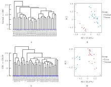
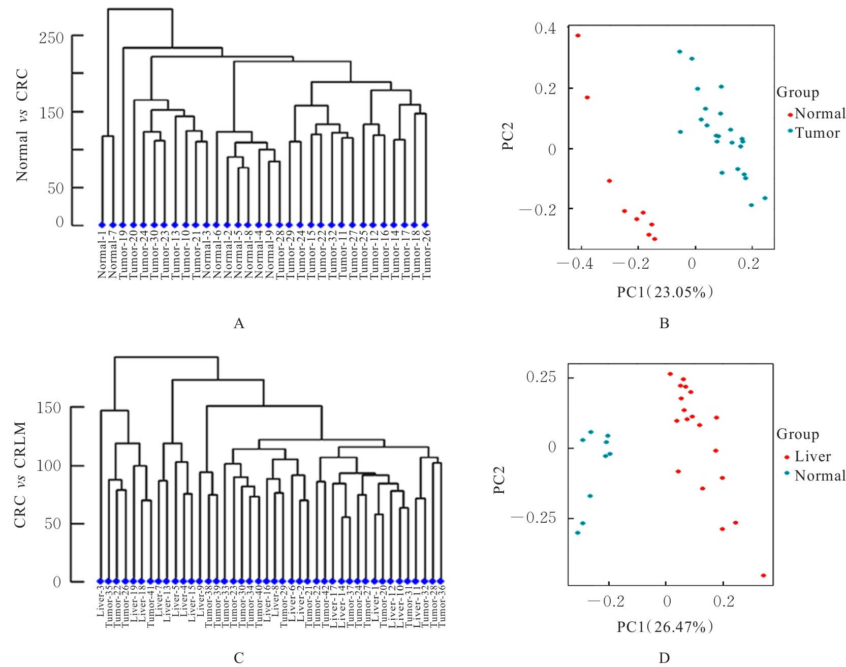
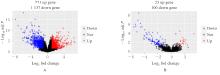
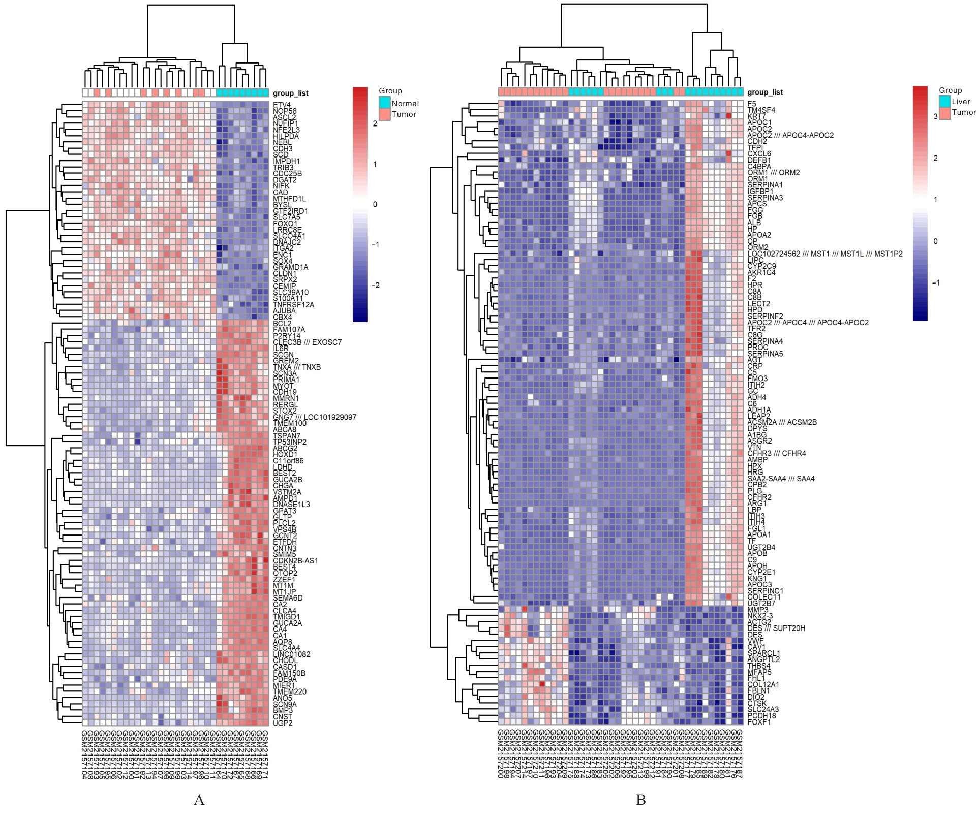
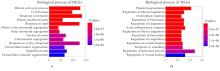
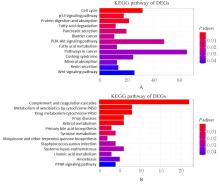
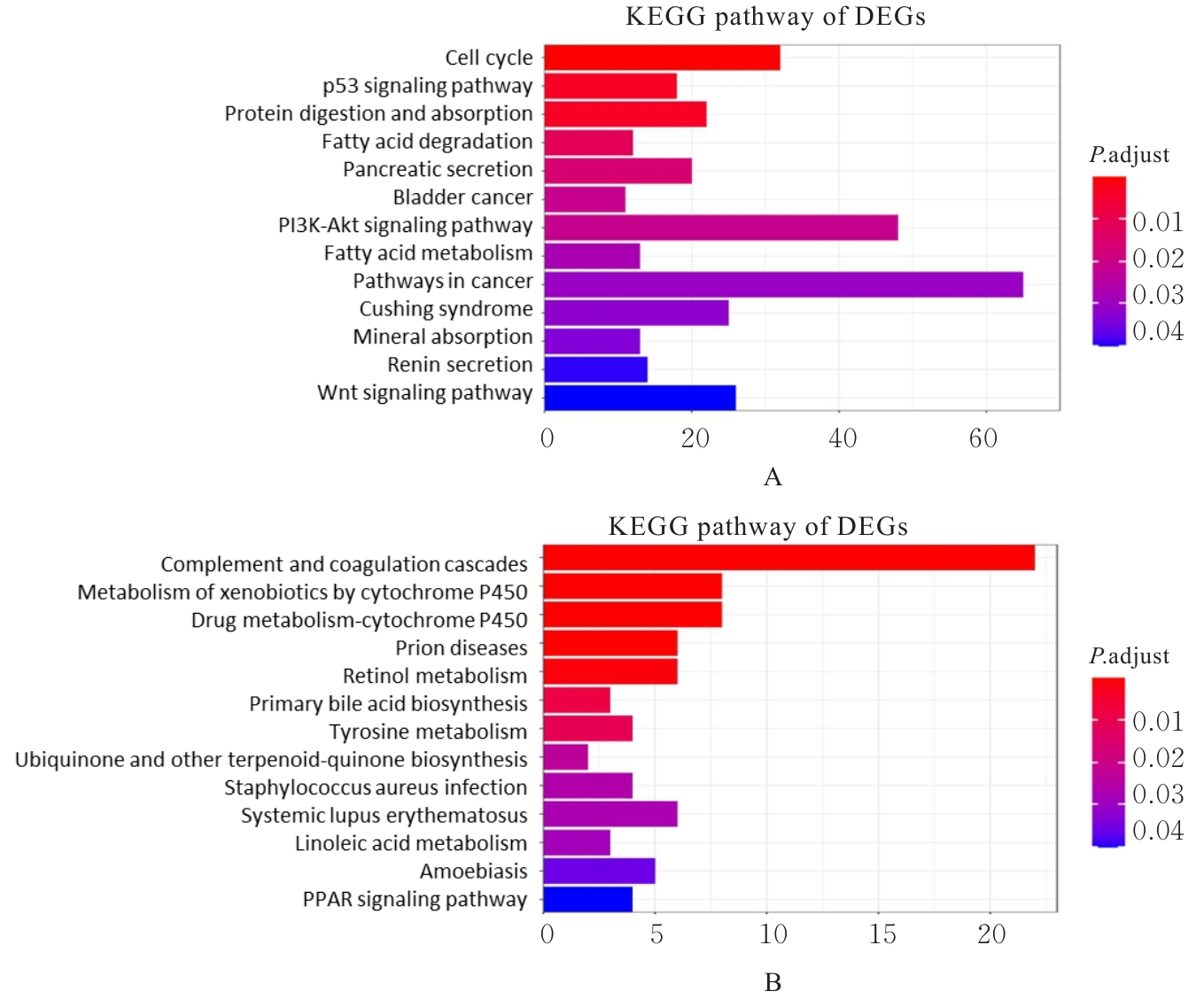
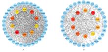
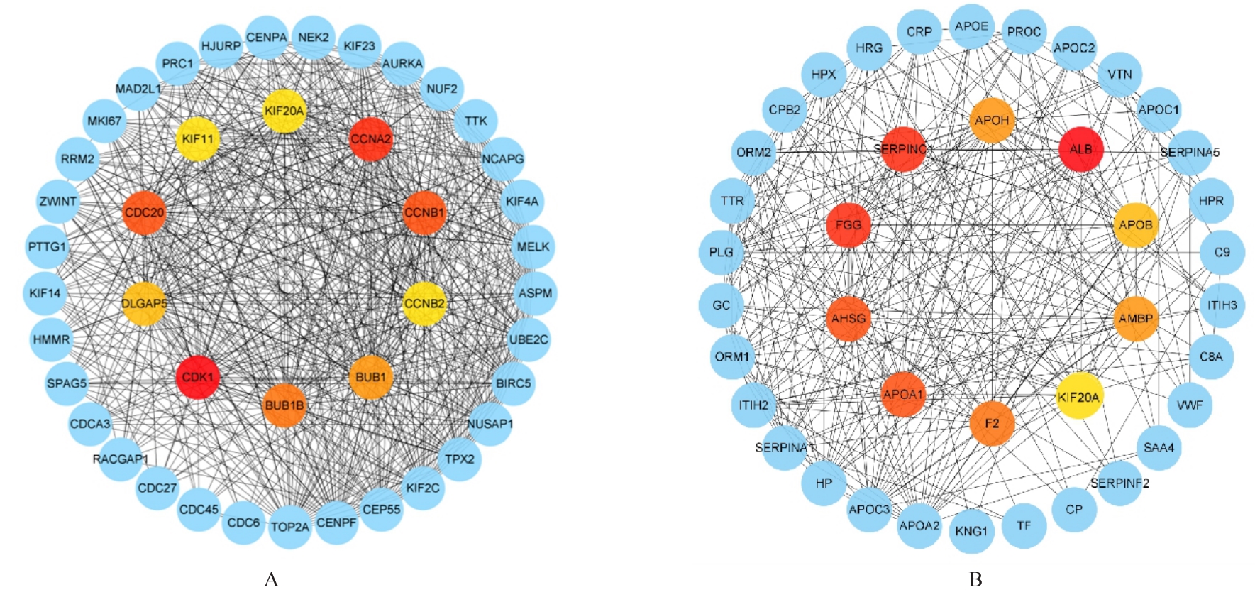
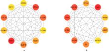
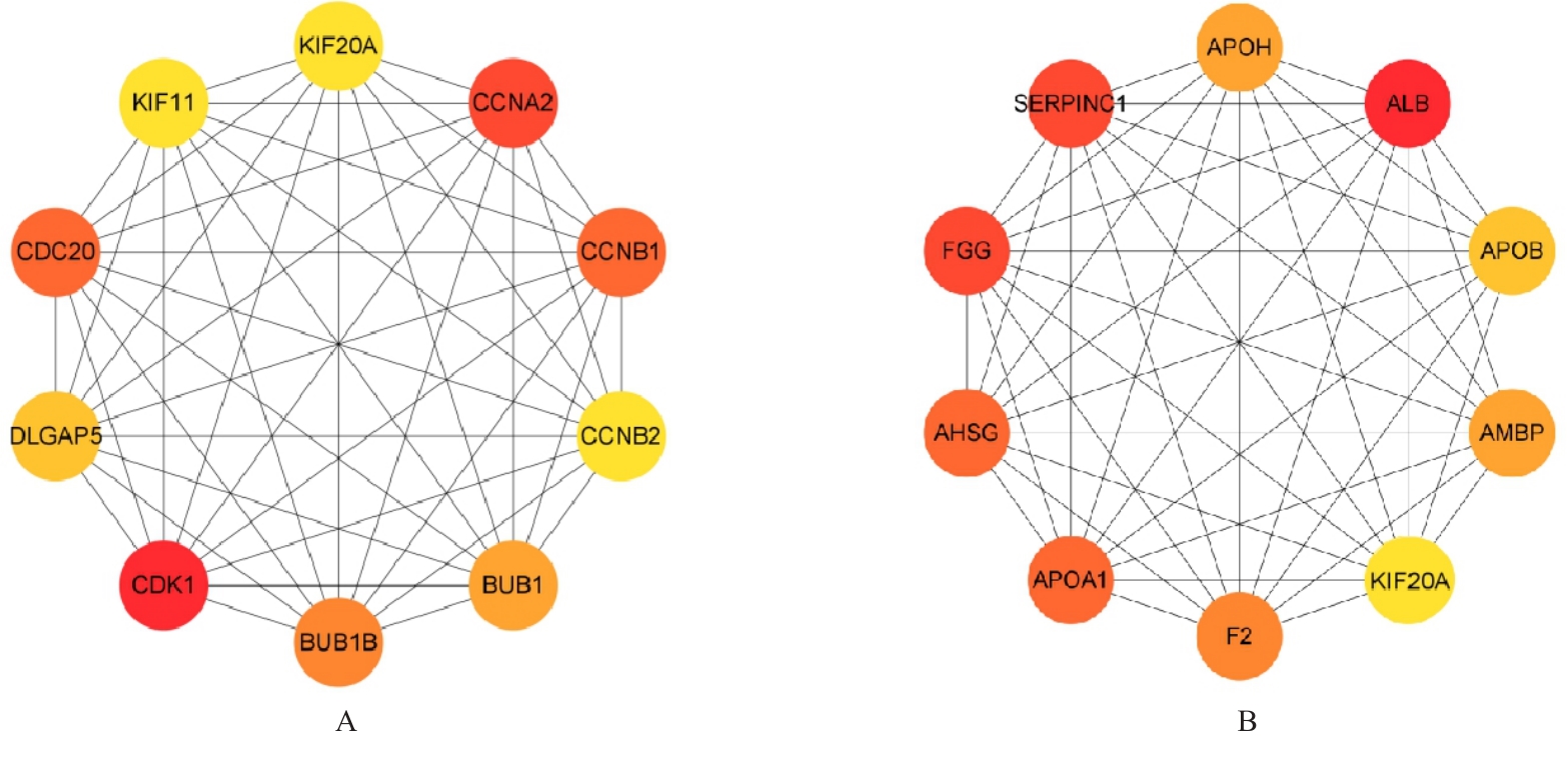
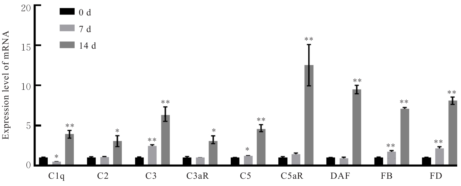
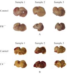
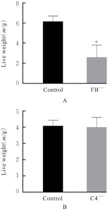
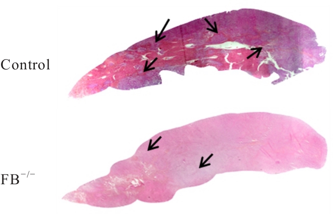
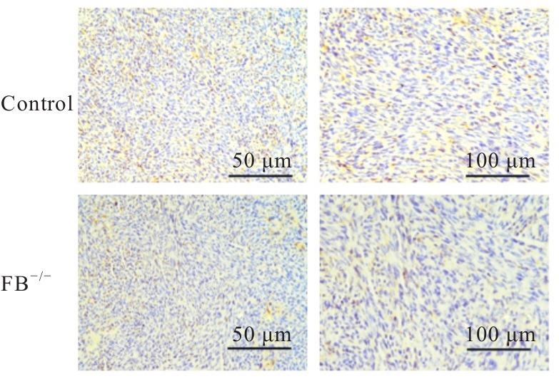
目的 基于生物信息学方法分析补体替代途径对小鼠结直肠癌(CRC)肝转移模型的调控作用,并进行实验验证。 方法 以“CRC肝转移”为关键词在基因表达综合(GEO)数据库中进行检索,获取GSE81558 GEO数据集,包括正常结肠组织样本、CRC组织样本和CRC肝转移组织样本,采用生物信息学方法分析,筛选差异表达基因(DEGs)。使用R和Cytoscape软件进行基因本体论(GO)功能富集分析和京都基因与基因组百科全书(KEGG)信号通路富集分析并进行可视化,利用检索相互作用基因/蛋白质(STRING)数据库对DEGs相关蛋白-蛋白相互作用(PPI)进行评估,绘制PPI网络图。12只C57BL/6小鼠脾脏注射SL4肿瘤细胞,并于0、7和14 d后采集小鼠肝组织,采用实时荧光定量PCR(RT-qPCR)法检测小鼠肝转移灶中补体途径相关基因表达水平。采用小鼠CRC肝转移模型验证补体信号通路,将小鼠分为对照组、B因子(FB)敲除组(FB-/-)和C4因子敲除组(C4-/-),每组6只,检测各组小鼠肝脏质量,HE染色观察对照组和FB-/-组小鼠CRC肝转移灶形态表现并计算肝转移灶面积百分率,免疫组织化学染色法观察对照组和FB-/-组小鼠肝组织中巨噬细胞浸润情况,计算巨噬细胞浸润百分率。 结果 正常结肠组织样本与CRC组织样本以及CRC组织样本与CRC肝转移组织样本之间距离较远,提示样本间差异性较大,可以对DEGs进行后续分析。在正常结肠组织样本与CRC组织样本数据集中共筛选出1 908个DEGs,其中771个DEGs上调,1 137个DEGs下调。在CRC与CRC肝转移数据集中共发现23个上调的DEGs及100个下调的DEGs。GO功能富集分析,与正常结肠组织样本比较,CRC组织样本的DEGs主要集中于有丝分裂细胞周期过程、细胞分裂、对激素的反应、有丝分裂核分裂和对脂质的反应等生物学过程(BP);与CRC组织样本比较,CRC肝转移组织样本的DEGs主要富集于凝血反应过程中,如血小板脱颗粒、凝血调节、急性期反应、止血调节和凝血调节等BP。KEGG信号通路富集分析,与正常结肠组织样本比较,CRC组织样本的DEGs主要富集的信号通路为细胞周期和P53信号通路;与CRC组织样本比较,CRC肝转移组织样本的DEGs主要富集于与补体、凝血级联和代谢相关的信号通路。DEGs的PPIs网络鉴定出的枢纽基因和血液蛋白有关。RT-qPCR法检测,与0 d组比较,7 d组小鼠CRC肝转移组织样本中补体相关基因补体1q(C1q)mRNA表达水平明显降低(P<0.05),补体3(C3)、补体5(C5)、FB和D因子(FD)mRNA表达水平均明显升高(P<0.05或P<0.01),14 d组小鼠CRC肝转移组织样本中补体通路相关基因C1q、补体2(C2)、C3、补体片段3a受体(C3aR)、C5、补体片段5a受体(C5aR)、衰变加速因子(DAF)、FB和FD mRNA表达水平均明显升高(P<0.05或P<0.01)。与对照组比较,FB-/-组小鼠肝脏质量明显降低(P<0.01),C4-/-组小鼠肝脏质量差异无统计学意义(P>0.05)。HE染色观察,与对照组比较,FB-/-组小鼠CRC肝转移灶明显减少,CRC肝转移面积百分率明显降低(P<0.01)。免疫组织化学染色法观察,与对照组比较,FB-/-组小鼠CRC肝转移灶中巨噬细胞浸润减少,巨噬细胞浸润百分率明显降低(P<0.01)。 结论 补体级联反应与CRC肝转移有关,替代补体通路调控CRC肝转移,提示该通路是CRC肝转移的潜在治疗靶点。

关键词: 结直肠癌, 肝转移, 基因表达综合数据库, 生物信息学, 差异表达基因

Abstract:

Objective To discuss the regulatory role of complement alternative pathway in mouse colorectal cancer (CRC) liver metastasis model based on bioinformatics methods, and to clarify its mechanism through experimental verification. Methods Using “CRC liver metastasis” as the keyword, the GSE81558 dataset was retrieved from Gene Expression Omnibus (GEO) database, including normal colon tissue samples, CRC tissue samples and CRC liver metastasis tissue samples. Bioinformatics methods were used to analyze and screen differentially expressed genes (DEGs). Gene Ontology (GO) functional enrichment analysis and Kyoto Encyclopedia of Genes and Genomes (KEGG) pathway enrichment analysis were performed using R and Cytoscape software, and the results were visualized. Search Tool for the Retrieval of Interacting Genes/Proteins (STRING) database was used to evaluate protein-protein interactions (PPIs) of DEGs and construct PPI network. Twelve C57BL/6 mice were injected with SL4 tumor cells into spleen, and the liver tissues were collected at 0, 7 and 14 d. Real-time fluorescence quantitative PCR (RT-qPCR) method was used to detect the expression levels of complement pathway-related genes in liver metastatic foci. The CRC liver metastasis mouse model was used to verify the complement signaling pathway. The mice were divided into control group, factor B knockout group (FB-/-) and C4 factor knockout group (C4-/-), and there were 6 mice in each group. The liver weights of the mice were measured; HE staining was used to detect the percentage of metastatic area in liver tissue in control group and FB-/- group; immunohistochemistry was used to detect macrophage infiltration in liver tissue in control group and FB-/- group, and the percentage of macrophage infiltration was calculated. Results The distances between normal colon tissue samples and CRC tissue samples, as well as between CRC tissue samples and CRC liver metastasis tissue samples were far, indicating significant differences between samples, allowing subsequent analysis of DEGs. A total of 1 908 DEGs were screened in the dataset comparing normal colon tissue samples and CRC tissue samples, including 771 up-regulated DEGs and 1 137 down-regulated DEGs. Twenty-three up-regulated DEGs and 100 down-regulated DEGs were identified in the dataset comparing CRC and CRC liver metastasis. The GO functional enrichment analysis results showed that compared with normal colon tissue samples, DEGs in CRC samples were mainly enriched in biological processes (BP) related to cell cycle and mitosis, including mitotic cell cycle process, cell division, response to hormone, mitotic nuclear division and response to lipid. Compared with CRC samples, the DEGs in CRC liver metastasis samples were mainly enriched in coagulation-related BP, including platelet degranulation, blood coagulation regulation, acute-phase response, hemostasis regulation and coagulation regulation. The KEGG pathway enrichment analysis results showed that compared with normal colon tissue samples, the DEGs in CRC tissue samples were mainly enriched in cell cycle and p53 signaling pathways. Compared with CRC tissue samples, the DEGs in CRC liver metastasis tissue samples were mainly enriched in complement, coagulation cascade and metabolism-related signaling pathways. The Hub genes identified in PPI network were related to blood proteins. The RT-qPCR results showed that compared with 0 d group, the mRNA expression level of complement related genes complement 1q (C1q) in liver metastatic foci tissue sampres in 7 d group was significantly decreased (P<0.05), the mRNA expression levels of complement 3 (C3), complement 5 (C5), FB, and factor D (FD) were significantly increased (P<0.05 or P<0.01), the mRNA expression levels of complement pathway-related genes C1q, complement 2 (C2), C3, complement fragment 3a receptor (C3aR), C5, complement fragment 5a receptor (C5aR), decay-accelerating factor (DAF), FB and FD in liver metastatic foci tissue sampres in 14 d group were significantly increased (P<0.05 or P<0.01). Compared with control group, the liver weight of the mice in FB-/- group was significantly decreased (P<0.01), while there was no significant difference was observed in C4-/- group (P>0.05). The HE staining results showed that compared with control group, the liver metastatic foci in FB-/- mice were significantly decreased, and the percentage of metastatic area was decreased (P<0.01). The immunohistochemistry results showed that compared with control group, the macrophage infiltration in liver metastatic foci of the mice in FB-/- group was reduced, and the percentage of macrophage infiltration was decreased (P<0.01). Conclusion Complement cascade is associated with CRC liver metastasis, and the alternative complement pathway regulates CRC liver metastasis, suggesting this pathway may serve as a potential therapeutic target for CRC liver metastasis.

Key words: Colorectal cancer, Liver metastasis, Gene Expression Omnibus database, Bioinformatics, Differentially expressed genes

中图分类号: 

  • R735.3