Journal of Jilin University(Medicine Edition) ›› 2024, Vol. 50 ›› Issue (6): 1499-1511.doi: 10.13481/j.1671-587X.20240603
• Research in basic medicine • Previous Articles
Inhibitory effect of Boschnikia rossica polysaccharides on THP-1 macrophage inflammation and its mechanism
Xinyue MA1,2,Hui XU2,Jiawen DIAO2,Aihua JIN1( ),Jishu QUAN2(
 )
)
- 1.Department of Clinical Laboratory,Affiliated Hospital,Yanbian University,Yanji 133000,China
2.Department of Biochemistry and Molecular Biology,School of Medical Sciences,Yanbian University,Yanji 133000,China
-
Received:2024-01-20Online:2024-11-28Published:2024-12-10 -
Contact:Aihua JIN,Jishu QUAN E-mail:aihua1028@sina.com;quanjs@ybu.edu.cn
CLC Number:
- R285.5
Cite this article
Xinyue MA,Hui XU,Jiawen DIAO,Aihua JIN,Jishu QUAN. Inhibitory effect of Boschnikia rossica polysaccharides on THP-1 macrophage inflammation and its mechanism[J].Journal of Jilin University(Medicine Edition), 2024, 50(6): 1499-1511.
share this article
Tab.1
Survival rates of THP-1 macrophages and IL-6 level in cell culture fluid after treated with different concentrations of LPS"
| Group | Survival rate(η/%) | IL-6 [ρB/(ng·L-1)] |
|---|---|---|
| 0 μg·L-1 LPS | 99.81±1.32 | 21.49±3.37 |
| 100 μg·L-1 LPS | 94.80±1.16 | 40.41±6.87* |
| 200 μg·L-1 LPS | 90.27±2.90 | 62.27±5.29* |
| 500 μg·L-1 LPS | 93.43±2.24 | 88.59±7.33* |
| 1 000 μg·L-1 LPS | 94.63±3.52 | 215.61±6.18* |
| 2 000 μg·L-1 LPS | 92.43±4.31 | 242.27±8.06* |
Tab. 2
Levels of inflammatory factors in culture fluid in THP-1 macrophages in various groups [n=3, xˉ±s, ρB/(ng·L-1)]"
| Group | IL-6 | TNF-α | IL-1β |
|---|---|---|---|
| Blank | 59.29±0.24 | 441.67±0.28 | 1.49±0.35 |
| Model | 215.13±0.62* | 1 200.33±1.23* | 29.93±0.83* |
| Low dose of BRPS | 171.38±0.39△ | 1 050.03±0.76△ | 26.61±0.44△ |
| Medium dose of BRPS | 127.58±0.43△ | 874.83±0.35△ | 23.88±0.46△ |
| High dose of BRPS | 78.88±0.80△ | 815.20±1.07△ | 12.21±0.46△ |
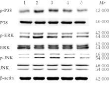
Tab.3
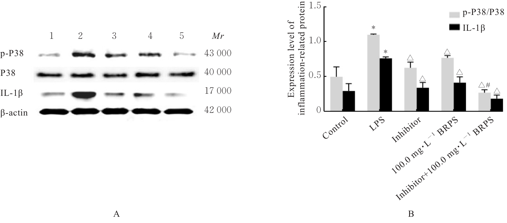
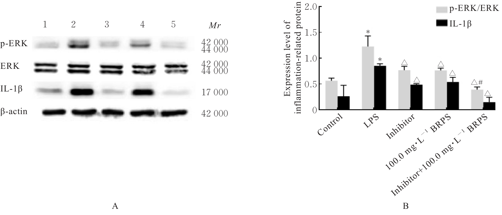
Ratios of p-P38/P38, p-ERK/ERK, p-JNK/JNK, and p-NF-κB/NF-κB in THP-1 macrophages in various groups"
| Group | p-P38/P38 | p-ERK/ERK | p-JNK/JNK | p-NF-κB/NF-κB |
|---|---|---|---|---|
| Blank | 0.59±0.05 | 0.70±0.10 | 0.52±0.05 | 0.67±0.18 |
| Model | 1.01±0.01* | 1.17±0.12* | 1.05±0.09* | 1.38±0.07* |
| Low dose of BRPS | 1.04±0.04 | 0.98±0.04 | 0.96±0.05 | 1.11±0.05 |
| Medium dose of BRPS | 0.79±0.08△ | 0.90±0.06△ | 0.83±0.02△ | 1.05±0.05△ |
| High dose of BRPS | 0.45±0.04△ | 0.81±0.06△ | 0.64±0.08△ | 0.82±0.12△ |
| 1 | 陈庆红, 张 睿, 冯秀春, 等. 长白山区草苁蓉研究现状与濒危机理[J]. 现代农业科技, 2015(14): 80-81. |
| 2 | QUAN J S, JIN M H, XU H X, et al. BRP, a polysaccharide fraction isolated from Boschniakia rossica, protects against galactosamine and lipopolysaccharide induced hepatic failure in mice[J]. J Clin Biochem Nutr, 2014, 54(3): 181-189. |
| 3 | 许长青, 许顺贵, 刘春光, 等. 草苁蓉的药理作用研究进展[J]. 武警医学, 2023, 34(1): 75-78. |
| 4 | 王丽华, 杨 弋, 周德生, 等. 川芎清脑颗粒治疗慢性脑缺血伴头痛的治疗研究[J]. 中国实用内科杂志, 2023, 43(1): 74-77. |
| 5 | 吴文龙, 王权成, 孟 强, 等. 小檗碱对肝癌细胞活性与增殖能力的影响及其机制[J]. 解放军医学杂志, 2023, 48(6): 653-662. |
| 6 | 华 龙, 王晨宇, 姚坤厚, 等. 草苁蓉多糖对人肝癌细胞侵袭迁移的影响及机制[J]. 解放军医学杂志, 2021, 46(8): 763-770. |
| 7 | 尹学哲, 王玉娇, 尹基峰, 等. 草苁蓉提取物对HepG2细胞氧化应激损伤的保护作用[J]. 食品科学, 2015, 36(15): 173-178. |
| 8 | 宋全胜. 草苁蓉根茎粗多糖的分离纯化及其部分性质的研究[D]. 延吉: 延边大学, 2005. |
| 9 | MONTELEONE G, MOSCARDELLI A, COLELLA A, et al. Immune-mediated inflammatory diseases: common and different pathogenic and clinical features[J]. Autoimmun Rev, 2023, 22(10): 103410. |
| 10 | YU P, ZHANG X, LIU N, et al. Pyroptosis: mechanisms and diseases[J]. Signal Transduct Target Ther, 2021, 6(1): 128. |
| 11 | ZHANG E L, HUANG J B, WANG K, et al. Pterostilbene protects against lipopolysaccharide/D-galactosamine-induced acute liver failure by upregulating the Nrf2 pathway and inhibiting NF-κB, MAPK, and NLRP3 inflammasome activation[J]. J Med Food, 2020, 23(9): 952-960. |
| 12 | HUANG Y, XU W, ZHOU R B. NLRP3 inflammasome activation and cell death[J]. Cell Mol Immunol, 2021, 18(9): 2114-2127. |
| 13 | 丁 杨, 胡 容. NLRP3炎症小体激活及调节机制的研究进展[J]. 药学进展, 2018, 42(4): 294-302. |
| 14 | CECCARELLI S, PANERA N, MINA M, et al. LPS-induced TNF-α factor mediates pro-inflammatory and pro-fibrogenic pattern in non-alcoholic fatty liver disease[J]. Oncotarget, 2015, 6(39): 41434-41452. |
| 15 | YU C P, ZHAO W, DUAN C J, et al. Poly-l-lysine-caused cell adhesion induces pyroptosis in THP-1 monocytes[J]. Open Life Sci, 2022, 17(1): 279-283. |
| 16 | DAIGNEAULT M, PRESTON J A, MARRIOTT H M, et al. The identification of markers of macrophage differentiation in PMA-stimulated THP-1 cells and monocyte-derived macrophages[J]. PLoS One, 2010, 5(1): e8668. |
| 17 | GIAMBELLUCA S, OCHS M, LOPEZ-RODRIGUEZ E. Resting time after phorbol 12-myristate 13-acetate in THP-1 derived macrophages provides a non-biased model for the study of NLRP3 inflammasome[J]. Front Immunol, 2022, 13: 958098. |
| 18 | 刘莉园, 张 钊, 葛乃嘉, 等. 草苁蓉多糖对脂多糖诱导的RAW264.7巨噬细胞炎症反应的影响[J]. 中国药学杂志, 2021, 56(18): 1479-1485. |
| 19 | NI Y J, ZHANG J, ZHU W J, et al. Echinacoside inhibited cardiomyocyte pyroptosis and improved heart function of HF rats induced by isoproterenol via suppressing NADPH/ROS/ER stress[J]. J Cell Mol Med, 2022, 26(21): 5414-5425. |
| 20 | CHEN L L, DENG H D, CUI H M, et al. Inflammatory responses and inflammation-associated diseases in organs[J]. Oncotarget, 2017, 9(6): 7204-7218. |
| 21 | HUANG Y, CHEN S P, YAO Y, et al. Ovotransferrin inhibits TNF-α induced inflammatory response in gastric epithelial cells via MAPK and NF-κB pathway[J]. J Agric Food Chem, 2023, 71(33): 12474-12486. |
| 22 | CUI J H, JIA J P. Natural COX-2 inhibitors as promising anti-inflammatory agents: an update[J]. Curr Med Chem, 2021, 28(18): 3622-3646. |
| 23 | 华湘黔, 张 漾. HMGB1及PD-L1在浸润性乳腺癌非特殊型中的表达及临床意义[J]. 现代肿瘤医学, 2024, 32(3): 477-481. |
| 24 | AN Y N, ZHANG H F, WANG C, et al. Activation of ROS/MAPKs/NF-κB/NLRP3 and inhibition of efferocytosis in osteoclast-mediated diabetic osteoporosis[J]. FASEB J, 2019, 33(11): 12515-12527. |
| 25 | LIN Q S, LI S, JIANG N, et al. PINK1-parkin pathway of mitophagy protects against contrast-induced acute kidney injury via decreasing mitochondrial ROS and NLRP3 inflammasome activation[J]. Redox Biol, 2019, 26: 101254. |
| 26 | AKTHER M, HAQUE M E, PARK J, et al. NLRP3 ubiquitination-a new approach to target NLRP3 inflammasome activation[J]. Int J Mol Sci, 2021, 22(16): 8780. |
| 27 | HEPWORTH E M W, HINTON S D. Pseudophosphatases as regulators of MAPK signaling[J]. Int J Mol Sci, 2021, 22(22): 12595. |
| 28 | ZHANG M, CHEN Y, YANG M J, et al. Celastrol attenuates renal injury in diabetic rats via MAPK/NF-κB pathway[J]. Phytother Res, 2019, 33(4): 1191-1198. |
| [1] | Xuejun JIN,Chuyuan LU. Inhibitory effect of leucovorin on growth and angiogenesis of subcutaneous transplanted tumors in mouse lung cancer cells and its mechanism [J]. Journal of Jilin University(Medicine Edition), 2024, 50(3): 612-619. |
| [2] | Shuang YANG,Na XU,Jianxu ZHANG,Chengbiao SUN,Yan WANG,Mingxin DONG,Wensen LIU. Improvement effect of rubusoside on motor dysfunction and neuroinflammation in mice with spinal cord injury and its mechanism [J]. Journal of Jilin University(Medicine Edition), 2024, 50(2): 326-335. |
| [3] | Shuang CHEN,Hong LI. Effect of silencing FOXK1 gene on proliferation, migration, and invasion of gastric cancer HGC-27 cells [J]. Journal of Jilin University(Medicine Edition), 2024, 50(2): 371-378. |
| [4] | Xiaoni WANG,Tao GUO,Qiyun LUO,Lifeng GUAN. Effect of usnic acid on biological behaviors of hypertrophic scar fibroblasts and JNK/MAPK signaling pathway [J]. Journal of Jilin University(Medicine Edition), 2023, 49(6): 1445-1451. |
| [5] | Haikang CUI,Xudong ZHANG,Xiaoning LI,Xi YANG,Lan YANG,Wenjie ZHANG. Bioinformatics analysis on predition effect of subtypes of cell pyroptosis and APOD on prognosis of gastric cancer patients [J]. Journal of Jilin University(Medicine Edition), 2023, 49(5): 1268-1279. |
| [6] | Haitao LI, Qin LI, Fei CAI, Guofu HU, Yunfei TENG. Effect of apigenin on polarization and inflammation of mouse RAW264.7 macrophages and its mechanism [J]. Journal of Jilin University(Medicine Edition), 2023, 49(3): 549-556. |
| [7] | Heran YANG,Xingjiang LI,Jiahang HU,Yanwei LI. Effect of Ghrelin on neural differentiation of adipose-derived mesenchymal stem cells by regulating MAPK/ERK pathway [J]. Journal of Jilin University(Medicine Edition), 2023, 49(3): 706-713. |
| [8] | Yan YU,Chengcheng YU,Yakun HAN. Analysis on phenotypes of plasma cells of gingiva and expression characteristics of RANKL in patients with periodontitis complicated with rheumatoid arthritis [J]. Journal of Jilin University(Medicine Edition), 2023, 49(3): 757-764. |
| [9] | Xinghong GAO,Hongmei TANG,Yuejiao LI,Xiaoyun WANG,Xing WANG,Xiefang YUAN,Min WU. Attenuating effect of translocator protein ligand XBD173 on pulmonary inflammatory response induced by cigarette smoke extraction in mice and its mechanism [J]. Journal of Jilin University(Medicine Edition), 2023, 49(2): 272-279. |
| [10] | Xiaojuan ZHU,Haitao DAI,Yan LI,Lingxin CUI,Ya WANG,Jiang XU,Nan WU. Improvement effect of grape seed proanthocyanidin extract on periodontal inflammation in diabetic periodontitis rats and its influence on expression levels of TLR4 and NF-κB in periodontal tissue [J]. Journal of Jilin University(Medicine Edition), 2023, 49(1): 31-38. |
| [11] | Hang YANG,Jiaqi CHEN,Shouhan WANG,Hongjun YANG,Bin WANG. Protective effect of ginsenoside Rg1 on cardiac injury in rats with severe acute pancreatitis and its mechanism [J]. Journal of Jilin University(Medicine Edition), 2022, 48(5): 1175-1181. |
| [12] | Ming xing YANG,Wen DONG,Ji LI. Inductive effect of peiminine on apoptosis of lung cancer A549 cells and its mechanism [J]. Journal of Jilin University(Medicine Edition), 2022, 48(3): 711-717. |
| [13] | Ming LI,Qiuting WANG,Shan CHEN,Huifang SHI. Improvement effect of p38 MAPK inhibitor on chronic obstructive pulmonary disease injury in mice through inhibiting cell pyrotosis mediated by NLRP3 pathway [J]. Journal of Jilin University(Medicine Edition), 2022, 48(3): 744-754. |
| [14] | Suxian CHEN,Zehui GU,Yangfei MA,Qi TAN,Qi LI,Yadi WANG. Promotion effect of rutin on apoptosis of human colon cancer SW480 cells and its mechanism [J]. Journal of Jilin University(Medicine Edition), 2022, 48(2): 356-363. |
| [15] | Haojie WU,Minghui ZHANG,Chengzhi HONG. Improvement effect of diosgenin on symptoms of synovitis rats and its reglatory effect on TLR2-NF-κB signaling pathway and mechanism [J]. Journal of Jilin University(Medicine Edition), 2021, 47(4): 943-950. |
|
||


