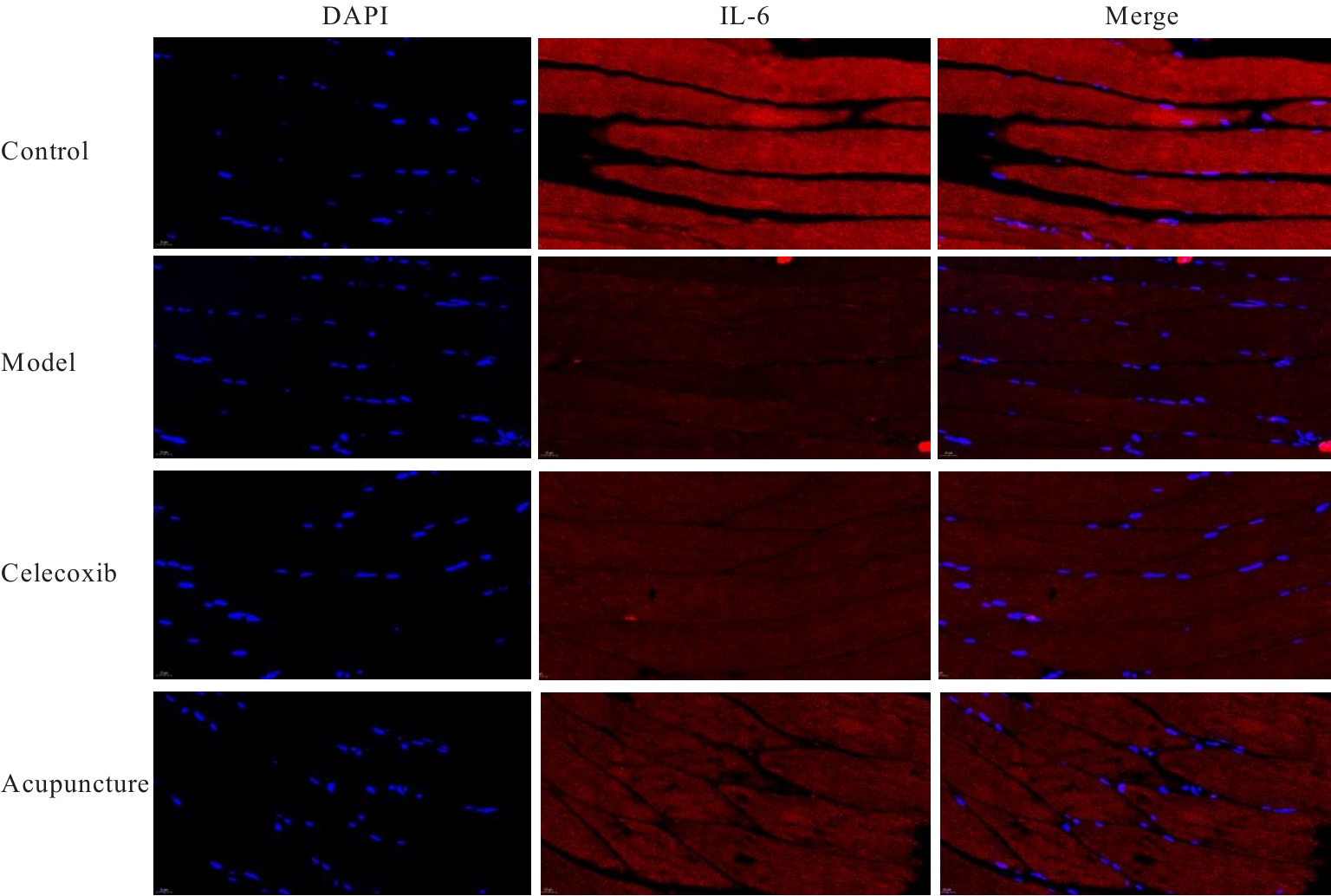
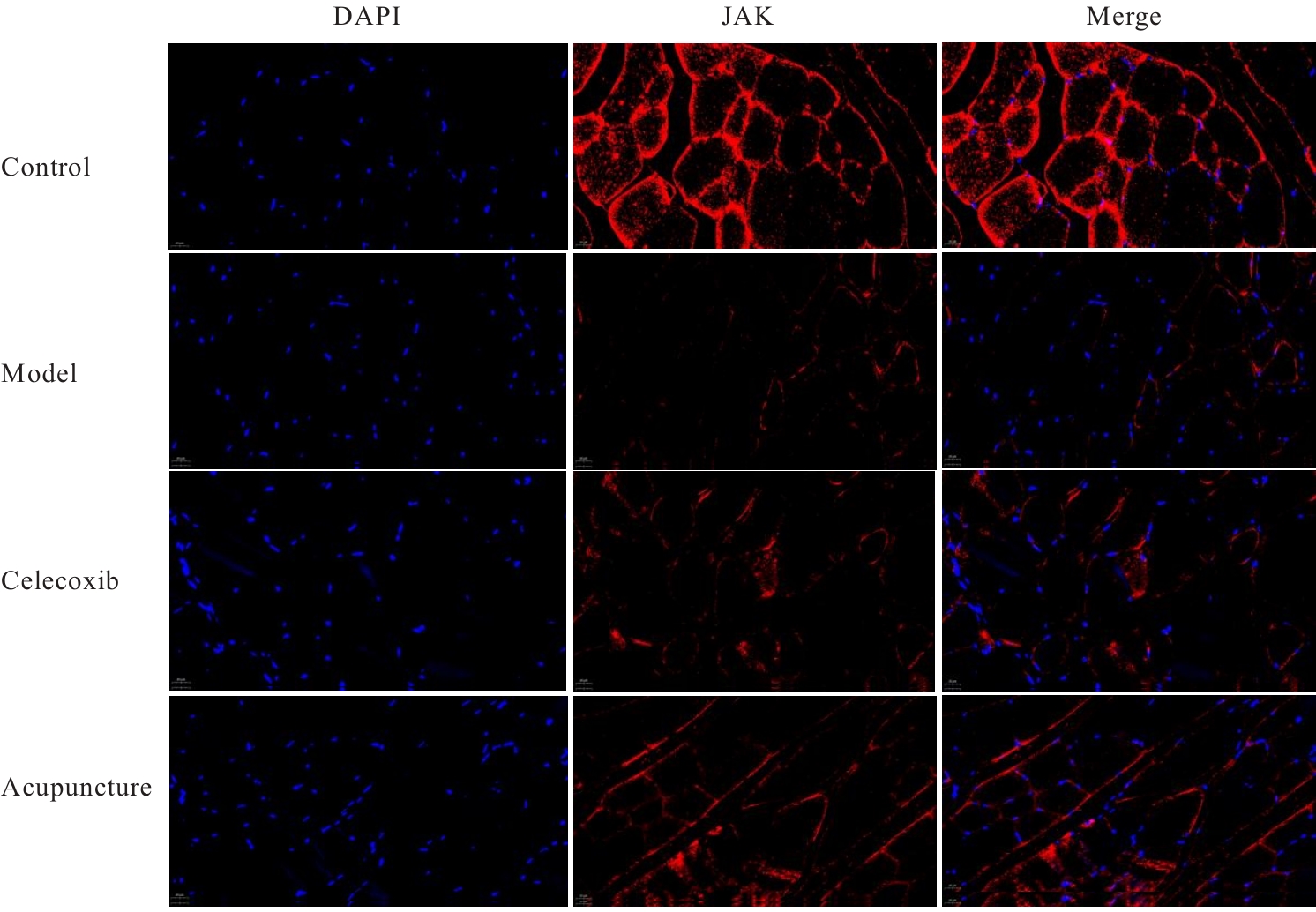
Journal of Jilin University(Medicine Edition) ›› 2025, Vol. 51 ›› Issue (6): 1475-1486.doi: 10.13481/j.1671-587X.20250604

• Research in basic medicine • Previous Articles    

Effect of acupuncture on differentiation and apoptosis of quadriceps muscle satellite cells in knee osteoarthritis model rats and its mechanism

Qu ZHENG1,2,3,Baoqiang DONG1,3(),Xingxing LIN1,3,Xuefeng GUAN1,Yu ZHANG4,Chaojie WANG1,Yiyan Han1()   

  1. 1.College of Acupuncture and massage,Liaoning University of Traditional Chinese Medicine,Shenyang 110847,China
    2.Key Laboratory of Acupuncture and Moxibustion Biology,Education of Liaoning Province,Shenyang 110847,China
    3.Key Laboratory of Acupuncture and Moxibustion Health and Rehabilitation,Shenyang 110847,China
    4.Shenyang Pharmaceutical University,Shenyang 110016,China
  • Received:2024-12-25 Accepted:2025-02-12 Online:2025-11-28 Published:2025-12-15
  • Contact: Baoqiang DONG,Yiyan Han E-mail:peterbaoqiang@163.com;hyy-zjtn@lnutcm.edu.cn

Abstract:

Objective To discuss the effect of acupuncture on the differentiation and apoptosis of quadriceps muscle satellite cells in model rats with knee osteoarthritis(KOA), and to clarify its related mechanism. Methods A total of 40 SPF-grade rats were selected and randomly divided into control group, model group, celecoxib group, and acupuncture group, with 10 rats in each group. The rats in control group only underwent joint cavity incision followed by suturing, while the rats in model group, celecoxib group, and acupuncture group were used to replicate the KOA models. The maximum circumference of the femoral segment of the affected limb, rat body mass, and quadriceps wet weight of the rats in various groups were measured; the quadriceps wet weight maintenance rate and quadriceps wet weight/body mass ratio of the rats in various groups were calculated. HE staining was used to observe the pathomorphology of articular cartilage and quadriceps muscle tissue of the rats in various groups; terminal deoxynucleotidyl transferase (TdT)-mediated dUTP nick end labeling (TUNEL) method was used to detect the apoptosis indexes in articular cartilage and quadriceps muscle tissue of the rats in various groups; immunofluorescence method was used to detect the protein expression levels of interleukin-6 (IL-6), Janus kinase (JAK), and signal transducer and activator of transcription 3 (STAT3) in quadriceps muscle tissue of the rats in various groups; Western blotting method was used to detect the expression levels of IL-6/JAK/STAT3 signaling pathway proteins, and muscle satellite cells, and apoptosis-related proteins in quadriceps muscle tissue of the rats in various groups. Results Compared with control group, the affected hind limb circumference, quadriceps wet weight, wet weight maintenance rate, and wet weight/body mass ratio of the rats in model group were significantly decreased (P<0.05); compared with model group, the affected hind limb circumference, quadriceps wet weight, wet weight maintenance rate, and wet weight/body mass ratio of the rats in celecoxib group and acupuncture group were significantly increased (P<0.05); compared with celecoxib group, the affected hind limb circumference, quadriceps wet weight, wet weight maintenance rate, and wet weight/body mass ratio of the rats in acupuncture group were significantly increased (P<0.05). The HE staining results showed that the knee articular cartilage of the rats in control group remained intact, chondrocytes were aggregated and horizontally arranged with smooth edges, and quadriceps muscle cells were long cylindrical, orderly arranged, and regular in shape; in model group, the knee articular cartilage was thinner with rough edges, reduced number of cartilage layers, and disordered arrangement, and the quadriceps muscle fibers were disorganized, with some muscle fiber dissolution and muscle cell membrane damage, accompanied by muscle fiber fragments and a large amount of inflammatory exudate; in celecoxib group, the morphology of knee articular cartilage was generally normal, occasionally with irregular cartilage arrangement and reduced thickness, sporadically visible necrotic chondrocytes, quadriceps muscle fibers and sarcolemma were relatively intact, new muscle fibers appeared, some muscle fiber edges were blurred, accompanied by a small amount of cell debris and mild inflammatory infiltration; in acupuncture group, the knee articular cartilage structure remained intact with smooth edges, occasionally rough edges, and chondrocytes were aggregated and orderly arranged. The TUNEL assay results showed that compared with control group, the apoptosis indexes in articular cartilage and quadriceps muscle tissue of the rats in model group were significantly increased (P<0.05); compared with model group, the apoptosis indexes in articular cartilage and quadriceps muscle tissue of the rats in celecoxib group and acupuncture group were significantly decreased (P<0.05); compared with celecoxib group, the apoptosis index in articular cartilage and quadriceps muscle tissue of the rats in acupuncture group were significantly decreased (P<0.05). The immunofluorescence assay results showed that compared with control group, the expression levels of IL-6, JAK, and STAT3 proteins in quadriceps muscle tissue of the rats in model group were significantly decreased (P<0.05); compared with model group, the expression levels of IL-6, JAK, and STAT3 proteins in quadriceps muscle tissue of the rats in celecoxib group and acupuncture group were significantly increased (P<0.05); compared with celecoxib group, the expression levels of IL-6, JAK, and STAT3 proteins in quadriceps muscle tissue of the rats in acupuncture group were significantly increased (P<0.05). The Western blotting results showed that compared with control group, the expression levels of IL-6, JAK, STAT3, paired box transcription factor 7 (Pax7), Desmin, Myosin, and Myogenin proteins in quadriceps muscle tissue of the rats in model group were significantly decreased (P<0.05); compared with model group, the expression levels of IL-6, JAK, STAT3, Pax7, Desmin, Myosin, and Myogenin proteins in quadriceps muscle tissue of the rats in celecoxib group and acupuncture group were significantly increased (P<0.05); compared with celecoxib group, the expression levels of IL-6, JAK, STAT3, Pax7, Desmin, Myosin, and Myogenin proteins in quadriceps muscle tissue of the rats in acupuncture group were significantly increased (P<0.05). Compared with control group, the expression levels of B-cell lymphoma 2 (Bcl-2), B-cell lymphoma-xl (Bcl-xl), and myeloid cell leukemia 1 (MCL1) proteins in quadriceps muscle tissue in model group were significantly decreased (P<0.05), and the expression levels of Bcl-2-associated X protein (Bax) and cysteinyl aspartate specific proteinase-3 (Caspase-3) proteins were significantly increased (P<0.05); compared with model group, the expression levels of Bcl-2, Bcl-xl, and MCL1 proteins in quadriceps muscle tissue of the rats in celecoxib group and acupuncture group were significantly increased (P<0.05), and the expression levels of Bax and Caspase-3 proteins were significantly decreased (P<0.05); compared with celecoxib group, the expression levels of Bcl-2, Bcl-xl, and MCL1 proteins in quadriceps muscle tissue of the rats in acupuncture group were significantly increased (P<0.05), and the expression levels of Bax and Caspase-3 proteins were significantly decreased (P<0.05). Conclusion Acupuncture can promote the differentiation of quadriceps muscle satellite cells and inhibit muscle cell apoptosis in the model rats with KOA, and the mechanism may be related to the up-regulation of expressions of IL-6, JAK, and STAT3 proteins in the quadriceps muscle tissue.

Key words: Acupuncture, Interleukin-6/Janus kinase/signal transducer and activator of transcription 3 signaling pathway, Muscle satellite cells, Knce osteoarthritis model, Quadriceps muscle

CLC Number: 

  • R684.3