Journal of Jilin University(Medicine Edition) ›› 2025, Vol. 51 ›› Issue (6): 1532-1541.doi: 10.13481/j.1671-587X.20250609
• Research in basic medicine • Previous Articles
Effects of heme-binding protein 1 gene knockdown on proliferation, migration, and inflammatory response of microglia BV2 and their mechanisms
Sifan FENG1,Yunfeng LI2,Jiaying WANG1,Fubin MA1,Yan WANG1( )
)
- 1.Institute of Neurology,Affiliated Hospital,Guangdong Medical University,Guangdong Provincial Key Laboratory of Age-Related Cardiac and Cerebral Diseases,Zhanjiang 524001,China
2.Department of Neurology,People’s Hospital,Gaozhou City,Guangdong Province,Gaozhou 525200,China
-
Received:2025-01-20Accepted:2025-02-22Online:2025-11-28Published:2025-12-15 -
Contact:Yan WANG E-mail:jwangyan@gdmu.edu.cn
CLC Number:
- R741.02
Cite this article
Sifan FENG,Yunfeng LI,Jiaying WANG,Fubin MA,Yan WANG. Effects of heme-binding protein 1 gene knockdown on proliferation, migration, and inflammatory response of microglia BV2 and their mechanisms[J].Journal of Jilin University(Medicine Edition), 2025, 51(6): 1532-1541.
share this article
Tab.1
Primer sequences of HEBP1 siRNA"
| Primer | Sequence(5'-3') |
|---|---|
| Si-HEBP1-1 | F: CUGUGGAAGUGACAGACAATT |
| R: UUGUCUGUCACUUCCACAGTT | |
| Si-HEBP1-2 | F: CUUUUGCCGUGUUUCCCAATT |
| R: UUGGGAAACACGGCAAAAGTT | |
| Si-HEBP1-3 | F: CCAGUGAUGAGAGUGUGAATT R: UUCACACUCUCAUCACUGGTT |
| Si-NC | F: UUCUCCGAACGUGUCACGUTT R: ACGUGACACGUUCGGAGAATT |
Tab.2
Primer sequences of RT-qPCR"
| Primer | Sequence(5'-3') |
|---|---|
| HEBP1 | F: CTGTTCGGGAGCGTGGAAA |
| R: CAGTAGCAAACTTGCCCCCTT | |
| IL-1β | F:GGGCCTCAAAGGAAAGAATCT |
| R:GAGGTGCTGATGTACCAGTTGG | |
| IL-6 | F: TAGTCCTTCCTACCCCAATTTCC R: TTGGTCCTTAGCCACTCCTTC |
| TNF-α | F:GACGTGGAACTGGCAGAAGAG R:TTGGTGGTTTGTGAGTGTGAG |
| GAPDH | F: AAGAGGGATGCTGCCCTTAC R: TACGGCCAAATCCGTTCACA |
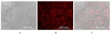
Fig. 1
Transfection efficiency of siRNA in BV2 cells with HEBP1-knockdown"
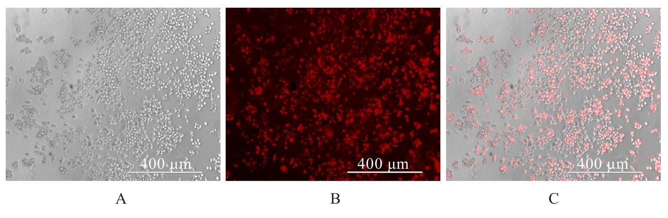
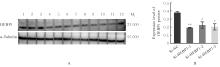
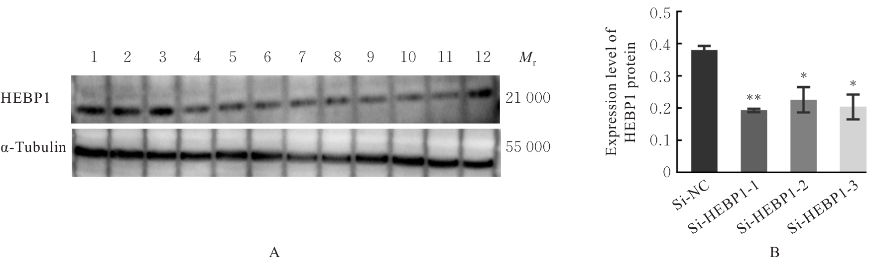
Fig. 2
Electrophoregram(A) and histogram(B) of expressions of HEBP1 proteins in BV2 cells with HEBP1-knockdown"
Tab.3
Expression levels of HEBP1 mRNA in BV2 cells in various groups"
| Group | HEBP1 mRNA |
|---|---|
| Si-NC | 1.00±0.05 |
| Si-HEBP1-1 | 0.01±0.03* |
| Si-HEBP1-2 | 0.30±0.14* |
| Si-HEBP1-3 | 0.38±0.12* |
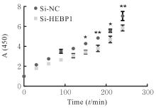
Fig. 3
Proliferation activities of BV2 cells in two groups"
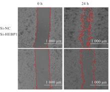
Fig. 4
Migration of cells in two groups detected by scratch assay"
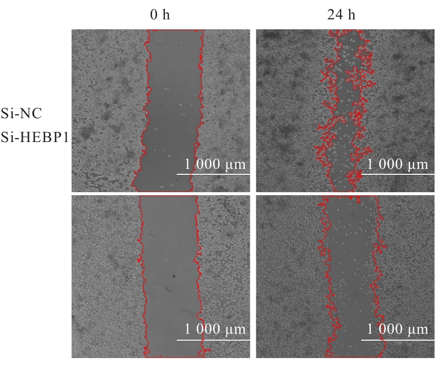
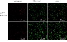
Fig. 5
Fluorescence images of mitochondrial membrane potential detection in BV2 cells in two groups"
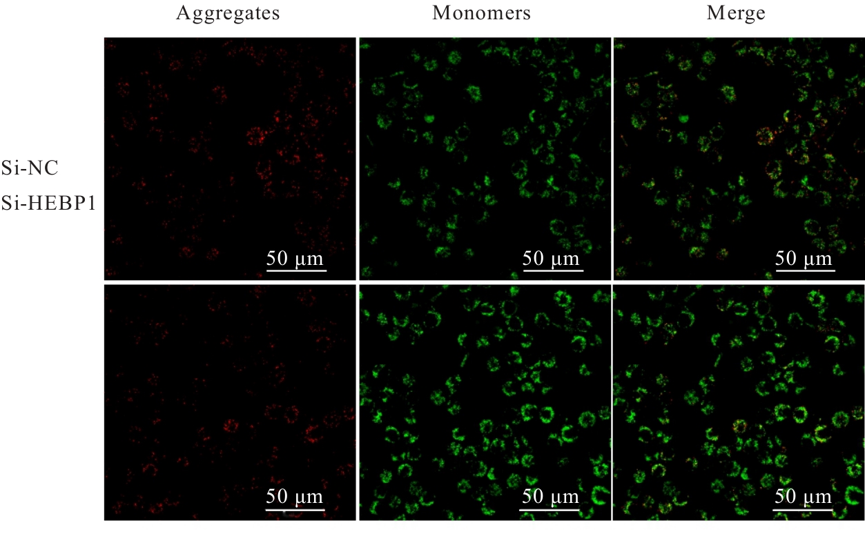
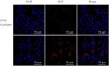
Fig. 6
Fluorescence images of ROS detection in BV2 cells in two groups"
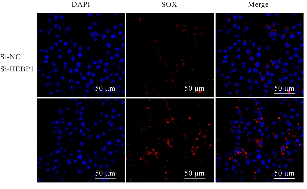
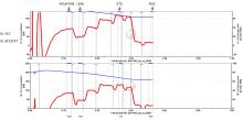
Fig. 7
Diagram of mitochondrial respiratory function changes of BV2 cells in two groups"
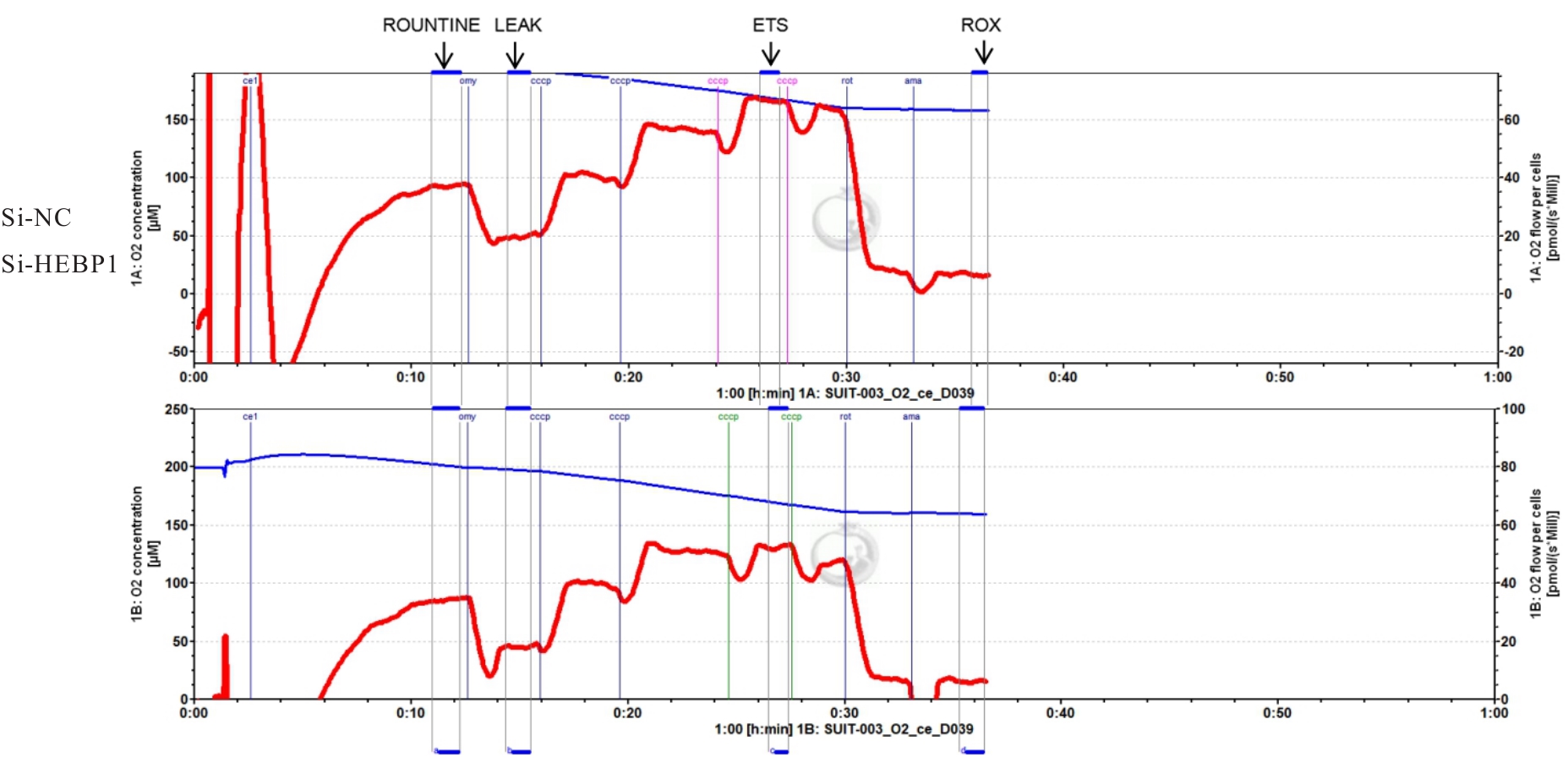
Tab.4
Mitochondrial respiratory oxygen consumption and ATP production of BV2 cells in two groups (n=3, x±s, pmol·s-1 per million cells)"
| Group | ROUNTINE | LEAK | EST | ROX | ATP |
|---|---|---|---|---|---|
| Si-NC | 36.44±0.80 | 19.31±0.31 | 72.35±18.08 | 5.57±1.35 | 17.06±0.57 |
| Si-HEBP1 | 32.99±1.37* | 17.94±0.40** | 54.06±2.16 | 5.16±0.73 | 15.04±1.11* |
Tab.5
Expression levels of IL-1β, TNF-α, and IL-6 mRNA in BV2 cells in various groups"
| Group | IL-1β | TNF-α | IL-6 |
|---|---|---|---|
| Si-NC | 1.00±0.05 | 1.00±0.05 | 1.00±0.05 |
| Si-HEBP1 | 0.94±0.48 | 1.22±0.38 | 2.21±1.15 |
| Si-NC+LPS | 1 139.35±211.34* | 10.22±3.58* | 174.96±37.05* |
| Si-HEBP1+LPS | 2 447.83±476.00△# | 19.15±2.38△# | 309.17±44.25△# |
| [1] | YAGENSKY O, KOHANSAL-NODEHI M, GUNASEELAN S, et al. Increased expression of heme-binding protein 1 early in Alzheimer’s disease is linked to neurotoxicity[J]. eLife, 2019, 8: e47498. |
| [2] | GOODFELLOW B J, FREIRE F, CARVALHO A L, et al. The SOUL family of heme-binding proteins: Structure and function 15 years later[J]. Coord Chem Rev, 2021, 448: 214189. |
| [3] | CHUA J J E. HEBP1 - An early trigger for neuronal cell death and circuit dysfunction in Alzheimer’s disease[J]. Semin Cell Dev Biol, 2023, 139: 102-110. |
| [4] | YIN G N. Pericyte-derived heme-binding protein 1 promotes angiogenesis and improves erectile function in diabetic mice[J]. Investig Clin Urol, 2022, 63(4): 464-474. |
| [5] | NICKEL W, RABOUILLE C. Mechanisms of regulated unconventional protein secretion[J]. Nat Rev Mol Cell Biol, 2009, 10(2): 148-155. |
| [6] | DEVOSSE T, DUTOIT R, MIGEOTTE I, et al. Processing of HEBP1 by cathepsin D gives rise to F2L, the agonist of formyl peptide receptor 3[J]. J Immunol, 2011, 187(3): 1475-1485. |
| [7] | GAO J L, GUILLABERT A, HU J Y, et al. F2L, a peptide derived from heme-binding protein, chemoattracts mouse neutrophils by specifically activating Fpr2, the low-affinity N-formylpeptide receptor[J]. J Immunol, 2007, 178(3): 1450-1456. |
| [8] | MIGEOTTE I, RIBOLDI E, FRANSSEN J D, et al. Identification and characterization of an endogenous chemotactic ligand specific for FPRL2[J]. J Exp Med, 2005, 201(1): 83-93. |
| [9] | CHEN K Q, IRIBARREN P, HU J Y, et al. Activation of Toll-like receptor 2 on microglia promotes cell uptake of Alzheimer disease-associated amyloid beta peptide[J]. J Biol Chem, 2006, 281(6): 3651-3659. |
| [10] | CUI Y H, LE Y Y, GONG W H, et al. Bacterial lipopolysaccharide selectively up-regulates the function of the chemotactic peptide receptor formyl peptide receptor 2 in murine microglial cells[J]. J Immunol, 2002, 168(1): 434-442. |
| [11] | CUI Y H, LE Y, ZHANG X, et al. Up-regulation of FPR2, a chemotactic receptor for amyloid beta 1-42 (a beta 42), in murine microglial cells by TNF alpha[J]. Neurobiol Dis, 2002, 10(3): 366-377. |
| [12] | GOMEZ PERDIGUERO E, KLAPPROTH K, SCHULZ C, et al. Tissue-resident macrophages originate from yolk-sac-derived erythro-myeloid progenitors[J]. Nature, 2015, 518(7540): 547-551. |
| [13] | BOTELLA LUCENA P, HENEKA M T. Inflammatory aspects of Alzheimer’s disease[J]. Acta Neuropathol, 2024, 148(1): 31. |
| [14] | FROST J L, SCHAFER D P. Microglia: architects of the developing nervous system[J]. Trends Cell Biol, 2016, 26(8): 587-597. |
| [15] | SCHAFER D P, LEHRMAN E K, KAUTZMAN A G, et al. Microglia sculpt postnatal neural circuits in an activity and complement-dependent manner[J]. Neuron, 2012, 74(4): 691-705. |
| [16] | CAI Y L, LIU J L, WANG B, et al. Microglia in the neuroinflammatory pathogenesis of Alzheimer’s disease and related therapeutic targets[J]. Front Immunol, 2022, 13: 856376. |
| [17] | HANISCH U K, KETTENMANN H. Microglia: active sensor and versatile effector cells in the normal and pathologic brain[J]. Nat Neurosci, 2007, 10(11): 1387-1394. |
| [18] | NAYAK D, ROTH T L, MCGAVERN D B. Microglia development and function[J]. Annu Rev Immunol, 2014, 32: 367-402. |
| [19] | XIE Z, MENG J, WU Z, et al. The dual nature of microglia in Alzheimer’s disease: a microglia-neuron crosstalk perspective[J]. Neuroscientist, 2023, 29(5): 616-638. |
| [20] | YAN H Y, WANG W, CUI T T, et al. Advances in the understanding of the correlation between neuroinflammation and microglia in Alzheimer’s disease[J]. Immunotargets Ther, 2024, 13: 287-304. |
| [21] | CALSOLARO V, EDISON P. Neuroinflammation in Alzheimer’s disease: Current evidence and future directions[J]. Alzheimers Dement, 2016, 12(6): 719-732. |
| [22] | TWAROWSKI B, HERBET M. Inflammatory processes in Alzheimer’s disease-pathomechanism, diagnosis and treatment: a review[J]. Int J Mol Sci, 2023, 24(7): 6518. |
| [23] | FITZGERALD K A, KAGAN J C. Toll-like receptors and the control of immunity[J]. Cell, 2020, 180(6): 1044-1066. |
| [24] | NABI S U, KHAN A, SIDDIQUI E M, et al. Mechanisms of mitochondrial malfunction in Alzheimer’s disease: new therapeutic hope[J]. Oxid Med Cell Longev, 2022, 2022: 4759963. |
| [25] | LI Y, XIA X H, WANG Y, et al. Mitochondrial dysfunction in microglia: a novel perspective for pathogenesis of Alzheimer’s disease[J]. J Neuroinflammation, 2022, 19(1): 248. |
| [26] | JOHANNSEN D L, RAVUSSIN E. The role of mitochondria in health and disease[J]. Curr Opin Pharmacol, 2009, 9(6): 780-786. |
| [27] | KAMATHAM P T, SHUKLA R, KHATRI D K, et al. Pathogenesis, diagnostics, and therapeutics for Alzheimer’s disease: Breaking the memory barrier[J]. Ageing Res Rev, 2024, 101: 102481. |
| [28] | WANG L X, PAVLOU S, DU X, et al. Glucose transporter 1 critically controls microglial activation through facilitating glycolysis[J]. Mol Neurodegener, 2019, 14(1): 2. |
| [29] | PRADEEPKIRAN J A, REDDY P H. Defective mitophagy in Alzheimer’s disease[J]. Ageing Res Rev, 2020, 64: 101191. |
| [1] | Lulu FU,Yinggang ZOU,Xiaoyu ZHENG,Xueying ZHANG,Jingshun ZHANG,Min WANG,Qiang ZHANG,Lianwen ZHENG. Expression of placenta expressed transcription factor 1 in ovarian tissue of polycystic ovary syndrome rats and its effect on proliferation of rat ovarian granulosa cells [J]. Journal of Jilin University(Medicine Edition), 2025, 51(5): 1177-1184. |
| [2] | Guang YANG,Zhifang ZHENG,Xinhua ZHANG. Research progress in protective effect of miRNA on neonatal hypoxic-ischemic brain injury [J]. Journal of Jilin University(Medicine Edition), 2025, 51(5): 1423-1428. |
| [3] | Pingsheng ZHU,Sitang GE,Lugen ZUO,Deli CHEN,Yangyang ZHANG. Effect of miR-325-3p targeting PRELID1 gene in regulation of EMT pathway on invasion and migration of colon cancer cells and their mechanisms [J]. Journal of Jilin University(Medicine Edition), 2025, 51(5): 1185-1193. |
| [4] | Zhongjun SHEN,Yao ZHAO,Mingbo JIA,Liyan ZHAO. Research progress in effects of hypoxia-inducible factors on cell migration and invasion during epithelial-mesenchymal transition in glioma cells [J]. Journal of Jilin University(Medicine Edition), 2025, 51(4): 1145-1154. |
| [5] | Yixuan GAO,Peng WANG,Silong ZHANG,Ruijuan GAO,Yingfang MA,Keke ZHANG,Dan FENG,Zongqi HUANG,Ketao MA,Li LI,Junqiang SI. Inhibitory effect of safranal on proliferation, migration and phenotypic transformation of vascular smooth muscle cells of rats induced by high glucose in vitro [J]. Journal of Jilin University(Medicine Edition), 2025, 51(4): 948-957. |
| [6] | Wenxuan LI,Minru ZONG. Research progress in role of migration of Schwann cells in repairment of peripheral nerve injury [J]. Journal of Jilin University(Medicine Edition), 2025, 51(4): 1137-1144. |
| [7] | Xiaoshuang HE,Lina XU,Mei CUI,Yu ZHAO,Bei WANG,Zheng HUANG,Yuchao WANG,Wenyan XIN,Chao WU. Effects of lncRNA DUXAP8 in lung cancer A549 cells-derived exosomes on lung cancer cell growth and its mechnism [J]. Journal of Jilin University(Medicine Edition), 2025, 51(4): 958-967. |
| [8] | Hongli LI,Mengyao WANG,Yangyang LIU,Hui ZHANG,Li LI. Effect of KHSRP on biological behavior of colorectal cancer cells through activation of JAK/STAT signaling pathway [J]. Journal of Jilin University(Medicine Edition), 2025, 51(4): 996-1006. |
| [9] | Yihui WANG,Qing ZHANG,Yingnan LI,Liping YE. Effect of KIAA1522 on proliferation, migration, and invasion of lung cancer cells and its mechanism [J]. Journal of Jilin University(Medicine Edition), 2025, 51(3): 727-739. |
| [10] | Donghui LIU,Yunzhe CI,Chunyan WANG,Wenyi MA. Effect of miR-199a-5p on expression of Caveolin-1, cell migration and apoptosis in glioma U251 cells [J]. Journal of Jilin University(Medicine Edition), 2025, 51(3): 663-671. |
| [11] | Fan WANG,Xin WEN,Yixuan WANG,Yuan WANG. Effect of gap junction β2 on prognosis of patients with lung adenocarcinoma and biological behavior of lung adenocarcinoma A549 cells [J]. Journal of Jilin University(Medicine Edition), 2025, 51(3): 716-726. |
| [12] | Mengyun LU,Yucheng HAN,Yihong HU,Minhui HE,Yanqun ZHANG,Xianqiong ZOU. Effects of glycolipid transfer protein on proliferation, migration,and invasion of pancreatic cancer PANC-1 cells and their mechanisms [J]. Journal of Jilin University(Medicine Edition), 2025, 51(2): 284-295. |
| [13] | Ying YANG,Liang ZHAO,Yong YOU,Qian XU,Zhenjun YANG. Influence of 17β-estradiol in proliferation and differentiation of hippocampal neural stem cells and its mechanism [J]. Journal of Jilin University(Medicine Edition), 2025, 51(2): 317-324. |
| [14] | Shuyan SUN,Huakun ZHANG,Ziru ZHOU,Feng LI,Xiaobin CUI. Expression of CRNN protein in esophageal squamous cell carcinoma tissue and influence of its overexpression in biological behavior of esophageal squamous cell carcinoma Eca9706 cells [J]. Journal of Jilin University(Medicine Edition), 2025, 51(2): 275-283. |
| [15] | Yaqi ZHANG,Jing MI,Jingrong YANG,Xinming LI,Li LI. Effect of up-regulation of miR-31 expression on osteogenic differentiation of dental pulp stem cells through Wnt-β/catenin signaling pathway [J]. Journal of Jilin University(Medicine Edition), 2025, 51(2): 412-419. |
|
||