Journal of Jilin University(Medicine Edition) ›› 2024, Vol. 50 ›› Issue (3): 728-738.doi: 10.13481/j.1671-587X.20240317

• Research in basic medicine • Previous Articles    

Inhibitory effect of miR-487a on M2-type polarization of gastric cancer tumor-associated macrophages by targeting TIA1

Yan QU1,Lin DAI1,Biao WANG1,Duji RUAN1,Yuchang ZHONG1,Xuefeng YANG1,2()   

  1. 1.Department of Gastrointestinal Surgery,Second Affiliated Hospital,Zunyi Medical University,Zunyi 563006,China
    2.Department of Gastrointestinal Surgery,Affiliated Hospital,Zunyi Medical University,Zunyi 563000,China
  • Received:2023-07-11 Online:2024-05-28 Published:2024-07-01
  • Contact: Xuefeng YANG E-mail:yangxuefeng1978@126.com

Abstract:

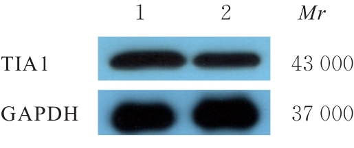
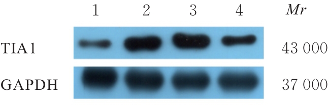
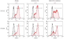
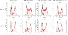
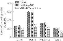
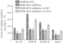
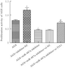
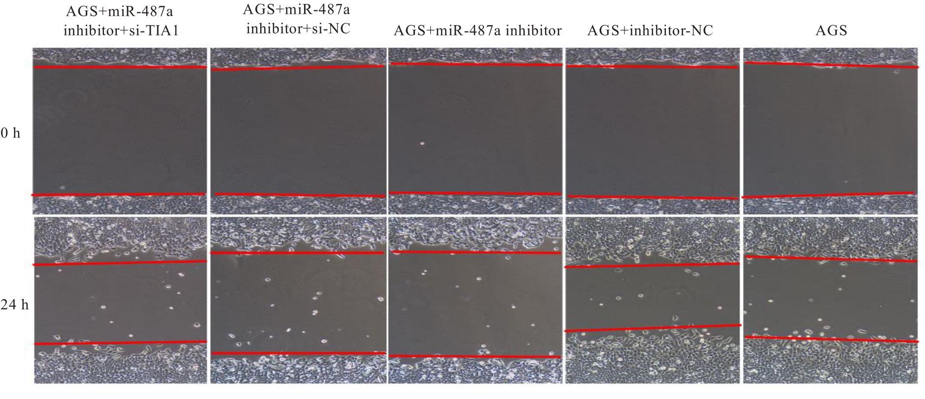
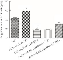
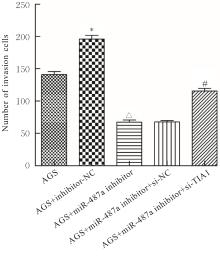
Objective To discuss the inhibitory effect of microRNA-487a (miR-487a) on the M2 polarization of tumor-associated macrophages (TAMs) in gastric cancer, and to clarify its effect on the proliferation, invasion, and migration of the gastric cancer AGS cells. Methods The TAMs from gastric cancer tissue and adjacent normal tissue macrophages (NTMs) from adjacent tissue of the primary gastric cancer patients were isolated and cultured. The human monocyte THP-1 cells were induced in vitro to differentiate into TAMs, and the differentiated M0, M1, and M2 macrophages were cultured for 24 h by conditioned medium (CM) to obtain the TAMs, M1-TAMs, and M2-TAMs respectively. The TAMs were transfected and then divided into blank group, inhibitor-NC group, miR-487a inhibitor group, miR-487a inhibitor+si-NC group, and miR-487a inhibitor+si-TIA1 group. The transfection efficiencies of the cells in various groups were detected by real-time fluorescence quantitative PCR (RT-qPCR) and Western blotting methods. The M2-TAMs were co-cultured with the AGS cells, and divided into AGS group, AGS+inhibitor-NC group, AGS+miR-487a inhibitor group, AGS+miR-487a inhibitor+si-NC group, and AGS+miR-487a inhibitor+si-TIA1 group. RT-qPCR method was used to detect the expression levels of miR-487a and lymphocyte intracytoplasmic antigen-1 (TIA1) mRNA in TAMs from gastric cancer tissue and NTMs from adjacent normal tissue in various groups; Western blotting method was used to detect the expression level of TIA1 protein in TAMs from gastric cancer tissue and NTMs from adjacent normal tissue and TAMs in various groups; flow cytometry was used to detect the levels of CD206 and CD163 in TAMs in various groups; enzyme-linked immunosorbent assay (ELISA) was used to detect the levels of interleukin-10 (IL-10), transforming growth factor-beta (TGF-β), vascular endothelial growth factor A (VEGF-A), and arginase-1 (Arg-1) in culture supernatant of the TAMs cells; CCK-8 assay was used to detect the proliferative activity of the AGS cells in various groups; wound healing assay was used to detect the migration rates of the AGS cells in various groups; Transwell assay was used to detect the number of invasion AGS cells in various groups. Results The RT-qPCR results shoued that compared with NTMs from adjacent tissue, the expression level of miR-487a in the TAMs from gastric cancer tissue was significantly increased (P<0.01) and the expression level of TIA1 mRNA was significantly decreased (P<0.01). Compared with TAMs, the expression level of miR-487a in M1-TAMs was significantly decreased (P<0.01), and the expression level of TIA1 mRNA was increased (P<0.01); the expression level of miR-487a in M2-TAMs was significantly increased (P<0.01), and the expression level of TIA1 mRNA was decreased (P<0.01). After transfection, compared with blank group and inhibitor-NC group, the expression level of miR-487a in the cells in miR-487a inhibitor group was significantly decreased (P<0.01), indicating successful transfection. The Western blotting results showed that compared with NTMs from adjacent normal tissue, the expression level of TIA1 protein in TAMs from gastric cancer tissue was decreased (P<0.01); compared with TAMs, the expression level of T1A1 protein in M1-TAMs was significantly increased (P<0.01), and the expression of TIA1 protein in M2-TAMs was significantly decreased (P<0.01); after co-transfection, compared with inhibitor-NC group, the expression level of TIA1 protein in the cells in miR-487a inhibitor group was significantly increased (P<0.01); compared with miR-487a inhibitor+si-NC group, the expression level of TIA1 protein in the cells in miR-487a inhibitor+si-TIA1 group was significantly decreased (P<0.01). The flow cytometry results showed that compared with blank group and inhibitor-NC group, the levels of CD206 and CD163 in the cells in miR-487a inhibitor group were significantly decreased (P<0.01); after co-transfection, compared with inhibitor-NC group, the levels of CD206 and CD163 in the cells in miR-487a inhibitor group were significantly decreased (P<0.01); compared with miR-487a inhibitor+si-NC group, the levels of CD206 and CD163 in the cells in miR-487a inhibitor+si-TIA1 group were significantly increased (P<0.01). The ELISA results showed that compared with blank group and inhibitor-NC group, the levels of IL-10, TGF-β, VEGF-A, and Arg-1 in culture supernatant of the TAMs in miR-487a inhibitor group were significantly decreased (P<0.01); after co-transfection, compared with inhibitor-NC group, the levels of IL-10, TGF-β, VEGF-A, and Arg-1 in culture supernatant of the TAMs in miR-487a inhibitor group were significantly decreased (P<0.01); compared with miR-487a inhibitor+si-NC group, the levels of IL-10, TGF-β, VEGF-A, and Arg-1 in culture supernatant of the TAMs in miR-487a inhibitor+si-TIA1 group were significantly increased (P<0.01). The CCK-8 assay results showed that compared with AGS group, the proliferation activity of the cells in AGS+inhibitor-NC group was significantly increased (P<0.01); compared with AGS+inhibitor-NC group, the proliferation activity of the cells in AGS+miR-487a inhibitor group was significantly decreased (P<0.01); compared with AGS+miR-487a inhibitor+si-NC group, the proliferation activity of the cells in AGS+miR-487a inhibitor+si-TIA1 group was significantly increased (P<0.01). The wound healing assay results showed that compared with AGS group, the migration rate of the cells in AGS+inhibitor-NC group was significantly (P<0.05); compared with AGS+inhibitor-NC group, the migration rate of the cells in AGS+miR-487a inhibitor group was significantly decreased (P<0.01); compared with AGS+miR-487a inhibitor+si-NC group, the migration rate of the cells in AGS+miR-487a inhibitor+si-TIA1 group was significantly increased (P<0.05). The Transwell assay results showed that compared with AGS group, the number of invasion AGS cells in AGS + inhibitor-NC group was significantly increased (P<0.01); compared with AGS + inhibitor-NC group, the number of invasion AGS cells in AGS+miR-487a inhibitor group was significantly decreased (P<0.01); compared with AGS+miR-487a inhibitor+si-NC group, the number of invasion AGS cells in AGS+miR-487a inhibitor+si-TIA1 group was significantly increased (P<0.01). Conclusion Silencing the miR-487a expression can inhibit the M2 polarization of the gastric cancer-associated macrophages by targeted upregulation of TIA1, and suppress the proliferation, migration, and invasion of the gastric cancer cells.

Key words: Gastric neoplasm, MicroRNA-487a, T-lymphocyte-restricted intracellular antigen-1, Tumor-associated macrophage, M2 polarization

CLC Number: 

  • R735.2