Journal of Jilin University(Medicine Edition) ›› 2024, Vol. 50 ›› Issue (4): 989-999.doi: 10.13481/j.1671-587X.20240413
• Research in basic medicine • Previous Articles Next Articles
Effect of Myod1 on proliferation and apoptosis of oxygen-glucose-deprived SHSY5Y cells by regulating lncRNA SNHG15 and miR-24-3p
Fangchao JI,Chenxin ZHANG,Zhanjun REN,Yunzhi PAN,Qi LU,Xingyuan SUN( )
)
- Department of Neurology,Third Affiliated Hospital,Qiqihar Medical University,Qiqihar 161000,China
-
Received:2023-07-14Online:2024-07-28Published:2024-08-01 -
Contact:Xingyuan SUN E-mail:sunxingyuandor@163.com
CLC Number:
- R743
Cite this article
Fangchao JI,Chenxin ZHANG,Zhanjun REN,Yunzhi PAN,Qi LU,Xingyuan SUN. Effect of Myod1 on proliferation and apoptosis of oxygen-glucose-deprived SHSY5Y cells by regulating lncRNA SNHG15 and miR-24-3p[J].Journal of Jilin University(Medicine Edition), 2024, 50(4): 989-999.
share this article
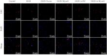
Tab. 3
Rates of EdU positive cells and rates of TUNEL positive cells in various groups after over-expression and knockdown of Myod1"
| Group | Rate of EdU positive cells | Rate of TUNEL positive cells |
|---|---|---|
Control OGD OGD+Vector OGD+Myod1 OGD+si-NC OGD+si-Myod1 | 42.14±2.01 | 3.17±0.29 |
| 12.34±1.11* | 12.05±1.05* | |
| 11.12±0.44 | 13.12±0.82 | |
3.56±0.96△ 13.89±1.31 32.98±1.46△ | 17.92±1.01△ 11.37±0.91 9.13±0.67△ |
Tab.4
Cell activities in various groups after knockdown of SNHG15 and over-expression of Myod1"
| Group | Cell activity | ||
|---|---|---|---|
| (t/h) 24 | 48 | 72 | |
| Control | 0.38±0.01 | 0.87±0.03 | 1.17±0.03 |
| OGD | 0.28±0.01 | 0.49±0.02* | 0.59±0.02* |
| OGD+si-NC | 0.30±0.01 | 0.52±0.02 | 0.64±0.02 |
| OGD+si-SNHG15 | 0.34±0.01 | 0.73±0.03△ | 0.96±0.04△ |
| OGD+si-SNHG15+Vector | 0.33±0.02 | 0.76±0.03 | 0.93±0.04 |
| OGD+si-SNHG15+Myod1 | 0.33±0.02 | 0.55±0.02# | 0.72±0.01# |
Fig.5 Rates of EdU positive cells and rates of TUNEL positive cells after knockdown of SNHG15 and over-expression of Myod1 (n=3,x±s,η/%)"
| Group | Rate of EdU positive cells | Rate of TUNEL positive cells |
|---|---|---|
Control OGD OGD+si-NC OGD+si-SNHG15 OGD+si-SNHG15+Vector OGD+si-SNHG15+Myod1 | 28.34±2.53 | 2.64±0.26 |
| 8.02±1.78* | 27.73±2.08* | |
| 7.54±1.22 | 29.18±2.95 | |
19.43±2.88△ 21.25±3.43 9.32±2.05# | 14.84±1.15△ 16.05±1.22 22.53±2.07# |
Tab. 6
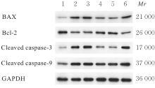
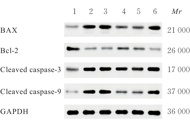
Expression levels of apoptosis-related proteins in cells in various groups after knockdown of SNHG15 and over-expression of Myod1"
| Group | Bax | Bcl-2 | Cleaved caspase-3 | Cleaved caspase-9 |
|---|---|---|---|---|
| Control | 1.06±0.13 | 0.98±0.09 | 3.13±0.39 | 0.98±0.09 |
| OGD | 4.21±0.29* | 0.33±0.05* | 17.21±1.80* | 2.57±0.37* |
| OGD+si-NC | 4.68±0.34 | 0.29±0.03 | 17.98±1.70 | 2.38±0.41 |
| OGD+si-SNHG15 | 2.23±0.18△ | 0.72±0.08△ | 8.83±1.01△ | 1.38±0.12△ |
| OGD+si-SNHG15+Vector | 2.15±0.14 | 0.82±0.05 | 9.91±1.16 | 1.69±0.20 |
| OGD+si-SNHG15+Myod1 | 3.77±0.29# | 0.43±0.04# | 22.18±2.8# | 2.11±0.16# |
Tab. 7
Cell activities in various groups after over-expression of miR-24-3p and SNHG15"
| Group | Cell activity | ||
|---|---|---|---|
| (t/h)24 | 48 | 72 | |
| Control | 0.80±0.03 | 1.32±0.03 | 1.64±0.05 |
| OGD | 0.68±0.04 | 0.93±0.05* | 1.10±0.04* |
| OGD+miR-NC | 0.77±0.01 | 1.03±0.01 | 1.24±0.05 |
| OGD+miR-mimics | 0.75±0.01 | 1.18±0.01△ | 1.43±0.04△ |
| OGD+miR-mimics +Vector | 0.79±0.01 | 1.22±0.02 | 1.39±0.05 |
| OGD+miR-mimics +SNHG15 | 0.81±0.01 | 0.99±0.02# | 1.16±0.02# |
Tab. 8
Rates of EdU positive cells and rates of TUNEL positive cells in various groups after over-expression of miR-24-3p and SNHG15"
| Group | Rate of EdU-positive cells | Rate of TUNEL- positive cells |
|---|---|---|
Control OGD OGD+miR-NC OGD+miR-mimics OGD+miR-mimics+Vector OGD+miR-mimics+SNHG15 | 24.57±3.16 | 3.18±0.46 |
| 6.42±1.11* | 27.49±2.74* | |
| 5.78±0.34 | 25.32±2.16 | |
14.85±1.83△ 15.72±1.37 8.21±1.42# | 13.18±1.21△ 14.67±2.02 20.30±2.57# |
Tab.9
Expression levels of apoptosis-related proteins in cells in various groups after over-expression of miR-24-3p and SNHG15"
| Group | Bax | Bcl-2 | Cleaved caspase-3 | Cleaved caspase-9 |
|---|---|---|---|---|
| Control | 1.04±0.05 | 0.99±0.09 | 1.01±0.09 | 0.98±0.09 |
| OGD | 4.82±0.51* | 0.34±0.05* | 2.88±0.28* | 3.52±0.28* |
| OGD+miR-NC | 4.17±0.38 | 0.37±0.07 | 2.77±0.29 | 3.40±0.37 |
| OGD+miR-mimics | 1.89±0.19△ | 0.47±0.09△ | 1.98±0.15△ | 1.89±0.16△ |
| OGD+miR-mimics+Vector | 2.14±0.25 | 0.43±0.06 | 1.83±0.20 | 2.03±0.24 |
| OGD+miR-mimics+SNHG15 | 3.88±0.42# | 0.39±0.05# | 3.00±0.42# | 2.95±0.27# |
| 1 | WANG Y J, LI Z X, GU H Q, et al. China Stroke Statistics: an update on the 2019 report from the National Center for Healthcare Quality Management in Neurological Diseases, China National Clinical Research Center for Neurological Diseases, the Chinese Stroke Association, National Center for Chronic and Non-communicable Disease Control and Prevention, Chinese Center for Disease Control and Prevention and Institute for Global Neuroscience and Stroke Collaborations[J]. Stroke Vasc Neurol, 2022, 7(5): 415-450. |
| 2 | STROKE COLLABORATORSGBD. Global, regional, and national burden of stroke and its risk factors, 1990-2019: a systematic analysis for the Global Burden of Disease Study 2019[J]. Lancet Neurol, 2021, 20(10): 795-820. |
| 3 | DING Q Q, LIU S W, YAO Y D, et al. Global, regional, and national burden of ischemic stroke, 1990-2019[J]. Neurology, 2022, 98(3): e279-e290. |
| 4 | SHEHJAR F, MAKTABI B, RAHMAN Z A, et al. Stroke: molecular mechanisms and therapies: update on recent developments[J]. Neurochem Int, 2023, 162: 105458. |
| 5 | BRIDGES M C, DAULAGALA A C, KOURTIDIS A. LNCcation: lncRNA localization and function[J]. J Cell Biol, 2021, 220(2): e202009045. |
| 6 | LI Y H, HU Y Q, WANG S C, et al. LncRNA SNHG5: a new budding star in human cancers[J]. Gene, 2020, 749: 144724. |
| 7 | ZHANG J, YUAN L, ZHANG X, et al. Altered long non-coding RNA transcriptomic profiles in brain microvascular endothelium after cerebral ischemia[J]. Exp Neurol, 2016, 277: 162-170. |
| 8 | YANG L, LU Z N. Long non-coding RNA HOTAIR promotes ischemic infarct induced by hypoxia through up-regulating the expression of NOX2[J]. Biochem Biophys Res Commun, 2016, 479(2): 186-191. |
| 9 | WANG J, CAO B, HAN D, et al. Long non-coding RNA H19 induces cerebral ischemia reperfusion injury via activation of autophagy[J]. Aging Dis, 2017, 8(1): 71-84. |
| 10 | QI X, SHAO M, SUN H J, et al. Long non-coding RNA SNHG14 promotes microglia activation by regulating miR-145-5p/PLA2G4A in cerebral infarction[J]. Neuroscience, 2017, 348: 98-106. |
| 11 | GUO T Z, LIU Y T, REN X L, et al. Errate: promoting role of long non-coding RNA small nucleolar RNA host gene 15 (SNHG15) in neuronal injury following ischemic stroke via the microRNA-18a/CXC chemokine ligand 13 (CXCL13)/ERK/MEK axis[J]. Med Sci Monit, 2022, 28: e938473. |
| 12 | DU H B, DING L Q, ZENG T, et al. LncRNA SNHG15 modulates ischemia-reperfusion injury in human AC16 cardiomyocytes depending on the regulation of the miR-335-3p/TLR4/NF-κB pathway[J]. Int Heart J, 2022, 63(3): 578-590. |
| 13 | LIU Z Z, ZHANG C Y, HUANG L L, et al. Elevated expression of lncRNA SNHG15 in spinal tuberculosis: preliminary results[J]. Eur Rev Med Pharmacol Sci, 2019, 23(20): 9017-9024. |
| 14 | DENG Q W, LI S, WANG H, et al. Differential long noncoding RNA expressions in peripheral blood mononuclear cells for detection of acute ischemic stroke[J]. Clin Sci, 2018, 132(14): 1597-1614. |
| 15 | WANG H F, WANG Z Q, DING Y, et al. Endoplasmic reticulum stress regulates oxygen-glucose deprivation-induced parthanatos in human SH-SY5Y cells via improvement of intracellular ROS[J]. CNS Neurosci Ther, 2018, 24(1): 29-38. |
| 16 | FU J D, HUANG Y B, XIAN L W. LncRNA SNHG15 regulates hypoxic-ischemic brain injury via miR-153-3p/SETD7 axis[J]. Histol Histopathol, 2022, 37(11): 1113-1125. |
| 17 | KANG M M, JI F C, SUN X Y, et al. LncRNA SNHG15 promotes oxidative stress damage to regulate the occurrence and development of cerebral ischemia/reperfusion injury by targeting the miR-141/SIRT1 axis[J]. J Healthc Eng, 2021, 2021: 6577799. |
| 18 | LI S, CAO Y Z, ZHANG H X, et al. Construction of lncRNA-mediated ceRNA network for investigating immune pathogenesis of ischemic stroke[J]. Mol Neurobiol, 2021, 58(9): 4758-4769. |
| 19 | 张亚杰, 尹立勇, 董晓娇, 等. lncRNA NEAT1沉默通过IL-10/STAT3信号通路调节小胶质细胞极化对脑缺血再灌注损伤大鼠的影响[J]. 中风与神经疾病杂志, 2023, 40(2): 118-123. |
| 20 | LIU W S, CHEN X S, ZHANG Y. Effects of microRNA-21 and microRNA-24 inhibitors on neuronal apoptosis in ischemic stroke[J]. Am J Transl Res, 2016, 8(7): 3179-3187. |
| 21 | KUAI F, ZHOU L, ZHOU J P, et al. Long non-coding RNA THRIL inhibits miRNA-24-3p to upregulate neuropilin-1 to aggravate cerebral ischemia-reperfusion injury through regulating the nuclear factor κB p65 signaling[J]. Aging, 2021, 13(6): 9071-9084. |
| 22 | HAN X, LI Q B, LIU C, et al. Overexpression miR-24-3p repressed Bim expression to confer tamoxifen resistance in breast cancer[J]. J Cell Biochem, 2019, 120(8): 12966-12976. |
| 23 | XU J, QIAN X Z, DING R. MiR-24-3p attenuates IL-1β-induced chondrocyte injury associated with osteoarthritis by targeting BCL2L12[J]. J Orthop Surg Res, 2021, 16(1): 371. |
| 24 | SHAN Q L, CHEN N N, MENG G Z, et al. Overexpression of lncRNA MT1JP mediates apoptosis and migration of hepatocellular carcinoma cells by regulating miR-24-3p[J]. Cancer Manag Res, 2020, 12: 4715-4724. |
| [1] | Wentao WANG,Xuguang MI,Yang ZHOU,Wenxing PU,Jiaxu GAO,Meng JING,Fankai MENG. Effect of autophagy induced by exosomes derived from bone marrow mesenchymal stem cells on survival of SH-SY5Y cells inhibited by MPP+ and its mechanism [J]. Journal of Jilin University(Medicine Edition), 2022, 48(3): 606-614. |
| [2] | BU Yi, ZHANG Shuo, QIAN Xudong, WANG Hongmei, DOU Zhijie. Expression of insulin-like growth factor binding protein-3 in ischemic brain tissue of cerebral infarction model rats and its relationship with angiogenesis [J]. Journal of Jilin University(Medicine Edition), 2020, 46(04): 759-764. |
| [3] | BU Yi, ZHANG Shuo, QIAN Xudong, WANG Hongmei, DOU Zhijie. Expressions of Galectin-3 and MMP-9 in brain tissue of mice with acute cerebral infarction and intervention effect of nimodipine [J]. Journal of Jilin University(Medicine Edition), 2020, 46(03): 551-556. |
| [4] | JIAO Guangmei, SHAN Hailei, ZHANG Xiaoxuan, ZHAO Liang, DOU Zhijie, KANG Lingling, MA Zheng, SUN Yanjun, YANG Ning. Protective effect of Shuxuening Injection on brain tissue of rats with acute cerebral infarction and its mechanism [J]. Journal of Jilin University(Medicine Edition), 2020, 46(03): 557-562. |
| [5] | BI Yongzhi, MA Fen, SHI Junxia, SONG Linlin, LI Linze. Protective effect of emodin on focal cerebral ischemia in rats and its mechanism [J]. Journal of Jilin University(Medicine Edition), 2020, 46(03): 569-574. |
| [6] | ZHONG Yue, LI Haichao, FENG Xinrui, CUI Yushu, ZHENG Zhonghua, WANG Weiyao, QI Ling. Inhibitory effect of foodborne procyanidins on growth of SH-SY5Y cells and its mechanism [J]. Journal of Jilin University(Medicine Edition), 2020, 46(01): 61-65. |
| [7] | JIAO Guangmei, SHAN Hailei, DOU Zhijie, ZHAO Liang, ZHANG Xiaoxuan, KANG Lingling, MA Zheng, YANG Ning. Effect of nerve growth factor on expression level of growth differentiation factor-15 in brain tissue of rats with cerebral infarction [J]. Journal of Jilin University(Medicine Edition), 2019, 45(03): 572-576. |
| [8] | ZHU Yunbo, LI Jiajia, MA Zheng, ZHAO Liang. Effect of alteplase on expressions of Claudin-1 and Claudin-5 proteins in vascular endothelial cells of rats with acute cerebral infarction [J]. Journal of Jilin University Medicine Edition, 2017, 43(06): 1137-1141. |
| [9] | GENG Yurong, LIU Yingjie, ZHANG Huili, WANG Hong. Clinical characteristics of patients with hyperthyroidism complicated with acute cerebral infarction and evaluation on prognosis and safety of intravenous thrombolysis treatment [J]. Journal of Jilin University Medicine Edition, 2017, 43(02): 369-374. |
| [10] | LIU Yingli, YANG Libin, ZHANG Shushi, ZHANG Shuyan. Therapeutic effect of ButylphthalideInjection inelderly patients with acute cerebral infarction and its influence incerebral hemodynamics andcerebral vascular reserve [J]. Journal of Jilin University Medicine Edition, 2017, 43(02): 344-348. |
| [11] | CHANG Yongxia, LI Jiao, MA Qiuyun, HOU Wenli, GE Lei, MENG Haichao, HU Jin, MA Chong, WANG Zhengtian. Improvement effect of electromyographic biofeedback on wrist dorsiflexion function of patients with cerebral infarction at different Brunnstrom stages [J]. Journal of Jilin University Medicine Edition, 2016, 42(05): 975-979. |
| [12] | YANG Qing, SUI Xin, WANG Baosen, YU Zhixin, ZHU Kai, LYU Guangfu, WANG Jianwei, YAN Wenhao, ZHANG Huiyuan, LIN Jianan, LIU Yingna, LI Na, QU Xiaobo. Protective effect of schizandra B on injury of SH-SY5Y cells induced by Glu and its mechanim [J]. Journal of Jilin University Medicine Edition, 2016, 42(01): 80-84. |
| [13] | YANG Yanyan, JIN Minghua, LI Yanbo, LI Yang, WANG Jiahui, ZHENG Tong, WANG Sihui, CHI Xiangyu, ZHANG Yujia, SUN Zhiwei. Induction effect of nano- SiO2 on apoptosis of neuroblastoma SH-SY5Y cells and its mechanism [J]. Journal of Jilin University Medicine Edition, 2015, 41(02): 249-254. |
| [14] | XIONG Yingqiong, PAN Jie, WU Xiaomu, CHENG Shaomin. Evaluation on clinical curative effect of myoelectricity triggering biofeedback in treatment of early hemiplegia of cerebral infarction patients [J]. Journal of Jilin University Medicine Edition, 2015, 41(01): 156-159. |
| [15] | WANG Ying-ying,GUO Na,HE Jin-ting,SHAO Yan-kun,BAO Xiao-qun,MANG Jing,XU Zhong-xin. Risk prediction values of different score models for cerebral infarction after transient ischemic attack [J]. Journal of Jilin University Medicine Edition, 2014, 40(04): 851-854. |
|


