Journal of Jilin University(Medicine Edition) ›› 2025, Vol. 51 ›› Issue (4): 996-1006.doi: 10.13481/j.1671-587X.20250416
• Research in basic medicine • Previous Articles Next Articles
Effect of KHSRP on biological behavior of colorectal cancer cells through activation of JAK/STAT signaling pathway
Hongli LI1,Mengyao WANG2,Yangyang LIU2,Hui ZHANG3( ),Li LI2()
- 1.Department of Oncology,Huaihe Hospital,Henan University,Kaifeng 475099,China
2.Department of,School of Nursing and Health,Henan University,Kaifeng 475004,China
3.Department of Gastroenterology,Huaihe Hospital,Henan University,Kaifeng 475099,China
-
Received:2024-08-08Accepted:2024-10-12Online:2025-07-28Published:2025-08-25 -
Contact:Hui ZHANG,Li LI E-mail:2998975895@qq.com;10210051@vip.henu.edu.cn
CLC Number:
- R735.3
Cite this article
Hongli LI,Mengyao WANG,Yangyang LIU,Hui ZHANG,Li LI. Effect of KHSRP on biological behavior of colorectal cancer cells through activation of JAK/STAT signaling pathway[J].Journal of Jilin University(Medicine Edition), 2025, 51(4): 996-1006.
share this article
Tab.1
Primer sequences of PCR"
| Gene | Primer sequences(5'-3') | |
|---|---|---|
| KHSRP | R | AATGAGTACGGATCTCGGATTGG |
| F | CCGTCATCTTGCTTGAACTGTA | |
| JAK1 | R | ACGCTCTGGGAAATCTGCTA |
| F | ATGATGGCTCGGAAGAAAGG | |
| STAT3 | R | CTGGCCTTTGGTGTTGAAAT |
| F | AAGGCACCCACAGAAACAAC | |
| GAPDH | R | GAAGGTGAAGGTCGGAGTC |
| F | GAAGATGGTGATGGGATTTC |
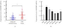
Fig.1
Expression levels of KHSRP mRNA in CRC tissue(A) and cells(B) detected by RT-qPCR method"
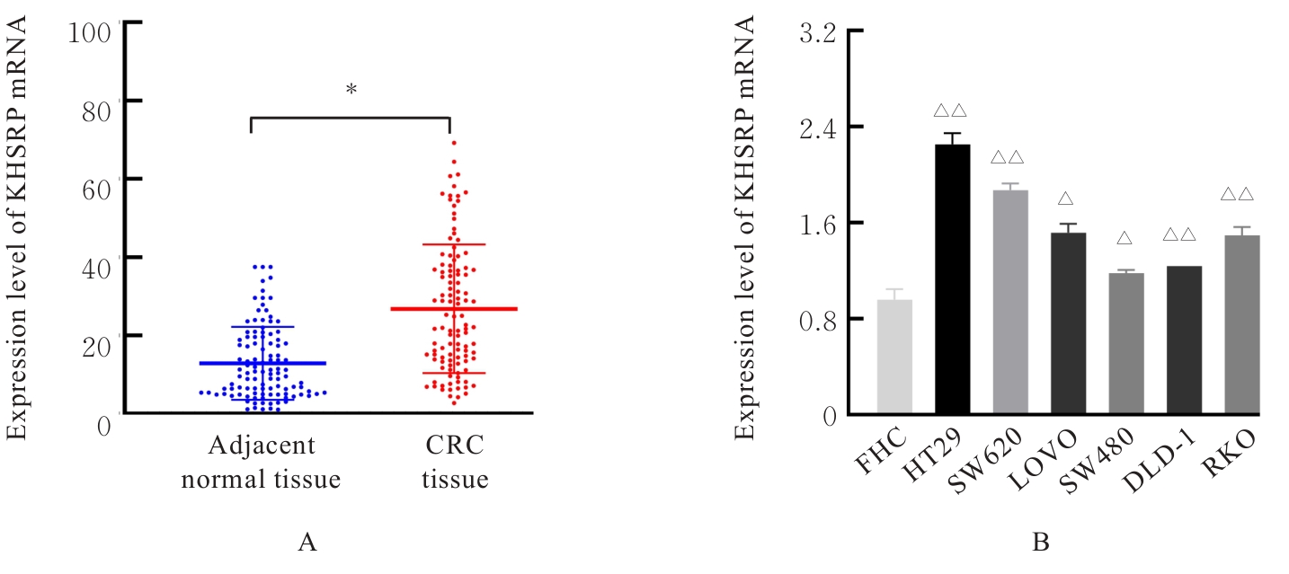
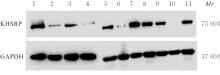
Fig. 2
Electrophoregrams of expression of KHSRP protein in CRC tissue and cells detected by Western blotting method"
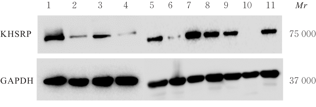
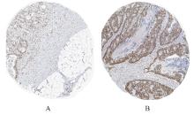
Fig.3
Expressions of KHSRP protein in CRC tissue(A) and adjacent normal tissue(B) detected by IHC staining method (×40)"
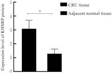
Fig.4
Expression levels of KHSRP protein in CRC tissue and adjacent normal tissue"


Fig. 5
Expression levels of KHSRP mRNA in CRC cells after knockdown or over-expression of KHSRP"
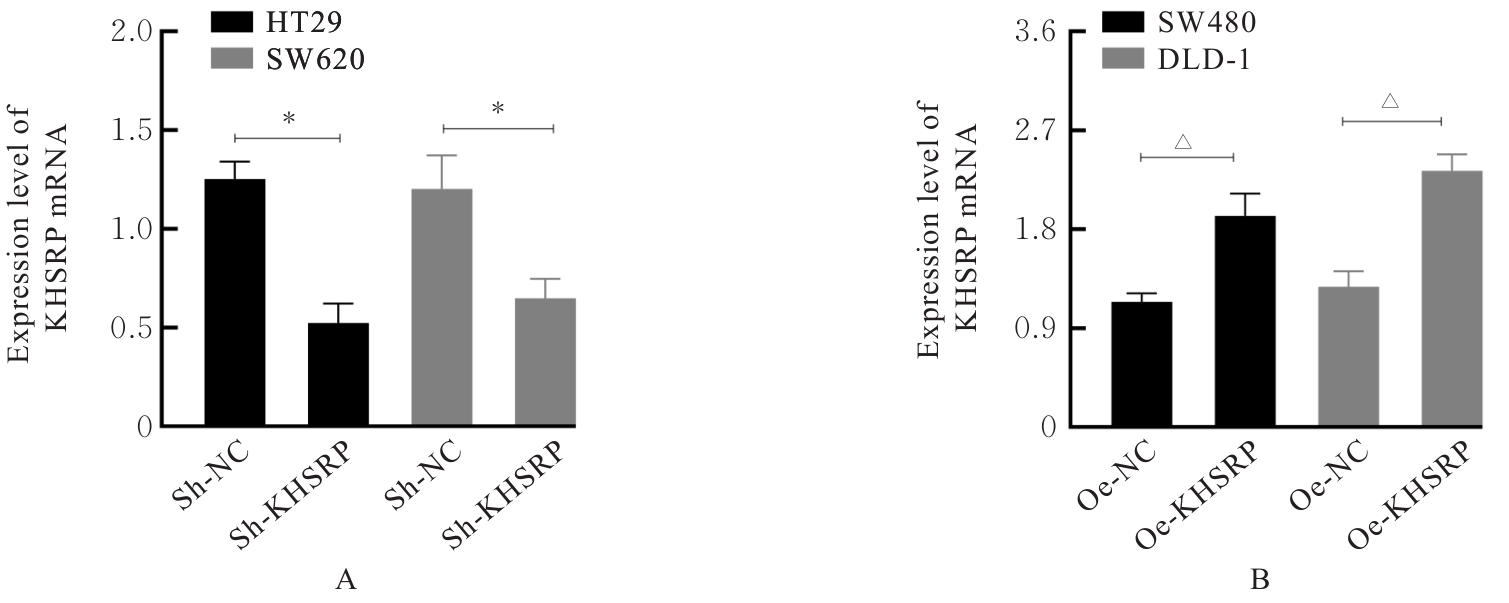
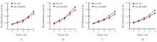
Fig. 6
Proliferation activities of CRC cells in various groups after knockdown and over-expression of KHSRP"
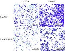
Fig. 7
Morphology of migration of HT29 and SW620 cells in two groups after knockdown of KHSRP (Crystal violet)"
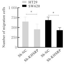
Fig. 8
Number of migration cells of HT29 and SW620 cells in two groups after knockdown of KHSRP"
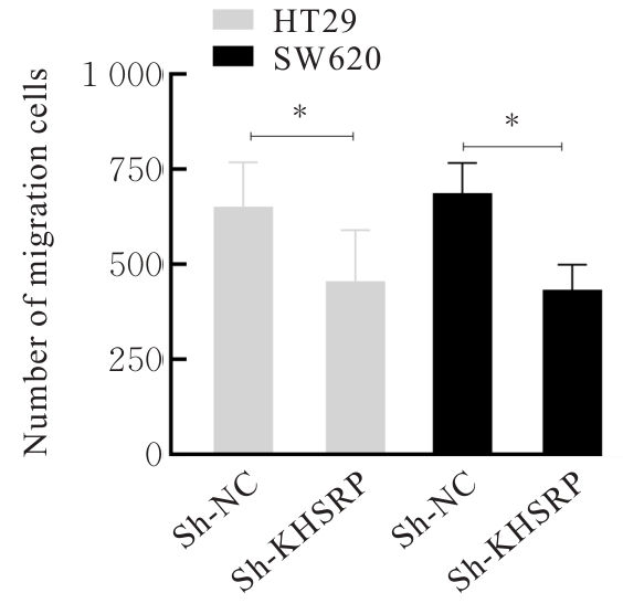
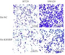
Fig. 9
Morphology of invasion of HT29 and SW620 cells in two groups after knockdown of KHSRP (Crystal violet)"

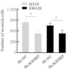
Fig. 10
Numbers of invasion HT29 and SW620 cells in two groups after knockdown of KHSRP"

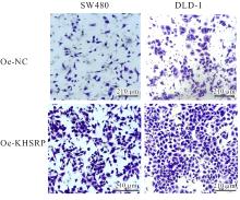
Fig. 11
Morphology of migration of SW480 and DLD-1 cells in two groups after over-expression of KHSRP (Crystal violet)"
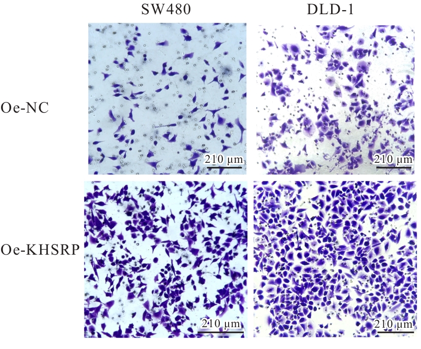
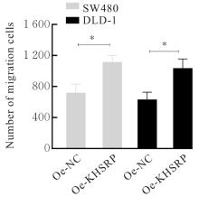
Fig. 12
Numbers of migration SW480 and DLD-1 cells in two groups after over-expression of KHSRP"

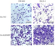
Fig. 13
Morphology of invasion of SW480 and DLD-1 cells in two groups after over-expression of KHSRP (Crystal violet)"
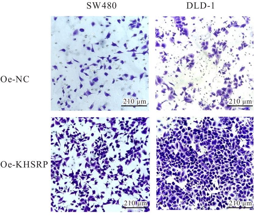
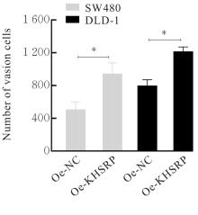
Fig. 14
Number of invasion SW480 and DLD-1 cells in two groups after over-expression of KHSRP"

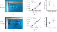
Fig. 15
In vivo tumor growth of mice in various groups"
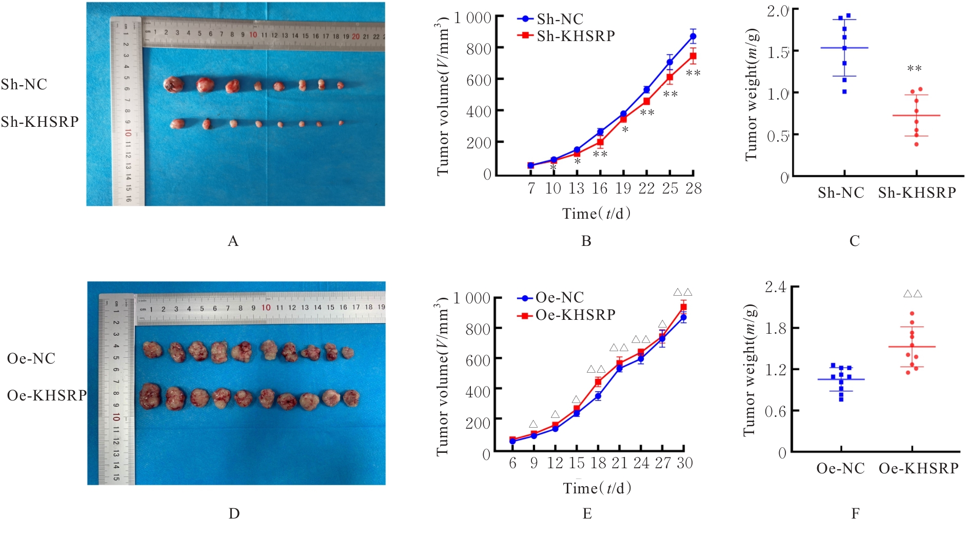
Fig. 16
Electrophoregram (A) and histograms (B-E) of expressions of JAK/STAT signaling pathway-related proteins in CRC cells in sh-NC and sh-KHSRP groups"
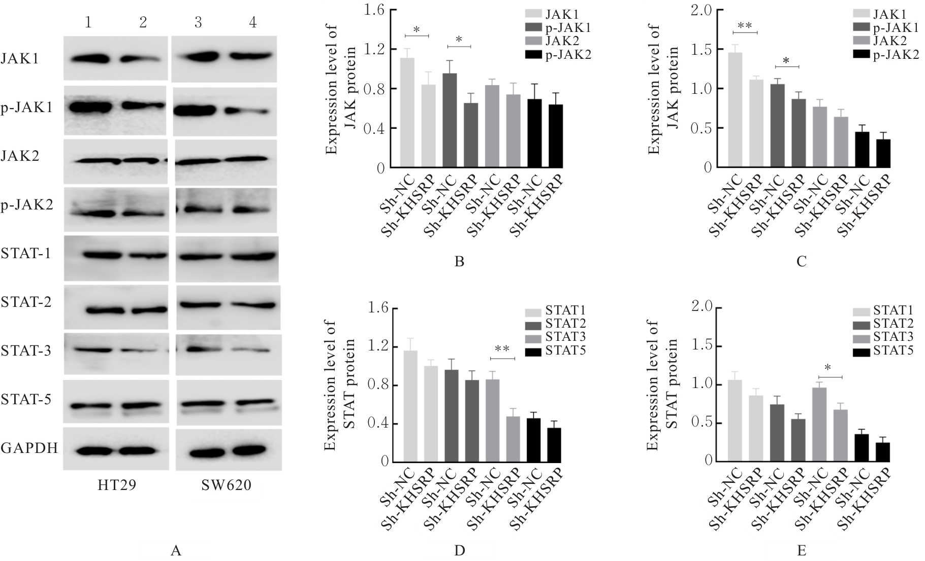
Fig. 17
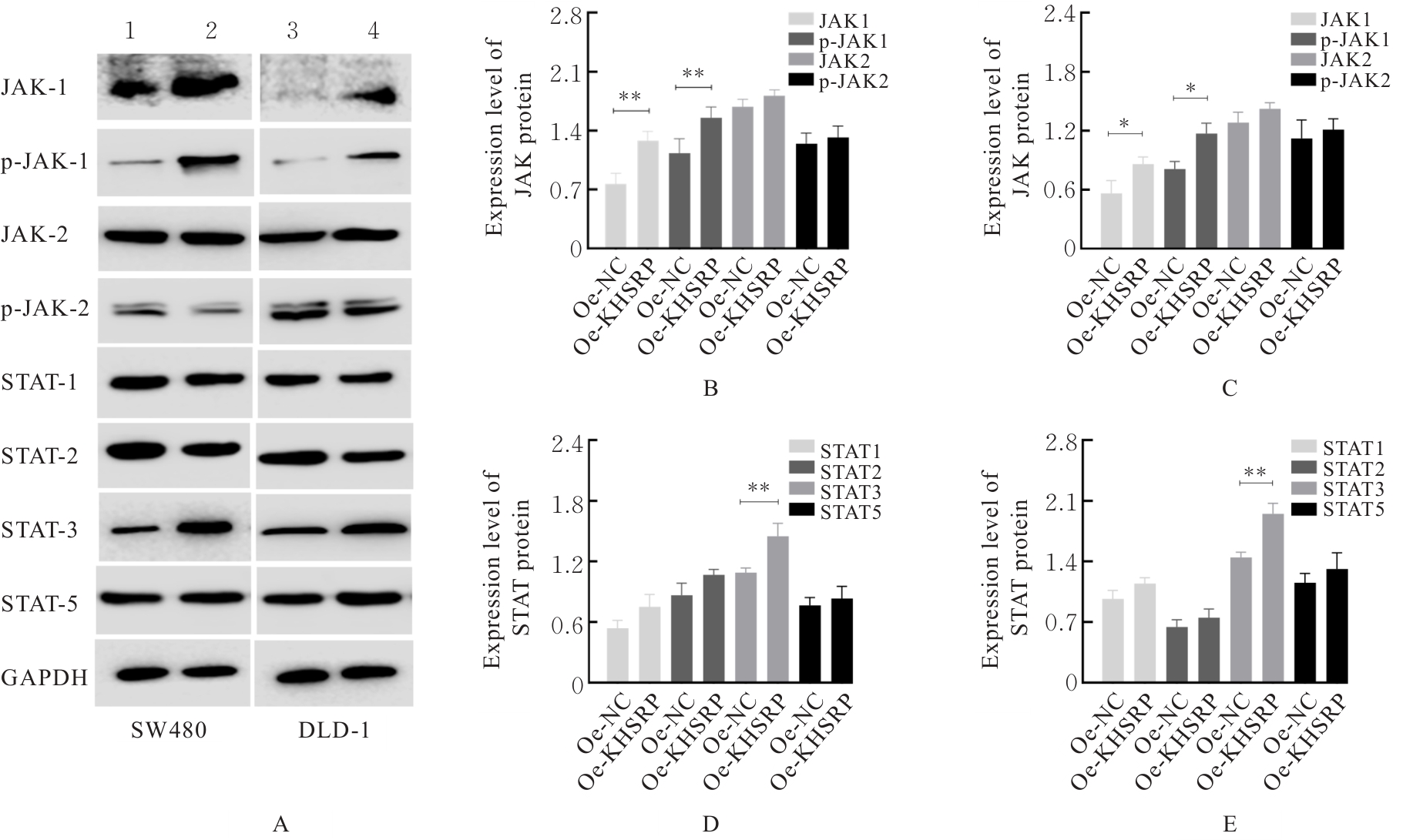
Electrophoregram (A) and histograms (B-E) of expressions of JAK/STAT signaling pathway-related proteins in CRC cells in oe-NC and oe-KHSRP groups"
| [1] | XI Y, XU P F. Global colorectal cancer burden in 2020 and projections to 2040[J]. Transl Oncol, 2021, 14(10): 101174. |
| [2] | XIA C F, DONG X S, LI H, et al. Cancer statistics in China and United States, 2022: profiles, trends, and determinants[J]. Chin Med J (Engl), 2022, 135(5): 584-590. |
| [3] | SUNG H, FERLAY J, SIEGEL R L, et al. Global cancer statistics 2020: GLOBOCAN estimates of incidence and mortality worldwide for 36 cancers in 185 countries[J]. CA Cancer J Clin, 2021, 71(3): 209-249. |
| [4] | MORGAN E, ARNOLD M, GINI A, et al. Global burden of colorectal cancer in 2020 and 2040: incidence and mortality estimates from GLOBOCAN[J]. Gut, 2023, 72(2): 338-344. |
| [5] | XU Y Q, BAO Y X, QIU G Z, et al. METTL3 promotes proliferation and migration of colorectal cancer cells by increasing SNHG1 stability[J]. Mol Med Rep, 2023, 28(5): 217. |
| [6] | TANIUCHI K, OGASAWARA M. KHSRP-bound small nucleolar RNAs associate with promotion of cell invasiveness and metastasis of pancreatic cancer[J]. Oncotarget, 2020, 11(2): 131-147. |
| [7] | CAIAZZA F, OFICJALSKA K, TOSETTO M, et al. KH-type splicing regulatory protein controls colorectal cancer cell growth and modulates the tumor microenvironment[J]. Am J Pathol, 2019, 189(10): 1916-1932. |
| [8] | JOHNSON H M, NOON-SONG E, AHMED C M. Noncanonical IFN signaling, steroids, and STATs: a probable role of V-ATPase[J]. Mediators Inflamm, 2019, 2019: 4143604. |
| [9] | XUE C, YAO Q F, GU X Y, et al. Evolving cognition of the JAK-STAT signaling pathway: autoimmune disorders and cancer[J]. Signal Transduct Target Ther, 2023, 8(1): 204. |
| [10] | HANAHAN D. Hallmarks of cancer: new dimensions[J]. Cancer Discov, 2022, 12(1): 31-46. |
| [11] | XU J Y, WANG D S, MA H L, et al. KHSRP combines transcriptional and posttranscriptional mechanisms to regulate monocytic differentiation[J]. Blood Sci, 2022, 4(3): 103-115. |
| [12] | CHEN L, ZHAO T J. Identification of KHSRP-regulated RNAs in esophageal cancer by integrated bioinformatics analysis[J]. Cancer Biother Radiopharm, 2021, 36(5): 412-424. |
| [13] | HUANG J G, SACHDEVA M, XU E, et al. The long noncoding RNA NEAT1 promotes sarcoma metastasis by regulating RNA splicing pathways[J]. Mol Cancer Res, 2020, 18(10): 1534-1544. |
| [14] | YAN M X, SUN L, LI J, et al. RNA-binding protein KHSRP promotes tumor growth and metastasis in non-small cell lung cancer[J]. J Exp Clin Cancer Res, 2019, 38(1): 478. |
| [15] | HE L, TIAN L. Downregulation of miR-409-3p suppresses LPS-induced inflammation in human bronchial epithelial cells through SOCS3/JAK1/STAT3 signaling: The implication for bronchopneumonia[J]. Mol Med Rep, 2021, 23(3): 190. |
| [16] | TANG J J, HAO T D K, LIM J J, et al. JAK/STAT signaling in hepatocellular carcinoma[J]. Hepat Oncol, 2020, 7(1): HEP18. |
| [17] | LIU C R, FENG H Q, SONG L H, et al. Synergistic effects of thalidomide and cisplatin are mediated via the PI3K/AKT and JAK1/STAT3 signaling pathways in cervical cancer[J]. Oncol Rep, 2022, 48(4): 169. |
| [18] | SARAPULTSEV A, GUSEV E, KOMELKOVA M, et al. JAK-STAT signaling in inflammation and stress-related diseases: implications for therapeutic interventions[J]. Mol Biomed, 2023, 4(1): 40. |
| [19] | LAMICHHANE S, MO J S, SHARMA G, et al. microRNA 452 regulates IL20RA-mediated JAK1/STAT3 pathway in inflammatory colitis and colorectal cancer[J]. Inflamm Res, 2021, 70(8): 903-914. |
| [20] | JIANG Y, XU C H, ZHAO Y, et al. LINC00926 is involved in hypoxia-induced vascular endothelial cell dysfunction via miR-3194-5p regulating JAK1/STAT3 signaling pathway[J]. Eur J Histochem, 2023, 67(1): 3526. |
| [21] | BROOKS A J, PUTOCZKI T. JAK-STAT signalling pathway in cancer[J]. Cancers (Basel), 2020, 12(7): 1971. |
| [22] | WANG F, WANG X C, LI J R, et al. CircNOL10 suppresses breast cancer progression by sponging miR-767-5p to regulate SOCS2/JAK/STAT signaling[J]. J Biomed Sci, 2021, 28(1): 4. |
| [23] | CHAN S, LIU Z X, CHEN Y Y, et al. The JAK-STAT signaling-related signature serves as a prognostic and predictive biomarker for renal cell carcinoma immunotherapy[J]. Gene, 2024, 927: 148719. |
| [1] | Yixuan GAO,Peng WANG,Silong ZHANG,Ruijuan GAO,Yingfang MA,Keke ZHANG,Dan FENG,Zongqi HUANG,Ketao MA,Li LI,Junqiang SI. Inhibitory effect of safranal on proliferation, migration and phenotypic transformation of vascular smooth muscle cells of rats induced by high glucose in vitro [J]. Journal of Jilin University(Medicine Edition), 2025, 51(4): 948-957. |
| [2] | Wenxuan LI,Minru ZONG. Research progress in role of migration of Schwann cells in repairment of peripheral nerve injury [J]. Journal of Jilin University(Medicine Edition), 2025, 51(4): 1137-1144. |
| [3] | Xiaoshuang HE,Lina XU,Mei CUI,Yu ZHAO,Bei WANG,Zheng HUANG,Yuchao WANG,Wenyan XIN,Chao WU. Effects of lncRNA DUXAP8 in lung cancer A549 cells-derived exosomes on lung cancer cell growth and its mechnism [J]. Journal of Jilin University(Medicine Edition), 2025, 51(4): 958-967. |
| [4] | Zhongjun SHEN,Yao ZHAO,Mingbo JIA,Liyan ZHAO. Research progress in effects of hypoxia-inducible factors on cell migration and invasion during epithelial-mesenchymal transition in glioma cells [J]. Journal of Jilin University(Medicine Edition), 2025, 51(4): 1145-1154. |
| [5] | Yihui WANG,Qing ZHANG,Yingnan LI,Liping YE. Effect of KIAA1522 on proliferation, migration, and invasion of lung cancer cells and its mechanism [J]. Journal of Jilin University(Medicine Edition), 2025, 51(3): 727-739. |
| [6] | Fan WANG,Xin WEN,Yixuan WANG,Yuan WANG. Effect of gap junction β2 on prognosis of patients with lung adenocarcinoma and biological behavior of lung adenocarcinoma A549 cells [J]. Journal of Jilin University(Medicine Edition), 2025, 51(3): 716-726. |
| [7] | Donghui LIU,Yunzhe CI,Chunyan WANG,Wenyi MA. Effect of miR-199a-5p on expression of Caveolin-1, cell migration and apoptosis in glioma U251 cells [J]. Journal of Jilin University(Medicine Edition), 2025, 51(3): 663-671. |
| [8] | Ying YANG,Liang ZHAO,Yong YOU,Qian XU,Zhenjun YANG. Influence of 17β-estradiol in proliferation and differentiation of hippocampal neural stem cells and its mechanism [J]. Journal of Jilin University(Medicine Edition), 2025, 51(2): 317-324. |
| [9] | Hanyue LI,Lian YANG,Jianfeng LIU,Shufei ZHANG,Li HONG. Effect of bone marrow mesenchymal stem cells of mice on proliferation and collagen expression levels of fibroblasts through JAK2/STAT3 signaling pathway [J]. Journal of Jilin University(Medicine Edition), 2025, 51(2): 325-332. |
| [10] | Yaqi ZHANG,Jing MI,Jingrong YANG,Xinming LI,Li LI. Effect of up-regulation of miR-31 expression on osteogenic differentiation of dental pulp stem cells through Wnt-β/catenin signaling pathway [J]. Journal of Jilin University(Medicine Edition), 2025, 51(2): 412-419. |
| [11] | Shuyan SUN,Huakun ZHANG,Ziru ZHOU,Feng LI,Xiaobin CUI. Expression of CRNN protein in esophageal squamous cell carcinoma tissue and influence of its overexpression in biological behavior of esophageal squamous cell carcinoma Eca9706 cells [J]. Journal of Jilin University(Medicine Edition), 2025, 51(2): 275-283. |
| [12] | Jing DENG,Xuan WANG,Changyu SHI,Siqi YANG,Qinling ZOU,Ming JIN. Effect of securinine on proliferation and apoptosis of human colon cancer SW620 cells and its mechanism [J]. Journal of Jilin University(Medicine Edition), 2025, 51(2): 307-316. |
| [13] | Mengyun LU,Yucheng HAN,Yihong HU,Minhui HE,Yanqun ZHANG,Xianqiong ZOU. Effects of glycolipid transfer protein on proliferation, migration,and invasion of pancreatic cancer PANC-1 cells and their mechanisms [J]. Journal of Jilin University(Medicine Edition), 2025, 51(2): 284-295. |
| [14] | Yan WANG,Zouyu ZHAO,Panpan YU,Ping YANG. Expression of I kappa B kinase-interacting protein in cervical cancer tissue and its effect on proliferation, migration and invasion of cervical cancer cells [J]. Journal of Jilin University(Medicine Edition), 2025, 51(2): 341-351. |
| [15] | Pengli WU,Fengyu LI,Bo LIU,Yang LYU. Effect of silencing DDX39A gene on proliferation, migration and invasion of esophageal cancer TE-1 cells and its mechanism [J]. Journal of Jilin University(Medicine Edition), 2025, 51(1): 115-123. |
|
||