Journal of Jilin University(Medicine Edition) ›› 2026, Vol. 52 ›› Issue (3): 602-611.doi: 10.13481/j.1671-587X.20260303
• Research in basic medicine • Previous Articles
Improvement effect of bone marrow mesenchymal stem cell-derived exosomes on airway resistance and lung tissue lesion in mice of asthmatic models and its mechanism
Lina XU,Xiaoshuang HE,Wenyan XIN,Chao WU( )
)
- Department of Respiratory and Critical Care Medicine,First Affiliated Hospital,Shihezi University,Shihezi 832008,China
-
Received:2025-06-28Accepted:2025-10-12Online:2026-05-28Published:2026-06-08 -
Contact:Chao WU E-mail:mail@healthy.vip

CLC Number:
- R56
Cite this article
Lina XU,Xiaoshuang HE,Wenyan XIN,Chao WU. Improvement effect of bone marrow mesenchymal stem cell-derived exosomes on airway resistance and lung tissue lesion in mice of asthmatic models and its mechanism[J].Journal of Jilin University(Medicine Edition), 2026, 52(3): 602-611.
share this article
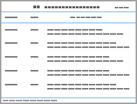
Tab.1
Primer sequences of miR-26a and U6"
| Gene | Primer sequence (5'-3') | |
|---|---|---|
| miR-26a | F:GCGCGCGTAACAGTCTACAGC R:GTCGTATCCAGTGCAGGGTCC | |
| U6 | F:CGCACTTTACGGCTACCTCT R:GCGACAAGGGAAGGGAACAA |
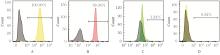
Fig. 1
Expressions of biomarkers of BMSCs"
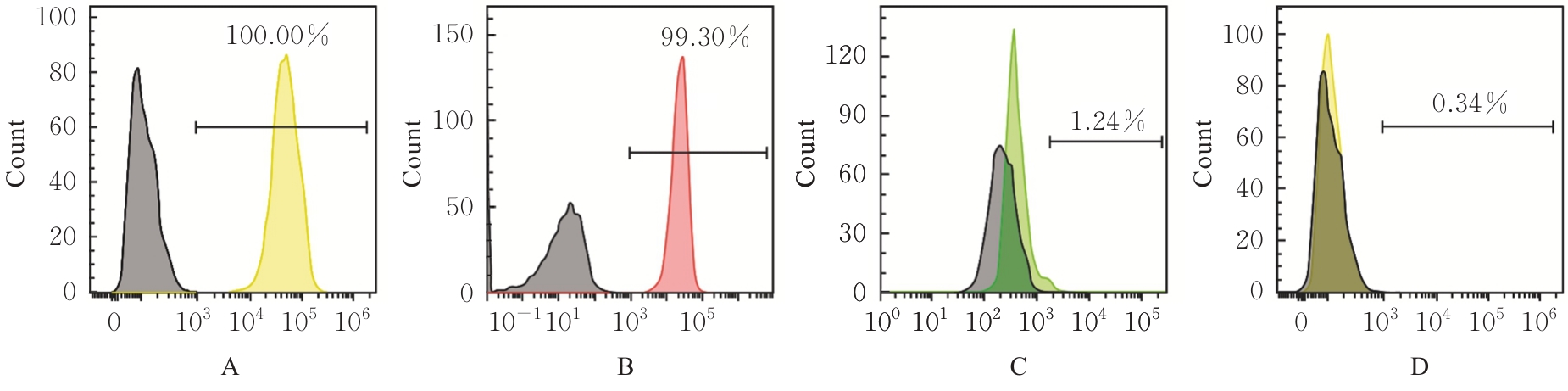
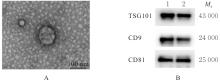
Fig. 2
Identification results of structure and expressions of markers of BMSC-exos"
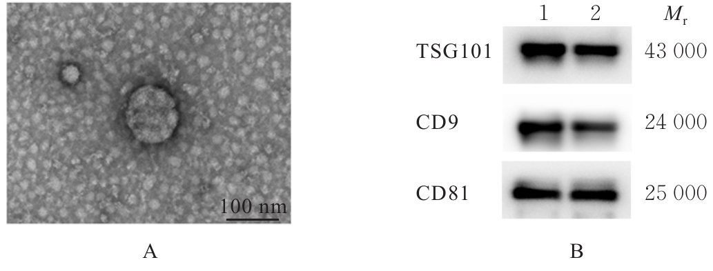
Fig. 3
Pathomorphology of lung tissue of mice in various groups(HE)"
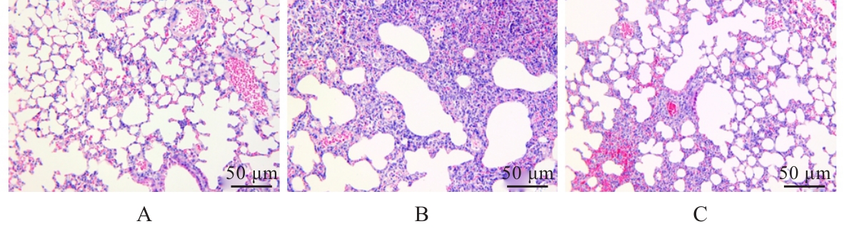

Fig. 4
Pathomorphology of lung tissue of mice in various groups after treatment of miR-26a BMSC-exos (HE)"

Tab.2
Airway resistances of mice in various groups [n=10, x±s, cmH?O·(L·s)-1]"
| Group | Airway resistance | ||||
|---|---|---|---|---|---|
| Mch[ρB/(g·L-1)] 0 | 6.25 | 12.50 | 25.00 | 50.00 | |
| Control | 2.50±0.50 | 3.20±0.20 | 8.27±0.31 | 9.00±0.20 | 12.13±0.15 |
| Model | 3.16±0.15* | 6.58±0.20* | 12.33±0.30* | 13.02±0.17* | 17.25±0.44* |
| BMSC-exos | 2.53±0.15△ | 5.65±0.14△ | 9.42±0.21△ | 11.60±0.23△ | 15.09±0.17△ |
Tab.3
Airway resistances of mice in various groups after treated with MiR-26a BMSC-exos [n=10, x±s, cmH?O·(L·s)-1]"
| Group | Airway resistance | ||||
|---|---|---|---|---|---|
| Mch[ρB/(g·L-1)] 0 | 6.25 | 12.50 | 25.00 | 50.00 | |
| Control | 2.60±0.47 | 3.50±0.22 | 4.35±0.30 | 6.05±0.16 | 10.07±0.16 |
| Model | 3.22±0.17* | 7.03±0.21* | 11.34±0.20* | 14.16±0.33* | 17.62±0.26* |
| MiR-NC exos | 2.55±0.12△ | 4.88±0.10△ | 8.55±0.23△ | 10.61±0.16△ | 14.62±0.22△ |
| Mimic-miR-26a exos | 2.05±0.11# | 3.33±0.12# | 5.25±0.15# | 8.47±0.19# | 12.01±0.20# |
| Inh-miR-26a exos | 3.03±0.12# | 8.01±0.18# | 10.83±0.25# | 14.7±0.33# | 17.5±0.26# |
Tab.4
Expression levels of miR-26a in miR-NC exos, mimic-miR-26a exos, inh-miR-26a exos"
| Exosome | Expression level of miR-26 |
|---|---|
| MiR-NC exos | 1.00±0.08 |
| Mimic-miR-26a exos | 3.45±0.16* |
| Inh-miR-26a exos | 0.58±0.18* |
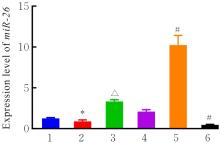
Fig. 5
Expression levels of miR-26a in lung tissue of mice in various groups"
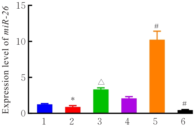
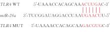
Fig. 6
Binding site of TLR4 and miR-26a predicted by Targetscan database"
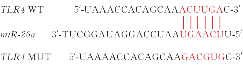
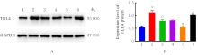
Fig. 7
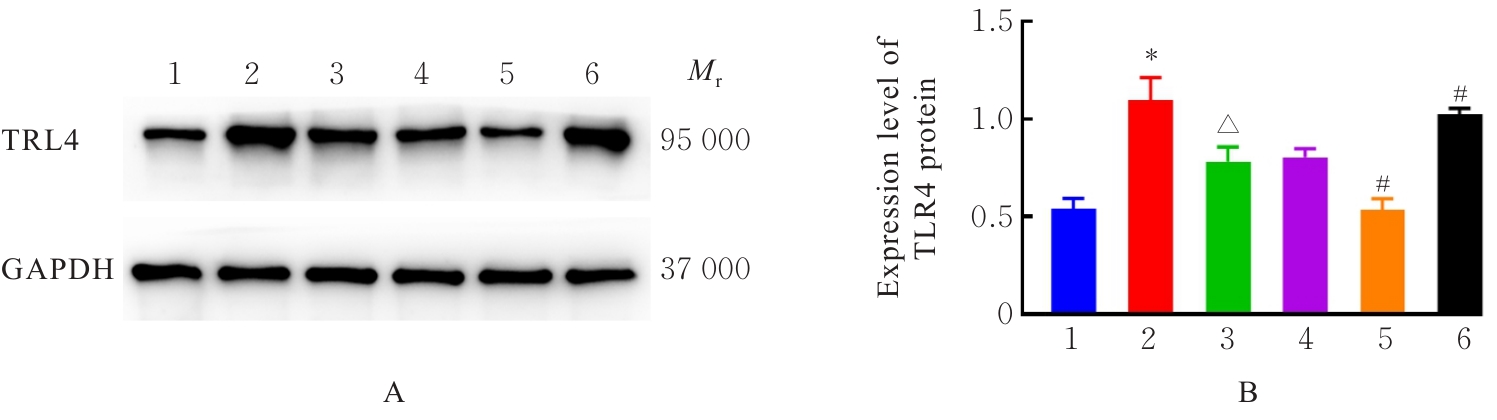
Electrephoregram(A) and histogram(B) of expressions of TLR4 protein in lung tissue of mice in various groups"
| [1] | VALENCIA-HERNÁNDEZ C A, DEL GRECO M F, SUNDARAM V, et al. Asthma and incident coronary heart disease: an observational and Mendelian randomisation study[J]. Eur Respir J, 2023, 62(5): 2301788. |
| [2] | RAJVANSHI N, KUMAR P, GOYAL J P. Global initiative for asthma guidelines 2024: an update[J]. Indian Pediatr, 2024, 61(8): 781-786. |
| [3] | NAKAGOME K, NAGATA M. Allergen immunotherapy in asthma[J]. Pathogens, 2021, 10(11): 1406. |
| [4] | HERRERA-LUIS E, MARTIN-ALMEIDA M, PINO-YANES M. Asthma-genomic advances toward risk prediction[J]. Clin Chest Med, 2024, 45(3): 599-610. |
| [5] | DESAI B, ADRISH M, MOHAN A, et al. Biologics in asthma: emerging biologics[J]. Immunol Allergy Clin North Am, 2024, 44(4): 751-763. |
| [6] | LI S J, LV D Q, YANG H, et al. A review on the current literature regarding the value of exosome miRNAs in various diseases[J]. Ann Med, 2023, 55(1): 2232993. |
| [7] | ROSZKOWSKI S. Therapeutic potential of mesenchymal stem cell-derived exosomes for regenerative medicine applications[J]. Clin Exp Med, 2024, 24(1): 46. |
| [8] | DONG B, WANG C, ZHANG J, et al. Exosomes from human umbilical cord mesenchymal stem cells attenuate the inflammation of severe steroid-resistant asthma by reshaping macrophage polarization[J]. Stem Cell Res Ther, 2021, 12(1): 204. |
| [9] | DEHNAVI S, DADMANESH M, HOSSEINI ROUZBAHANI N, et al. Mesenchymal stem cell-derived exosome; an interesting nanocarrier to improve allergen-specific intranasal immunotherapy[J]. Iran J Allergy Asthma Immunol, 2023, 22(6): 561-574. |
| [10] | CAO J Y, WANG B, TANG T T, et al. Exosomal miR-125b-5p deriving from mesenchymal stem cells promotes tubular repair by suppression of p53 in ischemic acute kidney injury[J]. Theranostics, 2021, 11(11): 5248-5266. |
| [11] | NTONTSI P, PHOTIADES A, ZERVAS E, et al. Genetics and epigenetics in asthma[J]. Int J Mol Sci, 2021, 22(5): 2412. |
| [12] | NGUYEN H, NASIR M. Management of chronic asthma in adults[J]. Med Clin North Am, 2024, 108(4): 629-640. |
| [13] | SHARMA S, GERBER A N, KRAFT M, et al. Asthma pathogenesis: phenotypes, therapies, and gaps: summary of the aspen lung conference 2023[J]. Am J Respir Cell Mol Biol, 2024, 71(2): 154-168. |
| [14] | WITTE A, TÜRK Y, BRAUNSTAHL G J. Obesity-related asthma: new insights leading to a different approach[J]. Curr Opin Pulm Med, 2024, 30(3): 294-302. |
| [15] | SANTILLÁN-GUAJÁN S M, SHAHI M H, CASTRESANA J S. Mesenchymal-stem-cell-based therapy against gliomas[J]. Cells, 2024, 13(7): 617. |
| [16] | XU H J, WANG B, LI A, et al. Mesenchymal stem cells-based cell-free therapy targeting neuroinflammation[J]. Aging Dis, 2024, 15(3): 965-976. |
| [17] | HUANG M M, MEHRABI NASAB E, ATHARI S S. Immunoregulatory effect of mesenchymal stem cell via mitochondria signaling pathways in allergic asthma[J]. Saudi J Biol Sci, 2021, 28(12): 6957-6962. |
| [18] | ZHAI Z Y, CUI T R, CHEN J L, et al. Advancements in engineered mesenchymal stem cell exosomes for chronic lung disease treatment[J]. J Transl Med, 2023, 21(1): 895. |
| [19] | SHAN L S, LIU S, ZHANG Q Z, et al. Human bone marrow-mesenchymal stem cell-derived exosomal microRNA-188 reduces bronchial smooth muscle cell proliferation in asthma through suppressing the JARID2/Wnt/β-catenin axis[J]. Cell Cycle, 2022, 21(4): 352-367. |
| [20] | ABBASZADEH H, GHORBANI F, ABBASPOUR-AGHDAM S, et al. Chronic obstructive pulmonary disease and asthma: mesenchymal stem cells and their extracellular vesicles as potential therapeutic tools[J]. Stem Cell Res Ther, 2022, 13(1): 262. |
| [21] | BHASKARA M, ANJORIN O, WANG M J. Mesenchymal stem cell-derived exosomal microRNAs in cardiac regeneration[J]. Cells, 2023, 12(24): 2815. |
| [22] | LIU X S, CHEN Q, JIANG S X, et al. microRNA-26a in respiratory diseases: mechanisms and therapeutic potential[J]. Mol Biol Rep, 2024, 51(1): 627. |
| [23] | SHI Z G, SUN Y, WANG K S, et al. Effects of miR-26a/miR-146a/miR-31 on airway inflammation of asthma mice and asthma children[J]. Eur Rev Med Pharmacol Sci, 2019, 23(12): 5432-5440. |
| [24] | LAI S, DENG L, LIU C, et al. Bone marrow mesenchymal stem cell-derived exosomes loaded with miR-26a through the novel immunomodulatory peptide DP7-C can promote osteogenesis[J]. Biotechnol Lett, 2023, 45(7): 905-919. |
| [25] | CAO Y, YANG H, HUANG Y, et al. Mesenchymal stem cell-derived exosomal miR-26a induces ferroptosis, suppresses hepatic stellate cell activation, and ameliorates liver fibrosis by modulating SLC7A11[J]. Open Med (Wars), 2024, 19(1): 20240945. |
| [26] | QIAN Y Y, LI Y F, JI J C, et al. Identification of two hub genes and miRNA-mRNA interactions in chronic obstructive pulmonary disease (COPD) plasma[J]. J Asthma, 2024, 61(9): 1058-1067. |
| [27] | LIN Z, BAO R, NIU Y, et al. KLF5-mediated pyroptosis of airway epithelial cells leads to airway inflammation in asthmatic mice through the miR-182-5p/TLR4 axis[J]. Mol Immunol, 2024, 170: 9-18. |
| [28] | WANG M L, QIAO H. Effect of miR-506-3p on proliferation and apoptosis of airway smooth muscle cells in asthmatic mice by regulating CCL2 gene expression and mediating TLR4/NF-κB signaling pathway activation[J]. Mol Biotechnol, 2021, 63(5): 410-423. |
| [1] | Di LI,Yang SU,Yong ZHANG,Jiaoqi WANG,Xiaofei ZHANG,Xuebing ZHAO. Expression levels of miR-328-3p and miR-671-5p in serum exosomes of Kawasaki disease model mice and their significance [J]. Journal of Jilin University(Medicine Edition), 2026, 52(2): 359-365. |
| [2] | Aimei LI,Jingfei HAN,Li DENG,Siyu CHEN. Protective effect of dexmedetomidine on myocardial injury in myocardial ischemia/reperfusion injury model rats [J]. Journal of Jilin University(Medicine Edition), 2026, 52(2): 451-459. |
| [3] | Huiying YANG,Weihong LIANG,Xinru XU,Haodan SUN,Xin JIAO,Haiping WANG. Research progress in effect of bone marrow mesenchymal stem cells on differentiation induction of cardiomyocyte-like cells, protection of cardiomyocytes, and myocardial-related diseases [J]. Journal of Jilin University(Medicine Edition), 2026, 52(1): 281-289. |
| [4] | Yu YAN,Dandan HUANG,Chunling HONG,Bowen WEI,Yuanyuan YANG,Guanghai YAN,Yilan SONG,Zhemin XIAN. Improvement effect of imperatorin on airway remodeling in bronchial asthma mice and its mechanism [J]. Journal of Jilin University(Medicine Edition), 2025, 51(6): 1508-1517. |
| [5] | Jing LIU,Yan WANG,Xu HUANG. Promoting effect of overexpressed circRNAs-modified dental pulp stem cell-derived exosomes on angiogenesis of human umbilical vein endothelial cells [J]. Journal of Jilin University(Medicine Edition), 2025, 51(6): 1561-1570. |
| [6] | Xiaoshuang HE,Lina XU,Mei CUI,Yu ZHAO,Bei WANG,Zheng HUANG,Yuchao WANG,Wenyan XIN,Chao WU. Effects of lncRNA DUXAP8 in lung cancer A549 cells-derived exosomes on lung cancer cell growth and its mechnism [J]. Journal of Jilin University(Medicine Edition), 2025, 51(4): 958-967. |
| [7] | Lieqian SUN,Mengyu GU,Jie YANG,Kaiyi WANG,Gaoshuai GUO,Hongbo ZHANG,Siyi ZHANG,Tanglong WANG,Zhiwei YANG,Yanni HE,Chao YANG. Effect of bone marrow-derived mesenchymal stem cell transplantation on mitochondrial autophagy in rats with vascular dementia through ROS/Nrf2 signaling and its mechanism [J]. Journal of Jilin University(Medicine Edition), 2025, 51(3): 610-620. |
| [8] | DILIXIATI·Dilidaer,Lin JIA. Improvement effect of exosomes derived from human adipose-derived stem cells and human dermal fibroblasts on ultraviolet-induced photoaging skin wrinkles in nude mice [J]. Journal of Jilin University(Medicine Edition), 2025, 51(3): 621-631. |
| [9] | Bo YUAN,Jiayi XIE,Siyu JIANG,Yajun MENG,Qinghua ZHU,Xiaofei LI,Xiumei FU,Lide XIE. Effect of adipose-derived stem cell-derived exosomes on migration ability of macrophages in vitro [J]. Journal of Jilin University(Medicine Edition), 2024, 50(3): 718-727. |
| [10] | Liping CHEN,Li HAN,Hua BIAN,Liye PANG. Bioinformatics analysis based on differentially expressed genes and screening of traditional Chinese medicine for treatment of severe bronchial asthma [J]. Journal of Jilin University(Medicine Edition), 2024, 50(2): 411-421. |
| [11] | Hongmei TANG,Yuejiao LI,Xing WANG,Zhibin WANG,Xiefang YUAN,Xiaoyun WANG. Effect of Jiegeng Yuanshen Tang on airway inflammation and mucus secretion in allergic asthmatic mice and its mechanism [J]. Journal of Jilin University(Medicine Edition), 2024, 50(1): 10-17. |
| [12] | Xiaolei XUE,Baomei XU. Expression of Klotho protein in placenta exosomes in patients with pre-eclampsia and its effect on oxidative stress in vascular endothelial cells [J]. Journal of Jilin University(Medicine Edition), 2023, 49(6): 1528-1538. |
| [13] | Xiaoshuang HE,Lina XU,Mei CUI,Wenyan XIN. Inhibitory effect of BMSCs over-expressing FOXO1 on pulmonary eosinophil infiltration and airway remodeling in asthmatic mice and its mechanism [J]. Journal of Jilin University(Medicine Edition), 2023, 49(5): 1192-1201. |
| [14] | Jiao ZHAGN,Baolian MA,Yonglan ZHANG. Antioxidant capacities of edible plant-exosomes-like nanoparticles in vitro and their protective effects on oxidative damage of PC12 cells induced by hydrogen peroxide [J]. Journal of Jilin University(Medicine Edition), 2023, 49(5): 1117-1124. |
| [15] | Hao ZHANG,Cuizhu WANG,Huimin HUANGFU,Xinwei ZHANG,Yidi ZHANG,Yanmin ZHOU. Effect of 20(S)-protopanaxadiol on osteogensis differentiation of bone marrow mesenchymal stem cells in rats and its mechanism [J]. Journal of Jilin University(Medicine Edition), 2023, 49(4): 867-874. |
|
||