Journal of Jilin University(Medicine Edition) ›› 2026, Vol. 52 ›› Issue (3): 612-620.doi: 10.13481/j.1671-587X.20260304
• Research in basic medicine • Previous Articles
Effect of selenoprotein N1 gene on senescence of L929 cells and its mechanism
Nuo JIANG,Shufei ZHANG,Li HONG( )
)
- Department of Obstetrics and Gynecology,People’s Hospital,Wuhan University,Wuhan 430060,China
-
Received:2025-09-27Accepted:2025-11-02Online:2026-05-28Published:2026-06-08 -
Contact:Li HONG E-mail:dr_hongli@whu.edu.cn

CLC Number:
- R711.4
Cite this article
Nuo JIANG,Shufei ZHANG,Li HONG. Effect of selenoprotein N1 gene on senescence of L929 cells and its mechanism[J].Journal of Jilin University(Medicine Edition), 2026, 52(3): 612-620.
share this article

Fig. 1
SA-β-gal staining of L929 cells in various groups (×200)"

Fig. 2
Electrophoregram(A) and histogram(B) of expressions of P21, P16, and SEPN1 proteins in L929 cells in two groups"
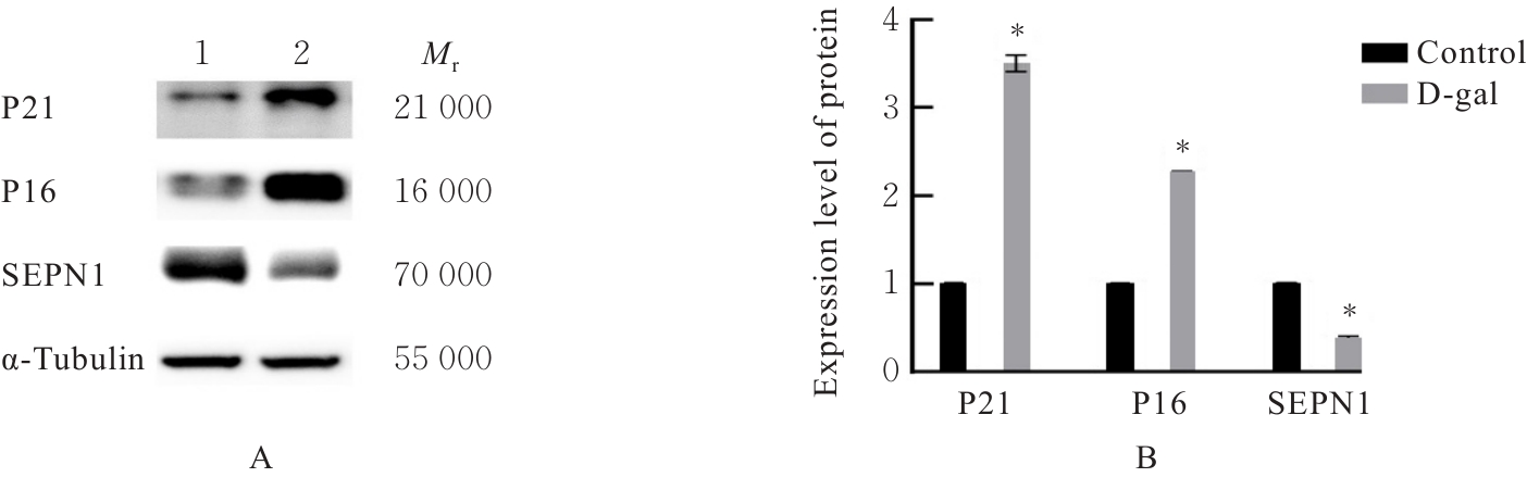
Fig. 3
Proliferation activities of L929 cells in two groups detected by EdU staining (×200)"
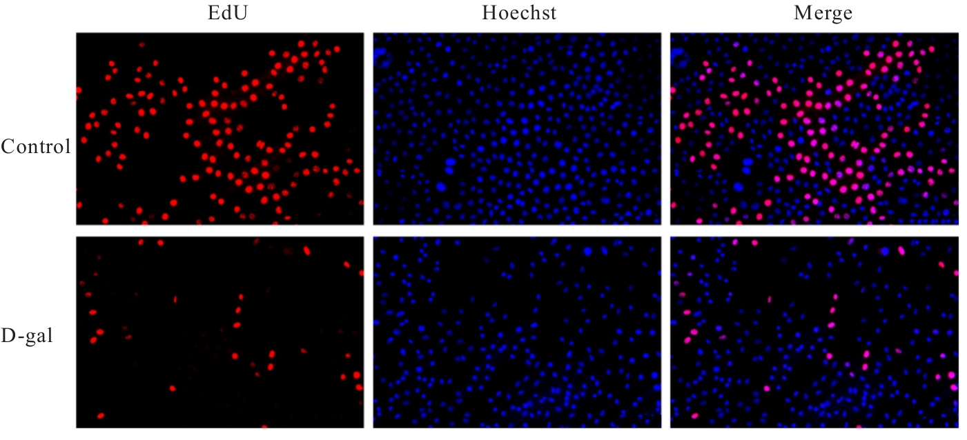
Fig. 4
Proliferation activities of L929 cells in two groups after transfection detected by EdU staining (×200)"
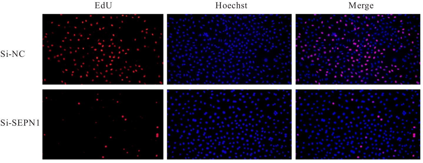
Fig. 5
Levels of ROS in L929 cells in various groups detected by DCFH-DA fluorescence probe method (×200)"
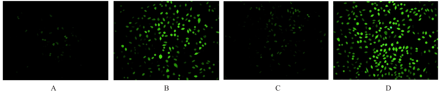
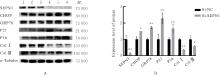
Fig. 6
Electrophoregram(A) and histogram(B) of expressions of SEPN1,CHOP,GRP78,P21,P16,Col Ⅰ,and Col Ⅲ proteins in L929 cells in two groups"
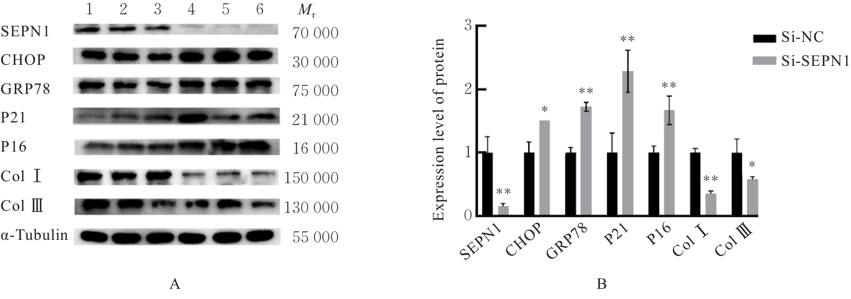
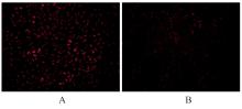
Fig. 7
Mitochondrial membrane potential level of L929 cells in two groups detected by TMRE kit (×200)"
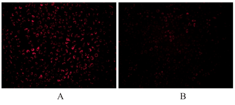
Fig. 8
Levels of Ca2+ in L929 cells in two groups detected by Fluo-4 AM fluorescence probe method (×200)"
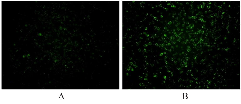
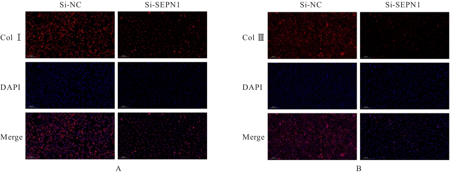
Fig. 9
Expressions of Col Ⅰ and Col Ⅲ proteins in L929 cells in two groups detected by immunofluorescence method(×200)"
| [1] | COLLINS S, LEWICKY-GAUPP C. Pelvic organ prolapse[J]. Gastroenterol Clin North Am, 2022, 51(1): 177-193. |
| [2] | SIVALINGARAJAH R, BALASINGAM B, URUTHIRAKUMAR P, et al. Gynecological morbidity in geriatric women: findings from a cross-sectional study in a tertiary care center in northern Sri Lanka[J]. Cureus, 2025, 17(6): e85416. |
| [3] | LI P P, MA H Y, ZHANG J, et al. Global trends in prevalence and future projections of pelvic organ prolapse: a 30-year epidemiological study[J]. Int Urogynecol J, 2025, 36(5): 991-1002. |
| [4] | GULER Z, ROOVERS J P. Role of fibroblasts and myofibroblasts on the pathogenesis and treatment of pelvic organ prolapse[J]. Biomolecules, 2022, 12(1): 94. |
| [5] | CHILDS B G, GLUSCEVIC M, BAKER D J, et al. Senescent cells: an emerging target for diseases of ageing[J]. Nat Rev Drug Discov, 2017, 16(10): 718-735. |
| [6] | HUANG G X, SUN S N, LIAO M D, et al. METTL3-mediated m6A modification regulates D-galactose-induced skin fibroblast senescence through miR-208a-5p[J]. Front Immunol, 2025, 16: 1577783. |
| [7] | LÓPEZ-OTÍN C, BLASCO M A, PARTRIDGE L, et al. Hallmarks of aging: an expanding universe[J]. Cell, 2023, 186(2): 243-278. |
| [8] | BARAZZUOL L, GIAMOGANTE F, CALÌ T. Mitochondria associated membranes (MAMs): architecture and physiopathological role[J]. Cell Calcium, 2021, 94: 102343. |
| [9] | MAKIO T, CHEN J S, SIMMEN T. ER stress as a sentinel mechanism for ER Ca2+ homeostasis[J]. Cell Calcium, 2024, 124: 102961. |
| [10] | GERMANI S, VAN HO A T, CHERUBINI A, et al. SEPN1-related myopathy depends on the oxidoreductase ERO1A and is druggable with the chemical chaperone TUDCA[J]. Cell Rep Med, 2024, 5(3): 101439. |
| [11] | RUI B J, RUI G H, YANG Y F. Unveiling pathogenesis of pelvic organ prolapse through transcriptomic and bioinformatic analyses in uterosacral ligament tissues of postmenopausal women[J]. Front Genet, 2025, 16: 1588278. |
| [12] | TAPPY E, SHI H L, PRUSZYNSKI J, et al. Use of the senolytics dasatinib and quercetin for prevention of pelvic organ prolapse in a mouse animal model[J]. Aging, 2024, 16(19): 12685-12696. |
| [13] | HUANG L W, ZHAO Z W, WEN J R, et al. Cellular senescence: a pathogenic mechanism of pelvic organ prolapse (Review)[J]. Mol Med Rep, 2020, 22(3): 2155-2162. |
| [14] | NAN L, GUO P C, HUI W, et al. Recent advances in dermal fibroblast senescence and skin aging: unraveling mechanisms and pioneering therapeutic strategies[J]. Front Pharmacol, 2025, 16: 1592596. |
| [15] | MA Z J, DING Y J, DING X F, et al. PDK4 rescues high-glucose-induced senescent fibroblasts and promotes diabetic wound healing through enhancing glycolysis and regulating YAP and JNK pathway[J]. Cell Death Discov, 2023, 9(1): 424. |
| [16] | MIAO Z R, WANG W, MIAO Z Y, et al. Role of Selenoprotein W in participating in the progression of non-alcoholic fatty liver disease[J]. Redox Biol, 2024, 71: 103114. |
| [17] | POTHION H, JEHAN C, TOSTIVINT H, et al. Selenoprotein T: an essential oxidoreductase serving as a guardian of endoplasmic reticulum homeostasis[J]. Antioxid Redox Signal, 2020, 33(17): 1257-1275. |
| [18] | OUYANG P, CAI Z Y, PENG J Y, et al. SELENOK-dependent CD36 palmitoylation regulates microglial functions and Aβ phagocytosis[J]. Redox Biol, 2024, 70: 103064. |
| [19] | KIM B S, JUNG J S, JANG J H, et al. Nuclear Argonaute 2 regulates adipose tissue-derived stem cell survival through direct control of miR10b and selenoprotein N1 expression[J]. Aging Cell, 2011, 10(2): 277-291. |
| [20] | VARONE E, POZZER D, DI MODICA S, et al. SELENON (SEPN1) protects skeletal muscle from saturated fatty acid-induced ER stress and insulin resistance[J]. Redox Biol, 2019, 24: 101176. |
| [21] | CHEN X, CUBILLOS-RUIZ J R. Endoplasmic reticulum stress signals in the tumour and its microenvironment[J]. Nat Rev Cancer, 2021, 21(2): 71-88. |
| [22] | REN J, BI Y G, SOWERS J R, et al. Endoplasmic reticulum stress and unfolded protein response in cardiovascular diseases[J]. Nat Rev Cardiol, 2021, 18(7): 499-521. |
| [23] | GAO S Y, CHEN T, DENG C L, et al. An endoplasmic reticulum stress-responsive nanocomposite hydrogel for diabetic wound healing through a fibroblast-immune cell dual regulation hub[J]. J Nanobiotechnology, 2025, 23(1): 689. |
| [24] | FARHAN H, ZAHOOR M, TAUER J T, et al. The endoplasmic reticulum proteostasis network and bone disease[J]. Trends Mol Med, 2026, 32(2): 119-132. |
| [25] | SANG M D, LI X D, CHEN M, et al. Role of mitochondria-associated ER in apoptosis[J]. Cell Biochem Funct, 2025, 43(7): e70105. |
| [26] | MADREITER-SOKOLOWSKI C T, THOMAS C, RISTOW M. Interrelation between ROS and Ca2+ in aging and age-related diseases[J]. Redox Biol, 2020, 36: 101678. |
| [27] | KONG M, WANG Z, HAO Y, et al. Role of the integrin-β1/TGF-β1 signaling pathway in the pathogenesis of pelvic organ prolapse: a study on vaginal wall tissue alterations and molecular dysfunction[J]. Mol Med Rep, 2025, 31(4): 104. |
| [28] | ZHENG Y J, LEI J D, ZHANG A, et al. Regulatory mechanisms of transforming growth factor-β in senescence of fibroblast associated with refractory skin diseases[J]. Exp Gerontol, 2025, 211: 112900. |
| [29] | YAN Y F, PENG J, HE W, et al. SGK1 inhibits oxidative injury and extracellular matrix degradation by activating the GSK-3β (Ser9)/Fyn/NRF2 pathway in pelvic organ prolapse[J]. Life Sci, 2025, 380: 123939. |
| [1] | Chunrong XIANG,Yong HE,Nuo JIANG,Shouqi HUANG,Li HONG. Ameliorative effect of Pueraria lobata-derived exosome-like nanovesicles on D-galactose-induced senescence in L929 mouse fibroblasts and its mechanism [J]. Journal of Jilin University(Medicine Edition), 2026, 52(3): 590-601. |
| [2] | Panxi SUN,Xue QIN,Chongyang ZHANG,Jia LUO,Yong CHEN,Lili WEI. Protective effect of TUG-891 on ischemic stroke induced by ischemia and hypoxia and its mechanism [J]. Journal of Jilin University(Medicine Edition), 2025, 51(4): 968-975. |
| [3] | DILIXIATI·Dilidaer,Lin JIA. Improvement effect of exosomes derived from human adipose-derived stem cells and human dermal fibroblasts on ultraviolet-induced photoaging skin wrinkles in nude mice [J]. Journal of Jilin University(Medicine Edition), 2025, 51(3): 621-631. |
| [4] | Jialiang CHEN,Qi LI,Xiangdong ZHOU,Xiaomei CHEN,Feng LIU,Chang LIU,Youqing ZHONG,Liang LI. Inhibitory effect of nobiletin on airway mucus hypersecretion in model rats with chronic obstructive pulmonary disease and its molecular mechanism [J]. Journal of Jilin University(Medicine Edition), 2024, 50(2): 295-302. |
| [5] | Xiaoni WANG,Tao GUO,Qiyun LUO,Lifeng GUAN. Effect of usnic acid on biological behaviors of hypertrophic scar fibroblasts and JNK/MAPK signaling pathway [J]. Journal of Jilin University(Medicine Edition), 2023, 49(6): 1445-1451. |
| [6] | Xiaoli TONG,Minghui FAN,Jia MENG,Jihong ZHU,Minjia SHENG. Effect of testosterone on apoptosis of human ovarian granulosa cells SVOG in vitro and its endoplasmic reticulum stress mechanism [J]. Journal of Jilin University(Medicine Edition), 2023, 49(5): 1154-1160. |
| [7] | AIKEREMUJIANG·Aerken, RIXIATI·Paerhati,Zheng ZHANG,Sheng ZHAI. Effect of miR-7 on osteogenic differentiation of steroid-induced avascular necrosis of femoral head cell model and mechanism of endoplasmic reticulum stress-mediated apoptosis [J]. Journal of Jilin University(Medicine Edition), 2023, 49(4): 947-957. |
| [8] | Dongming TAN,Hongying YIN,Xiangmin DENG,Xu DING. Antidepressant effect of carnosic acid on chronic unpredictable mild stress-induced depression in rats and its SIRT1/ERS regulatory mechanism [J]. Journal of Jilin University(Medicine Edition), 2023, 49(2): 360-368. |
| [9] | Shuwei QIAO,Baosheng LI,Xiaoyu LI,Zixuan WANG,Birong LI,Bo DONG,Weiyan MENG. Expression levels of SLC7A11 and GPX4 in human periodontal ligament fibroblasts under inflammation and their significance [J]. Journal of Jilin University(Medicine Edition), 2022, 48(4): 922-928. |
| [10] | Ying DONG,Jianyu GUO,Siyi WANG,Dan GUO,Like WANG,Xu WEN,Lifeng LIU,Meng QU,Chunyan YU,Nannan LIU,Dan WANG,Changjie CHEN. Effect of endoplasmic reticulum stress PERK-eIF2α-ATF4 signaling pathway on delaying transplanted tumor growth in APP/PS1 mice [J]. Journal of Jilin University(Medicine Edition), 2022, 48(2): 324-330. |
| [11] | Zhaohui WAN,Liang ZENG,Hui ZHOU. Effect of overexpression of Bax inhibitor 1 on cardiomyocyte apoptosis in rats with acute myocardial infarction and its mechanism [J]. Journal of Jilin University(Medicine Edition), 2022, 48(1): 74-81. |
| [12] | WANG Yingying, SUN Lijuan, FAN Ziwei, GU Hong, YOU Xianmei, GUAN Tianhao, ZHANG Chengyi, CHEN Xi. Inhibitory effect of baicalin on autophagy of synovial RSC-364 cells of rats induced by lipopolysaccharide [J]. Journal of Jilin University(Medicine Edition), 2020, 46(03): 498-503. |
| [13] | WAN Qi, YU Baogang. Effects of miR-125b on proliferation and migration of cardiac fibroblasts by TLR4/NF-κB signaling pathway [J]. Journal of Jilin University(Medicine Edition), 2020, 46(02): 286-291. |
| [14] | WANG Ye, WANG Hong, ZHANG Shujian, JI Huayi, JING Zhengyong. Protective effect of glutamine on hyperoxic lung injury of neonatal rats through endoplasmic reticulum stress pathway [J]. Journal of Jilin University(Medicine Edition), 2020, 46(01): 7-13. |
| [15] | WANG Ye, WANG Hong, PIAO Lizhen, YANG Shan, ZHANG Shujian, JIN Zhengyong. Pathogenesis of brain injury induced by hyperoxia in newborn rats and intervention of prostaglandin E1 [J]. Journal of Jilin University(Medicine Edition), 2019, 45(06): 1206-1211. |
|
||