| [1] |
Yuanying SONG,Jing KAN,Kun PENG,Yue LI.
Effect of honeysuckle extract on proliferation and apoptosis of airway smooth muscle cells in asthmatic model mice
[J]. Journal of Jilin University(Medicine Edition), 2023, 49(4): 1001-1007.
|
| [2] |
Meng LIU,Xiaodong HUANG,Zheng HAN,Qingxi ZHU,Jie TAN,Xia TIAN.
Effect of cadherin-17 on proliferation and apoptosis of colorectal cancer cells and its PI3K/AKT/mTOR signaling pathway regulatory mechanism
[J]. Journal of Jilin University(Medicine Edition), 2023, 49(4): 1008-1017.
|
| [3] |
Xuying WANG,Mingzhen JING,Jin YU,Rong FU,Ru YANG.
Effects of miR-181a-5p and BACH2 expressions on apoptosis and invasion of leukemic CCRF-CEM cells
[J]. Journal of Jilin University(Medicine Edition), 2023, 49(4): 840-849.
|
| [4] |
Guan LIU,Guizhou TAO,Hongxin WANG.
Effect of baicalin on myocardial hypertrophy and apoptosis induced by abdominal aorta ligation in rats and its mechanism
[J]. Journal of Jilin University(Medicine Edition), 2023, 49(4): 850-857.
|
| [5] |
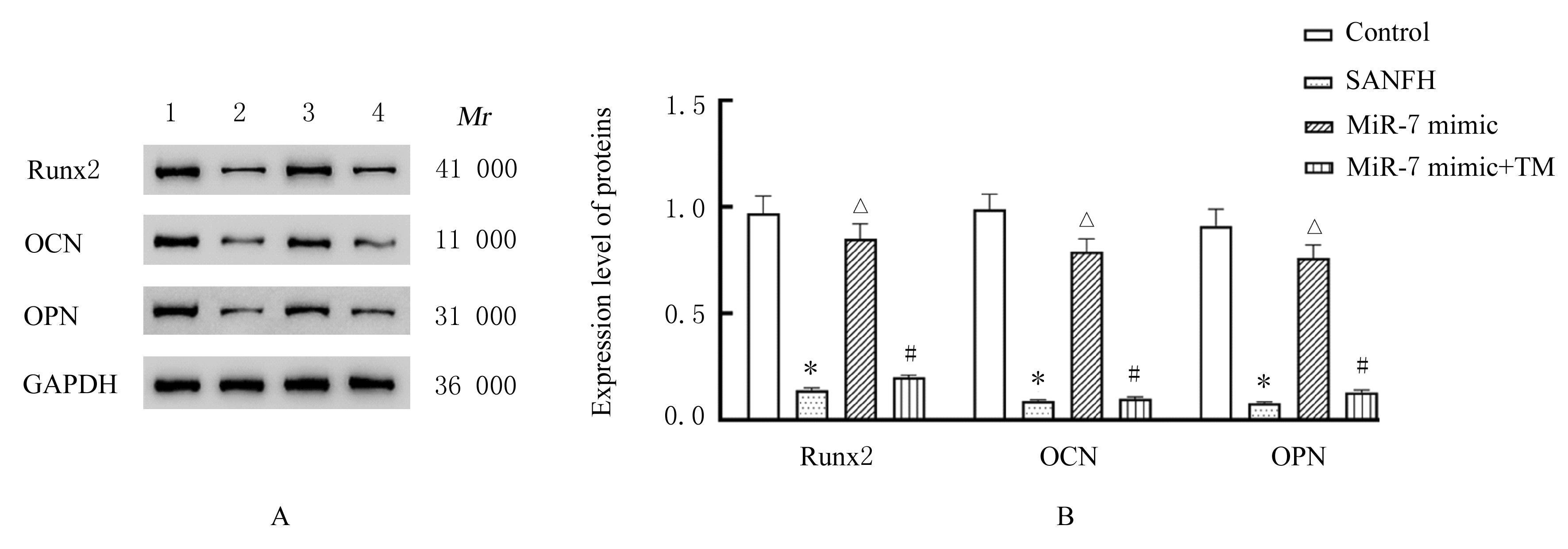
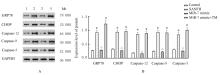
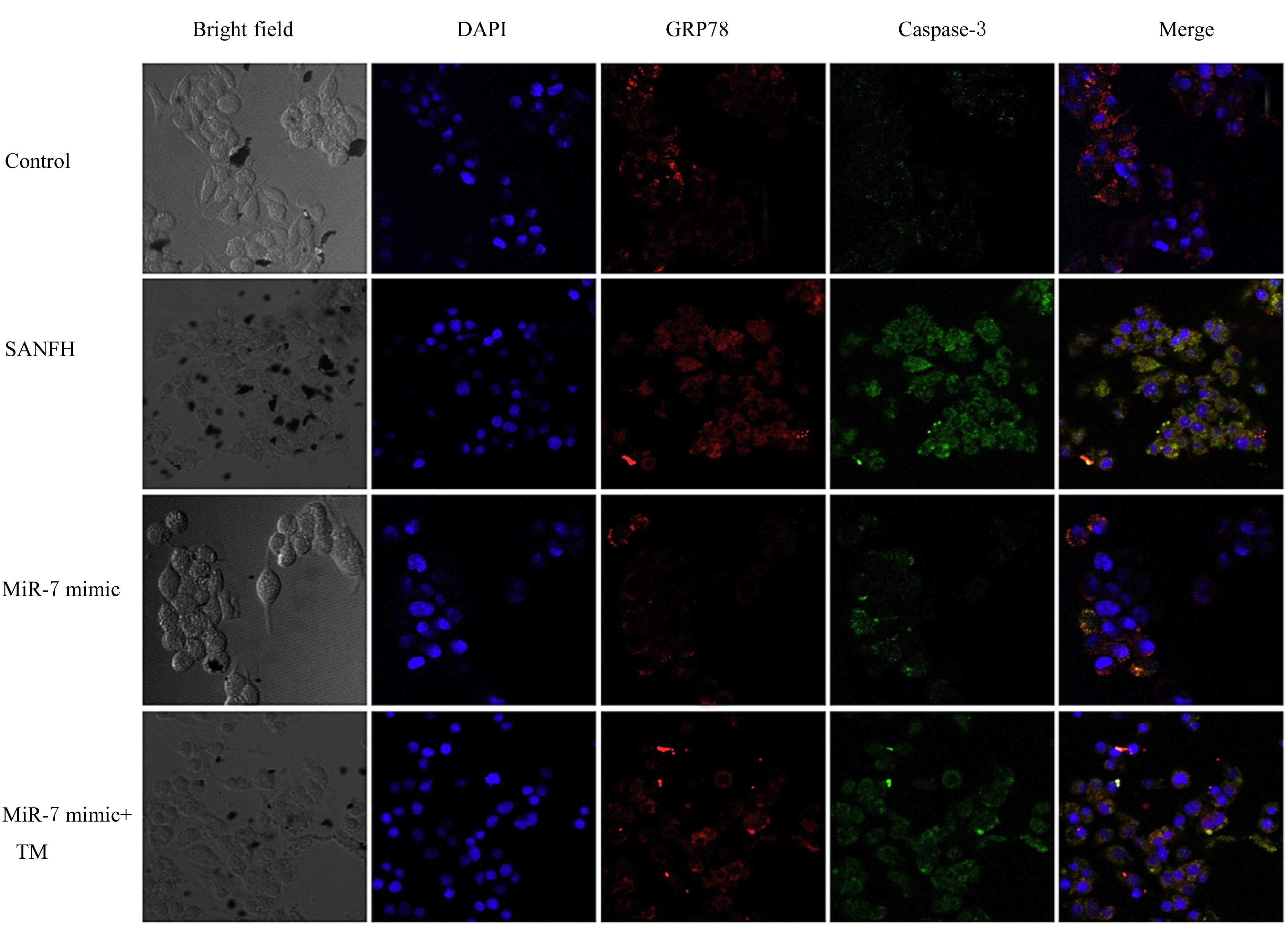
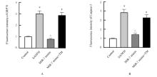
Hao ZHANG,Cuizhu WANG,Huimin HUANGFU,Xinwei ZHANG,Yidi ZHANG,Yanmin ZHOU.
Effect of 20(S)-protopanaxadiol on osteogensis differentiation of bone marrow mesenchymal stem cells in rats and its mechanism
[J]. Journal of Jilin University(Medicine Edition), 2023, 49(4): 867-874.
|
| [6] |
Yuanyuan LIANG,Song ZHAO,Jing HU,Ni AN,Yanlu WEI,Rongjian SU.
Effect of motesanib combined with EZH2 inhibitor GSK126 on proliferation and apoptosis of liver cancer Huh7 cells and its mechanism
[J]. Journal of Jilin University(Medicine Edition), 2023, 49(4): 896-904.
|
| [7] |
Hongli CUI,Siqi FAN,Wenfei GUAN,E MENG,Jiatong LIU,Xuetong SUN,Chunxu CAO,Lixin LIU,Yali QI,Fang FANG,Zhicheng WANG.
Inhibitory effect of irradiation enhanced by gallic acid-lecithin complex-induced oxidative stress on proliferation of A549 cells
[J]. Journal of Jilin University(Medicine Edition), 2023, 49(4): 941-946.
|
| [8] |
Hongyuan TIAN,Caiyun YIN,Li WANG,Peiyun HU,Chenyang ZHANG,Qiuyue LI,Qingzhao ZHENG,Yali QI,Fang FANG,Zhicheng WANG.
Effects of hydroxyurea combined with radiation on cell cycle and apoptosis of cells after silencing ATRX
[J]. Journal of Jilin University(Medicine Edition), 2023, 49(3): 590-598.
|
| [9] |
Xingye WANG,Xiangri KONG,Mengli JIN,Bingmei WANG,Mingquan LI.
Improvement effect of β-sitosterol on cognitive function in Alzheimer’s disease model mice and its mechanism
[J]. Journal of Jilin University(Medicine Edition), 2023, 49(3): 599-607.
|
| [10] |
Shengyu YAN,Changhua LIU,Zhijie XU,Yating DING,Yafeng XIE,Qiao ZHANG,Wanying LIU.
Effect of lncRNA PAX8-AS1 on proliferation, apoptosis and invasion of colorectal cancer cells and its mechanism
[J]. Journal of Jilin University(Medicine Edition), 2023, 49(3): 656-664.
|
| [11] |
Shuya ZHANG,Hongying SUN,Jian MAO,Chengxi MENG,Gelong BA.
Expression of circ_EFCAB2 in epileptic cell model and its mechanism
[J]. Journal of Jilin University(Medicine Edition), 2023, 49(3): 691-696.
|
| [12] |
Kai WANG,Han HUANG.
Effects of atorvastatin on proliferation, apoptosis, and migration of human tongue squamous cell carcinoma CAL-27 cells and their mechanisms
[J]. Journal of Jilin University(Medicine Edition), 2023, 49(2): 324-331.
|
| [13] |
Yifei SUN,Dinuo LI,Yubin WANG.
Inhibitory effect of curcumin on proliferation and invasion of gastric cancer MGC-803 cells by down-regulating PI3K/Akt/mTOR signaling pathway protein expression
[J]. Journal of Jilin University(Medicine Edition), 2023, 49(2): 332-340.
|
| [14] |
Dongming TAN,Hongying YIN,Xiangmin DENG,Xu DING.
Antidepressant effect of carnosic acid on chronic unpredictable mild stress-induced depression in rats and its SIRT1/ERS regulatory mechanism
[J]. Journal of Jilin University(Medicine Edition), 2023, 49(2): 360-368.
|
| [15] |
Junxiong ZHAO,Qian WU,Liangui NIE,Shengquan LIU,Zhentao JIANG,Jian CHEN,Ting XIAO,Jun YANG.
Ameliorative effect of SO2 on myocardial fibrosis in type 2 diabetes mellitus rats and its mechanism
[J]. Journal of Jilin University(Medicine Edition), 2023, 49(1): 8-14.
|
 )
)