吉林大学学报(医学版) ›› 2021, Vol. 47 ›› Issue (2): 249-256.doi: 10.13481/j.1671-587X.20210201

• 基础研究 •    下一篇

PGE2对非小细胞肺癌组织浸润T淋巴细胞中PD-1表达的影响及其机制

袁野1,庄金宝1,石旭1,李长远2()   

  1. 1.吉林大学第一医院二部检验科,吉林 长春 130021
    2.吉林大学第一医院二部胸外科,吉林 长春 130031
  • 收稿日期:2020-09-09 出版日期:2021-03-28 发布日期:2021-03-25
  • 通讯作者: 李长远 E-mail:lichangyuanjlu@163.com
  • 作者简介:袁 野(1986-),女,吉林省长春市人,主管技师,生物化学与分子生物学硕士,主要从事肺癌发病机制方面的研究。
  • 基金资助:
    国家自然科学基金项目(81800557)

Influence of PGE2 on PD-1 expression in infiltrating T lymphocytes in non-small cell lung cancer tissue and its mechanism

Ye YUAN1,Jinbao ZHUANG1,Xu SHI1,Changyuan LI2()   

  1. 1.Department of Laboratory,First Hospital,Jilin University,Changchun 130021,China
    2.Department of Thoracic Surgery,First Hospital,Jilin University,Changchun 130031,China
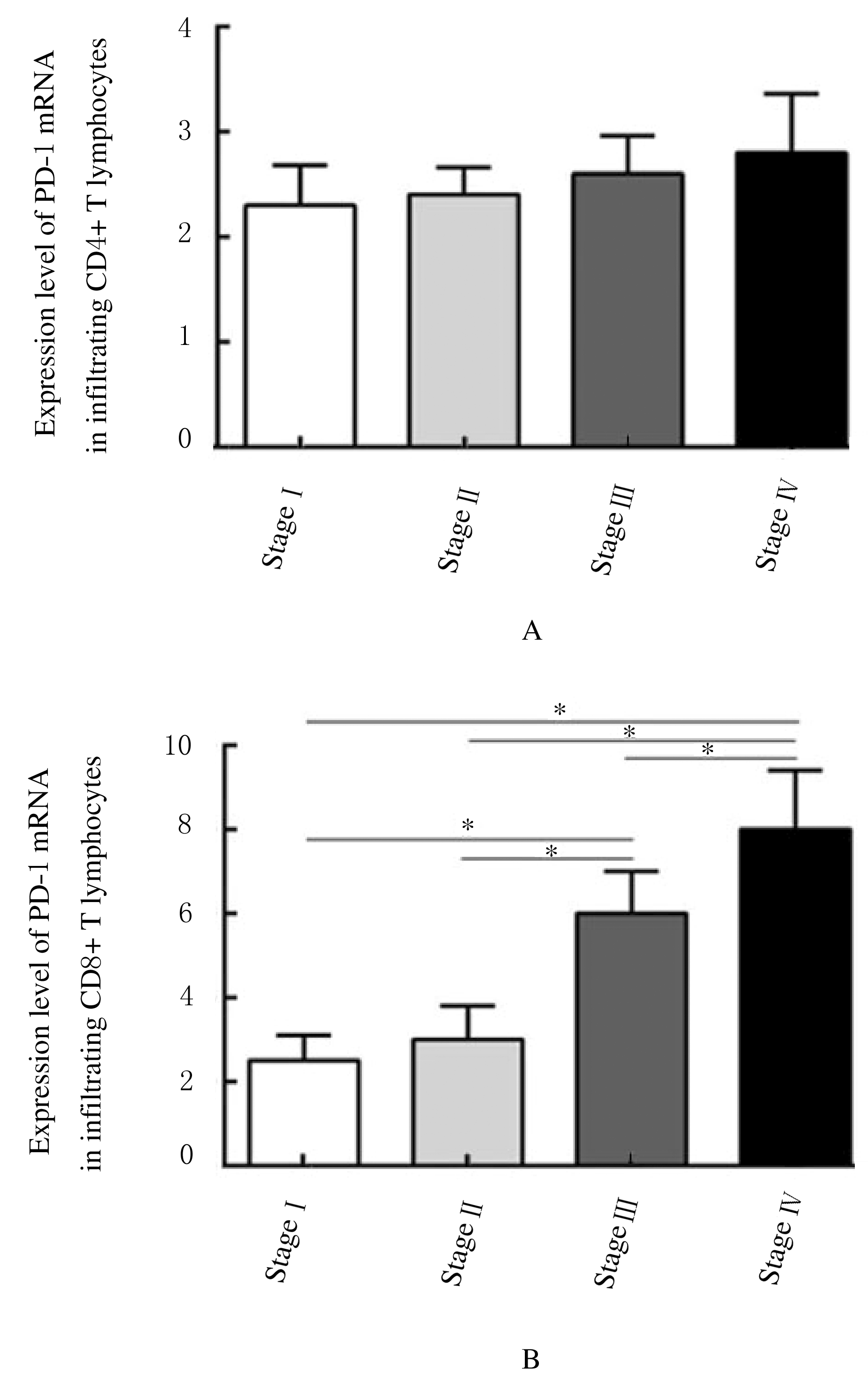
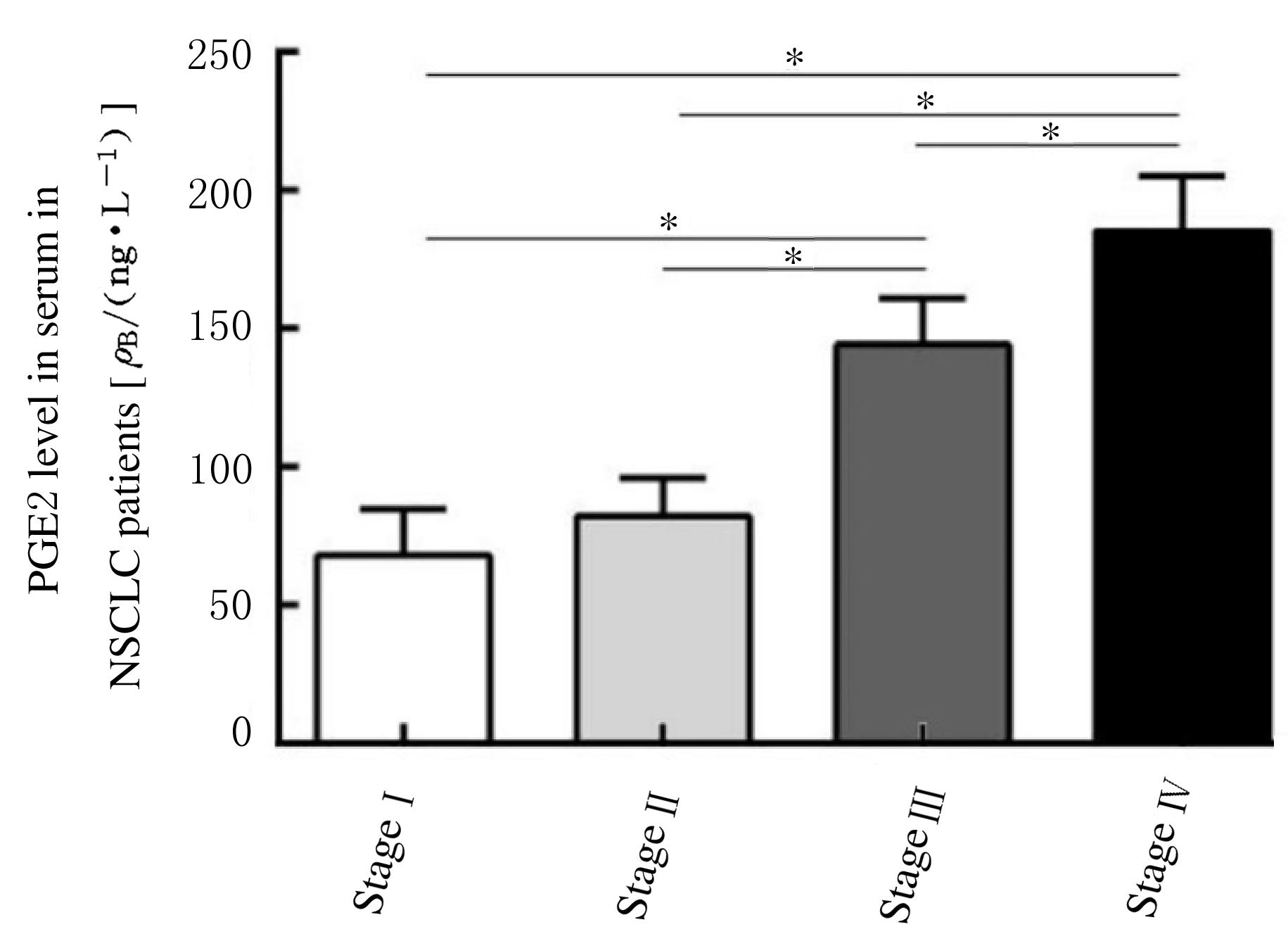
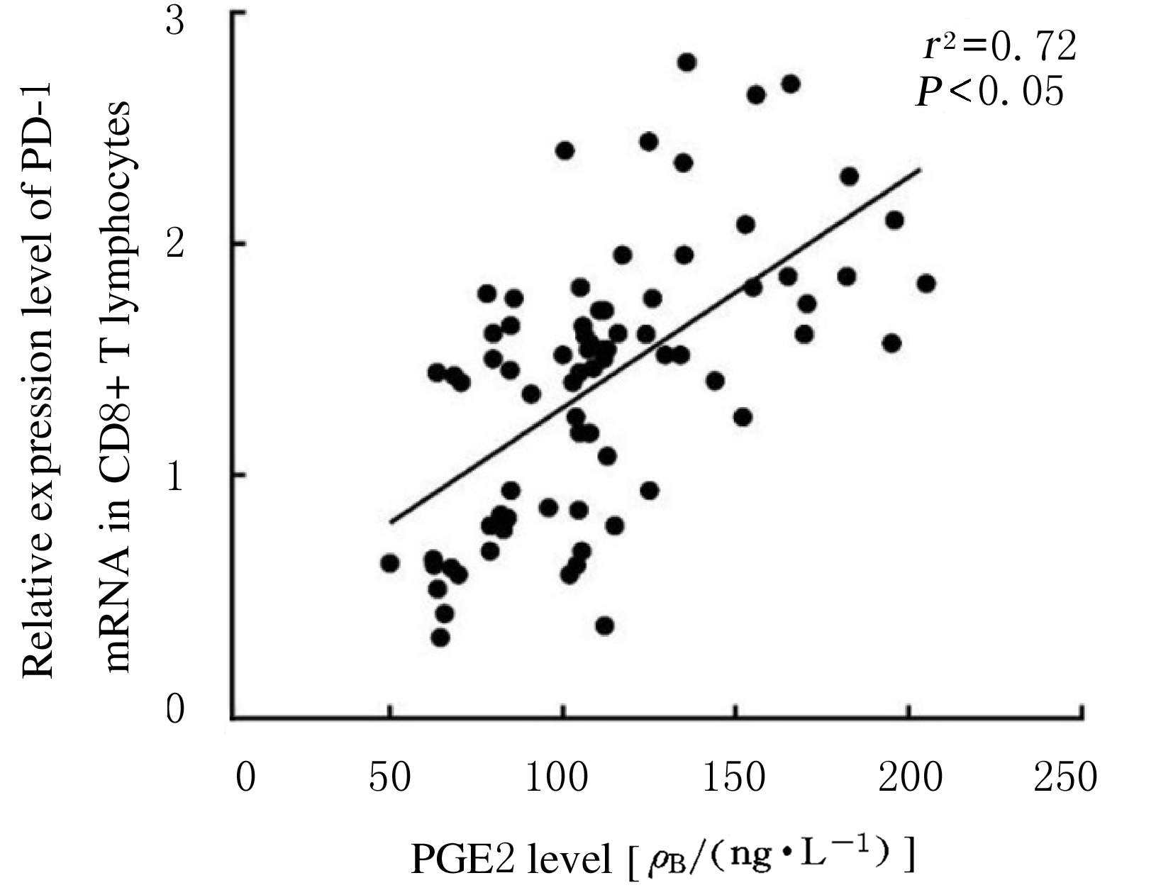
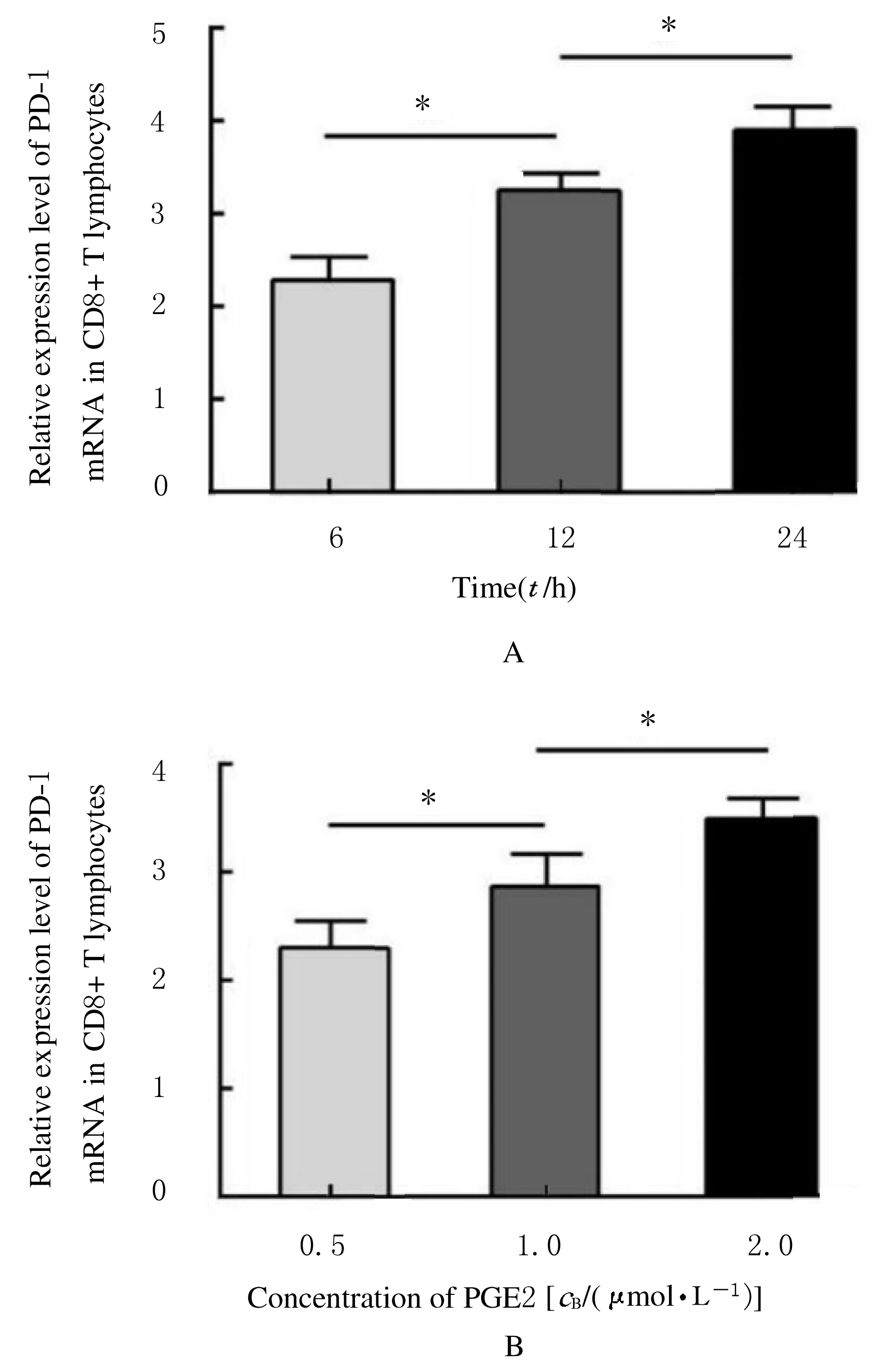
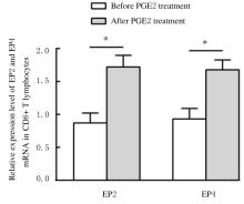
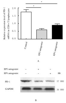
  • Received:2020-09-09 Online:2021-03-28 Published:2021-03-25
  • Contact: Changyuan LI E-mail:lichangyuanjlu@163.com

摘要: 目的

探讨程序性死亡受体1(PD-1)在非小细胞肺癌(NSCLC)组织浸润CD4+和CD8+T淋巴细胞中的表达情况,阐明前列腺素E2(PGE2)对PD-1的影响及其相关机制。

方法

选取确诊的75例NSCLC患者作为研究对象,根据国际抗癌联盟(UICC)颁布的NSCLC TNM分期标准,将患者分为I~Ⅳ期。应用密度梯度离心法分离获得NSCLC组织匀浆中浸润的单个核细胞。采用流式细胞术分析浸润淋巴细胞中CD4+T淋巴细胞和CD8+T淋巴细胞所占百分率。采用实时荧光定量聚合酶链式反应(RT-qPCR)和Western blotting法检测浸润T淋巴细胞中PD-1 mRNA和蛋白表达水平。利用酶联免疫吸附试验(ELISA)法检测血清中PGE2水平。

结果

Ⅲ期和Ⅳ期NSCLC患者局部浸润CD8+T淋巴细胞百分率明显高于Ⅰ期和Ⅱ期NSCLC患者(P<0.05),而CD4+T淋巴细胞百分率在不同TNM分期患者组间比较差异无统计学意义(P>0.05)。在Ⅳ期NSCLC患者CD8+T淋巴细胞中PD-1 mRNA表达水平高于其他分期患者(P<0.05),在不同TNM分期患者CD4+T淋巴细胞中PD-1 mRNA表达水平比较差异无统计学意义(P>0.05)。在Ⅲ期和Ⅳ期NSCLC患者血清中PGE2水平明显高于Ⅰ期和Ⅱ期NSCLC患者(P<0.05)。PGE2刺激CD8+T淋巴细胞后EP2和EP4 mRNA表达水平明显升高(P<0.05),而加入EP2和EP4拮抗剂后CD8+T淋巴细胞中PD-1 mRNA表达水平明显降低(P<0.05),PD-1蛋白表达量降低。

结论

PGE2能够通过PGE2/EP2和PGE2/EP4信号通路促进NSCLC组织浸润CD8+T淋巴细胞中PD-1的表达,从而抑制CD8+T淋巴细胞的免疫功能。

关键词: 癌,非小细胞肺, T淋巴细胞, 程序性死亡受体1, 前列腺素E2

Abstract: Objective

To explore the expression of programmed death-1 (PD-1) in the infiltrating CD4+ and CD8+ T lymphocytes in non-small cell lung cancer (NSCLC) tissue, and to illuminate the influence of prostaglandin E2(PGE2) on the PD-1 expression and its related mechanism.

Methods

A total of 75 patients diagnosed as NSCLC were enrolled and divided into stages Ⅰ-Ⅳ according to the TNM staging criteria for NSCLC issued by the Union Internationale Against cancer (UICC).The infiltrating mononuclear cells in NSCLC tissue homogenate were separated by density gradient centrifugation. The percentages of CD4+ and CD8+ T lymphocytes in the infiltrating lymphocytes were analyzed by flow cytometry. Real-time fluorescence quantitative polymerase chain reaction(RT-qPCR) and Western blotting methods were used to detect the expression levels of PD-1 mRNA and protein in the infiltrating T lymphocytes. The PGE2 level in serum was detected by ELISA.

Results

The percentages of infiltrating CD8+ T lymphocytes in the NSCLC patients at stages Ⅲ and Ⅳ were obviously higher than those of the NSCLC patients at stages Ⅰ and Ⅱ(P<0.05), but the percentages of CD4+ T lymphocytes showed no significant differences among the patients at different stages(P>0.05). The expression level of PD-1 mRNA in the CD8+ T lymphocytes in the NSCLC patients at stage Ⅳ was higher than those of the patients at other stages (P<0.05), but the expression levels of PD-1 mRNA in the CD4+ T lymphocytes had no significant differences among the patients at different stages (P>0.05). The serum levels of PGE2 in the NSCLC patients at stages Ⅲ and Ⅳ were obviously higher than those in the NSCLC patients at stages Ⅰ and Ⅱ (P<0.05). The expression levels of EP2 and EP4 mRNA in the CD8+ T lymphocytes were obviously increased after PGE2 stimulating(P<0.05);the PD-1 mRNA expression levels in the CD8+ T lymphocytes were significantly decreased after adding EP2 and EP4 antagonists (P<0.05),and the PD-1 protein expression amounts were decreased.

Conclusion

PGE2 can promote the PD-1 expression in infiltrating CD8+ T lymphocytes in NSCLC tissue through PGE2/EP2 and PGE2/EP4 signaling pathways, resulting in the decreased immune function of CD8+ T lymphocytes.

Key words: cancer,non-small cell lung, T lymphocytes, programmed death-1, prostaglandin E2

中图分类号: 

  • R34