吉林大学学报(医学版) ›› 2021, Vol. 47 ›› Issue (3): 615-622.doi: 10.13481/j.1671-587X.20210310

• 基础研究 • 上一篇    下一篇

桦木酸通过调控JAK2/STAT3信号通路对骨髓瘤细胞增殖和凋亡的影响及其机制

冯佳1,徐海婵1,温健2,吴泽华1,陈琦1()   

  1. 1.北京大学深圳医院血液内科, 北京 518035
    2.北京大学深圳医院脊柱外科, 北京 518035
  • 收稿日期:2020-10-28 出版日期:2021-05-28 发布日期:2021-05-28
  • 通讯作者: 陈琦 E-mail:hongjiahongjia1981@163.com
  • 作者简介:冯 佳(1981-),女,广东省清远市人,副主任医师,医学硕士,主要从事淋巴瘤和骨髓瘤发病机制的分子生物学方面的研究。
  • 基金资助:
    广东省卫健委医学科研项目(A2019540);广东省深圳市医疗卫生三名工程项目(SZSM201612004)

Effect of betulinic acid on proliferation and apoptosis of myeloma cells by regulating JAK2/STAT3 signaling pathway and its mechanism

Jia FENG1,Haichan XU1,Jian WEN2,Zehua WU1,Yi CHEN1()   

  1. 1.Department of Hematology,Shenzhen Hospital,Peking University,Beijing 518035,China
    2.Department of Spinal Surgery,Shenzhen Hospital,Peking University,Beijing 518035,China
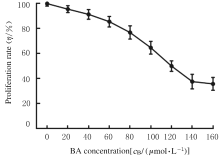
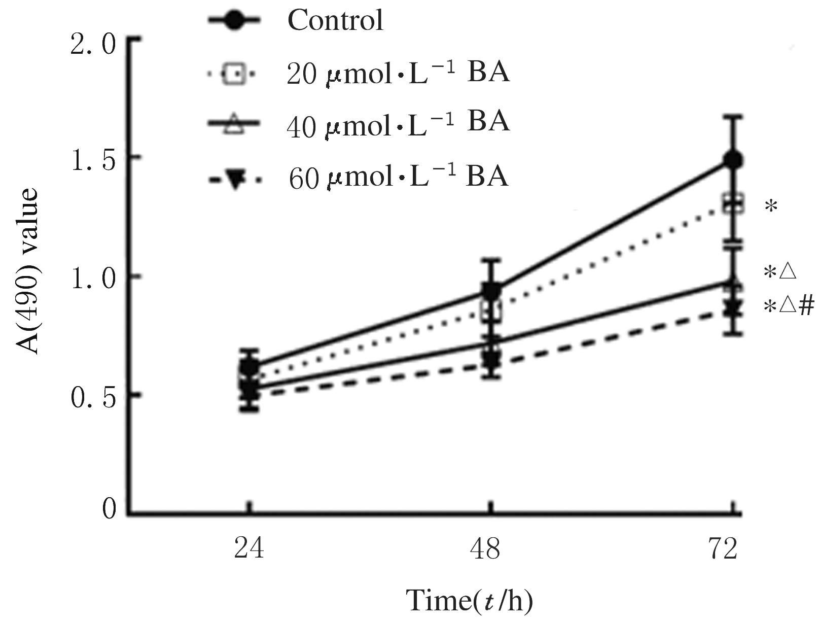
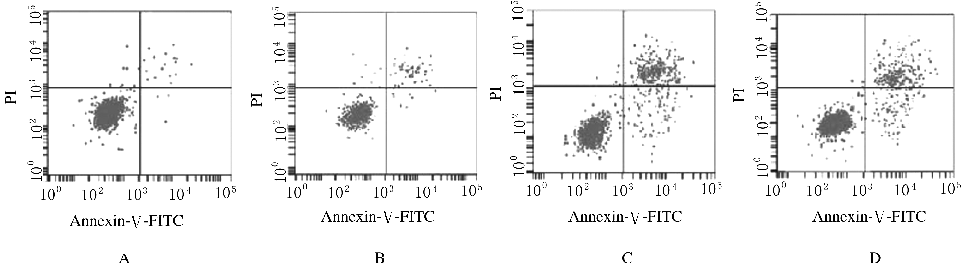
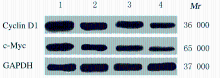
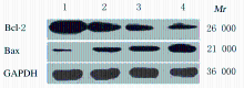
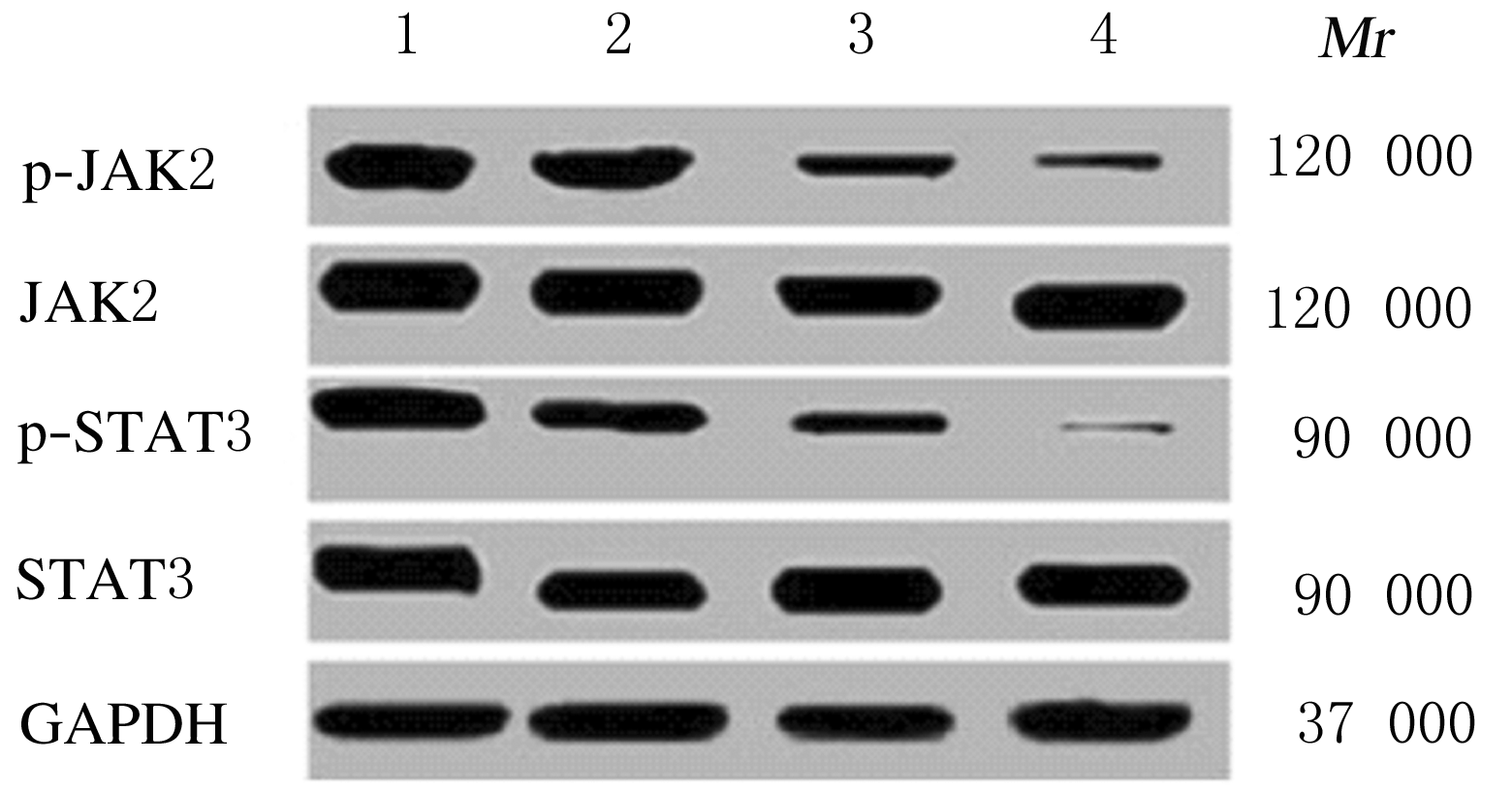
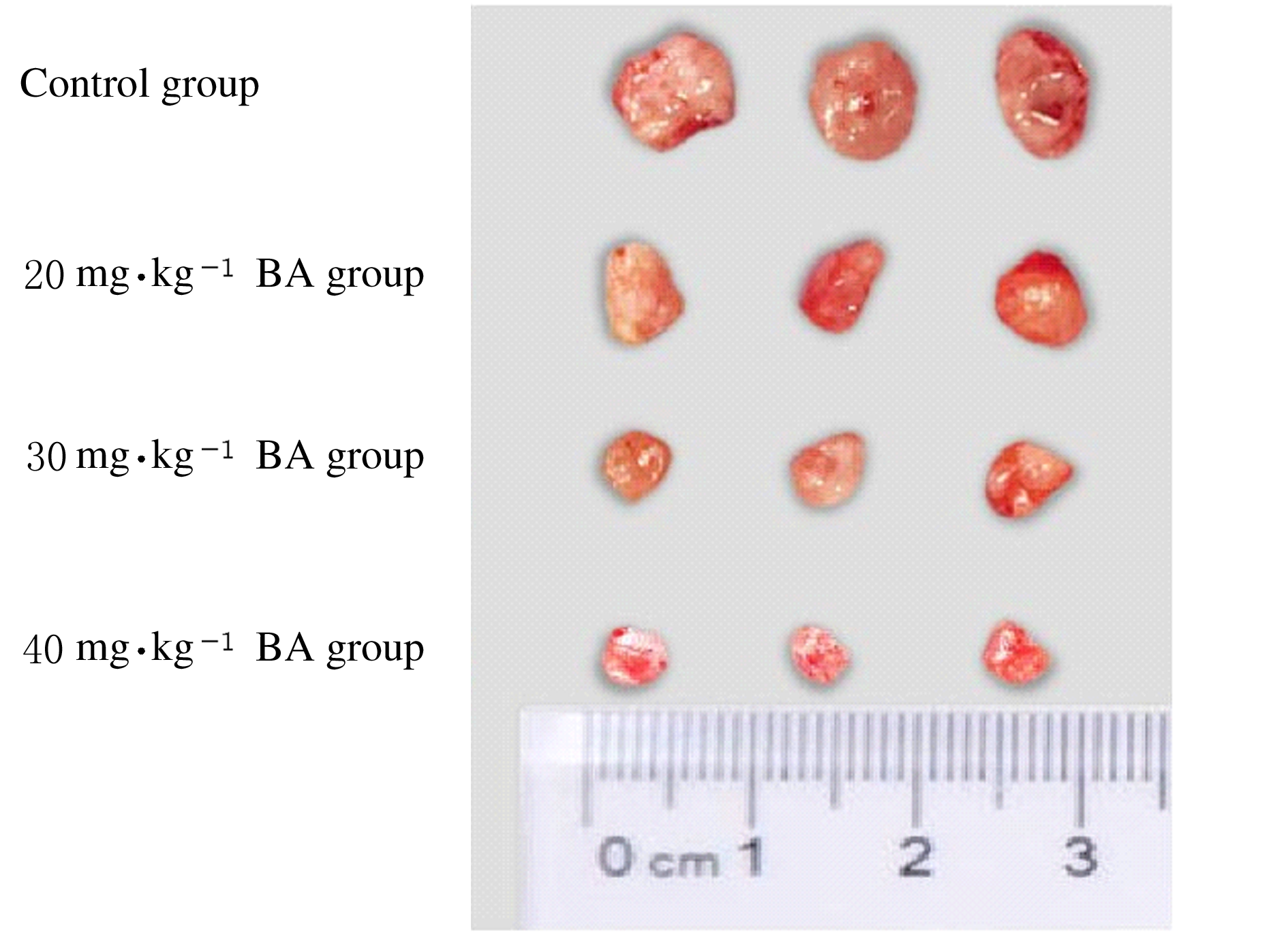
  • Received:2020-10-28 Online:2021-05-28 Published:2021-05-28
  • Contact: Yi CHEN E-mail:hongjiahongjia1981@163.com

摘要: 目的

探讨桦木酸(BA)通过介导Janus激酶2(JAK2)/信号转导与转录激活因子(STAT3)信号通路对多发性骨髓瘤(MM)细胞增殖和凋亡的影响及其机制。

方法

骨髓瘤U266细胞给予不同浓度(0、20、40、60、80、100、120和160 μmol·L-1)BA作为不同浓度BA组,同时设置空白组(不加入BA且无细胞),以未经BA处理的U266细胞作为对照组。检测各组骨髓瘤U266细胞增殖率。将U266细胞分为对照组和20、40、60 μmol·L-1 BA组,CCK-8法检测各组细胞增殖率,Annexin Ⅴ-FITC/PI双染法检测各组细胞凋亡率,Western blotting法检测各组细胞中原癌基因(c-Myc)、细胞周期蛋白D1(cyclin D1)、B细胞淋巴瘤2(Bcl-2)、Bcl-2相关X蛋白(Bax)和JAK2/STAT3信号通路蛋白表达水平。将裸鼠分为对照组(给予100 μL DMSO)和20、30和40 mg·kg-1 BA组(给予20、30及40 mg·kg-1 BA),观察各组裸鼠瘤体质量和体积。

结果

与对照组比较,作用48和72 h时,20、40和60 μmol·L-1 BA组细胞增殖率和细胞中c-Myc、cyclin D1、Bcl-2及JAK2/STAT3信号通路蛋白表达水平明显降低(P<0.05),细胞凋亡率和细胞中Bax蛋白表达水平明显升高(P<0.05)。作用48和72 h时,与20 μmol·L-1 BA组比较,40和60 μmol·L-1 BA组细胞增殖率、细胞中c-Myc、cyclin D1、Bcl-2及JAK2/STAT3信号通路蛋白表达水平明显降低(P<0.05),细胞凋亡率和细胞中Bax蛋白表达水平明显升高(P<0.05);与40 μmol·L-1 BA组比较,60 μmol·L-1BA组细胞增殖率、细胞中c-Myc、cyclin D1、Bcl-2及JAK2/STAT3信号通路蛋白表达水平明显降低(P<0.05),细胞凋亡率和细胞中Bax蛋白表达水平明显升高(P<0.05)。与对照组比较,20、30和40 mg·kg-1 BA组裸鼠瘤质量及体积明显降低(P<0.05);与20 mg·kg-1 BA比较,30和40 mg·kg-1 BA组裸鼠瘤质量及体积明显降低(P<0.05),与30 mg·kg-1BA组比较,40 mg·kg-1 BA组裸鼠瘤质量及体积明显降低(P<0.05)。

结论

BA可促进骨髓瘤细胞凋亡、抑制骨髓瘤细胞增殖及裸鼠移植瘤生长,其作用机制可能与BA抑制JAK2/STAT3信号通路激活有关。

关键词: 桦木酸, JAK2/STAT3信号通路, 骨髓瘤细胞, 细胞增殖, 细胞凋亡

Abstract: Objective

To investigate the effect of betulinic acid(BA) on the proliferation and apoptosis of the myeloma cells through the Janus kinase 2(JAK2)/signal transducers and activators of transcription 3(STAT3) signaling pathway and its mechanism.

Methods

The myeloma U266 cells were treated with different concentrations(0,20,40,60,80,100,120,and 160 μmol·L-1) of BA as different concentrations of BA groups, at the same time,blank group (without BA and without cells) was set up,and the U266 cells without BA treatment were used as control group.The proliferation rates of the U266 cells in various groups were detected. The U266 cells were divided into control group and 20, 40,and 60 μmol·L-1 BA groups. The proliferation rates of the cells in various groups were detected by CCK-8 method;the apoptotic rates of the cells in various groups were detected by Annexin Ⅴ-FITC/PI double staining method; the expression levels of c-myc oncogene(c-Myc),cell cycle proteins D1(cyclin D1),B cell lymphoma-2(Bcl-2),Bcl-2 associated X protein (Bax) and JAK2/STAT3 signaling pathway proteins in the cells in various groups were detected by Western blotting method.The nude mice were divided into control group(given 100 μL DMSO)and 20,30 and 40 mg·kg-1 BA group(given 20,30,40 mg·kg-1 BA);the weights and volumes of tumor of the nude mice in various groups were observed.

Results

Compared with control group, the proliferation rate of the myeloma U266 cells at 48 and 72 h after treatment,the expression levels of C-MYc, cyclin D1, Bcl-2, and JAK2/STAT3 signaling pathway proteins in the cells and the tumor weight of nude mice in 20,40, and 60 μmol·L-1 BA groups were significantly decreased(P<0.05), while the apoptotic rates and the expression levels of Bax protein were significantly increased (P<0.05).Forty-eight and 72 h after treatment,compared with 20 μmol·L-1 BA group,the proliferation rates of the myeloma U266 cells in 40 and 60 μmol·L-1 BA groups, and the expression levels of c-MYc, cyclin D1, Bcl-2,and JAK2/STAT3 signaling pathway proteins in the cells in 40 and 60 μmol·L-1 BA groups were significantly decreased(P<0.05), while the apoptotic rates and the expression levels of Bax protein were significantly increased (P<0.05);compared with 40 μmol·L-1 BA group, the proliferation rates of the cells in 60 μmol·L-1 BA group,the expression levels of c-MYc, cyclin D1, Bcl-2, and JAK2/STAT3 signaling pathway proteins in the cells in 60 μmol·L-1 BA group were significantly decreased(P<0.05), while the apoptotic rates and the expression level of Bax protein were significantly increased (P<0.05).Compared with control group, the tumor weights and volumes of the nude mice in 20, 30,and 40 mg·kg-1 BA groups were significantly decreased (P<0.05). Compared with 20 mg·kg-1 BA group, the tumor weight and volume of the nude mice in 30 and 40 mg·kg-1 BA groups were significantly decreased (P<0.05).Compared with 30 mg·kg-1 BA group, the tumor weight and volume in 40 mg·kg-1 BA group were significantly decreased (P<0.05).

Conclusion

BA can promote the apoptosis of myeloma cells and inhibit the proliferation of myeloma cells and the growth of transplanted tumor in the nude mice. The mechanism may be related to the inhibition of JAK2/STAT3 signaling pathway activation by BA.

Key words: betulinic acid, Janus kinase2/signal transducers and activators of transcription3, myeloma cells, proliferation, apoptosis

中图分类号: 

  • R733.3