吉林大学学报(医学版) ›› 2021, Vol. 47 ›› Issue (4): 958-964.doi: 10.13481/j.1671-587X.20210419

• 基础研究 • 上一篇    下一篇

香叶木素对异丙肾上腺素诱导的小鼠心肌肥大的抑制作用及其机制

王平丽1,张申伟2(),李江1   

  1. 1.河南医学高等专科学校外科教研室,河南 郑州 451191
    2.河南省郑州市第七人民医院心内科,河南 郑州 455000
  • 收稿日期:2020-11-28 出版日期:2021-07-28 发布日期:2021-07-22
  • 通讯作者: 张申伟 E-mail:zhangsw214@163.com
  • 作者简介:王平丽(1979-),女,河南省鹤壁市人,主治医师,医学硕士,主要从事心肌缺血和心肌肥大相关方面的研究。
  • 基金资助:
    河南省卫健委医学科技攻关计划项目(2018020467)

Inhibitory effect of diosmetin on myocardial hypertrophy induced by isoproterenol in mice and its mechanism

Pingli WANG1,Shenwei ZHANG2(),Jiang LI1   

  1. 1.Department of Surgery,Henan Medical College,Zhengzhou 451191,China
    2.Department of Cardiology
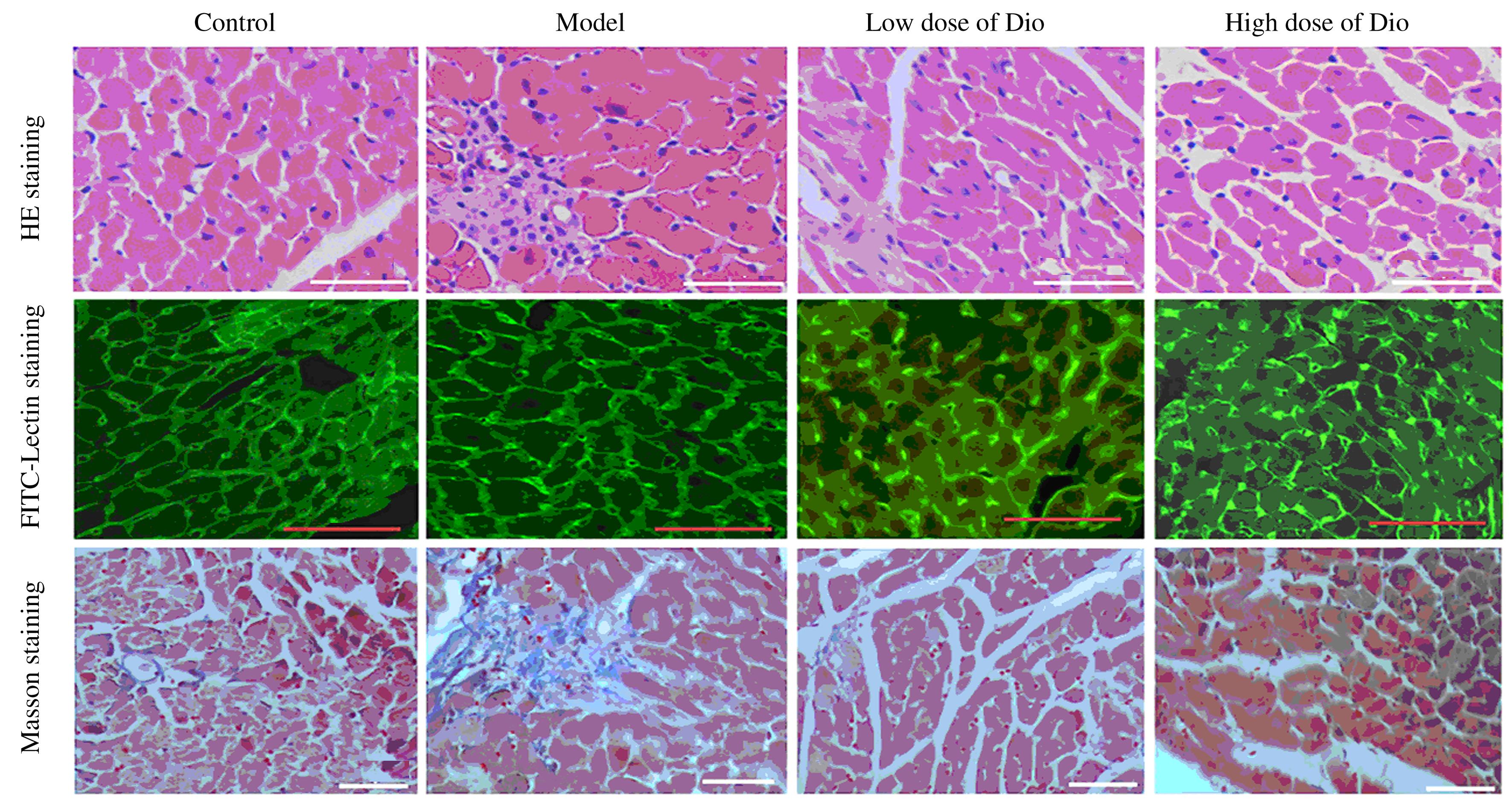
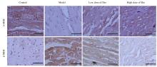
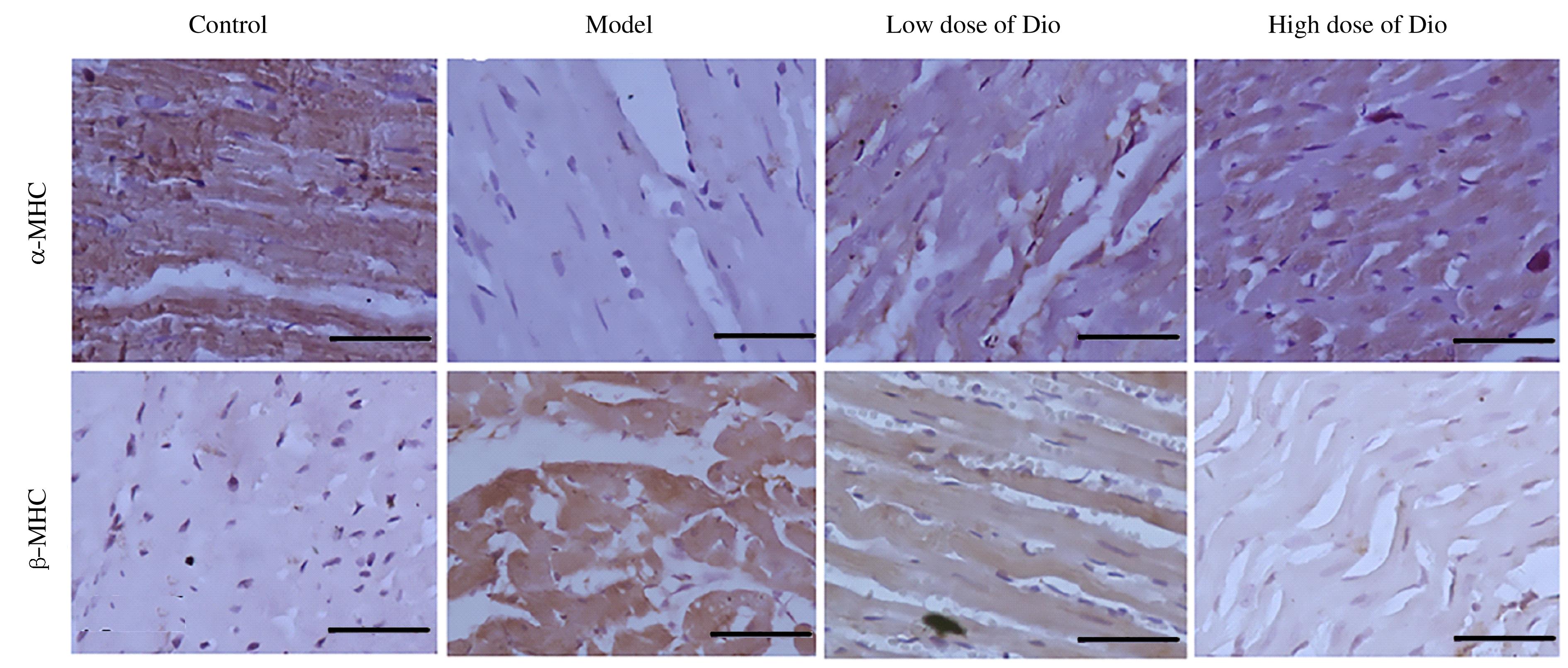
  • Received:2020-11-28 Online:2021-07-28 Published:2021-07-22
  • Contact: Shenwei ZHANG E-mail:zhangsw214@163.com

摘要: 目的

探讨香叶木素(Dio)对异丙肾上腺素(Iso)诱导的小鼠心肌肥大的抑制作用,并阐明其生物学机制。

方法

28只小鼠随机分为对照组、模型组、低剂量Dio组和高剂量Dio组。对照组和模型组小鼠分别连续14 d皮下注射生理盐水或Iso(5 mg·kg-1·d-1);低和高剂量Dio组小鼠连续14 d皮下注射Iso(5 mg·kg-1·d-1),同时连续14 d腹腔注射Dio(5和20 mg·kg-1·d -1)。14 d后处死小鼠,收集心脏和胫骨,称量心脏质量和左心室质量,测量胫骨长度,并计算心脏质量指数和左心室质量指数。采用HE、FITC-Lectin和Masson染色法观察小鼠左心室心肌组织病理形态表现,采用免疫组织化学法检测小鼠左心室心肌组织中α-肌球蛋白重链(α-MHC)和β-肌球蛋白重链(β-MHC)蛋白表达水平,采用Western blotting法检测大鼠左心室心肌组织中细胞外调节蛋白激酶1/2(ERK1/2)、磷酸化ERK1/2(p-ERK1/2)、c-Jun氨基末端激酶1/2(JNK1/2)和磷酸化JNK1/2(p-JNK1/2)蛋白表达水平。

结果

与对照组比较,模型组小鼠心脏质量指数和左心室质量指数均明显升高(P<0.01),左心室心肌组织形态发生明显改变,心肌纤维破坏,出现炎性细胞浸润,心肌细胞横截面积明显增大(P<0.01),心肌组织微循环灌注降低,间质纤维化程度明显增加(P<0.01);左心室心肌组织中α-MHC蛋白表达水平明显降低(P<0.01),β-MHC、p-ERK1/2和p-JNK1/2蛋白表达水平明显升高(P<0.01)。与模型组比较,不同剂量Dio组小鼠心脏质量指数和左心室质量指数明显降低(P<0.05或P<0.01),心肌组织损伤明显减轻,心肌细胞横截面积明显缩小(P<0.05或P<0.01),心肌组织微循环灌注增强,间质纤维化程度明显降低(P<0.05或P<0.01);左心室心肌组织中α-MHC蛋白表达水平明显升高(P<0.05或P<0.01),β-MHC、p-ERK1/2和p-JNK1/2蛋白表达水平明显降低(P<0.05或P<0.01),且呈剂量依赖性。

结论

Dio能改善Iso诱导的小鼠心肌肥大,其机制可能与其抑制ERK1/2和JNK1/2信号通路有关。

关键词: 香叶木素, 异丙肾上腺素, 心肌肥大, α-肌球蛋白重链, β-肌球蛋白重链

Abstract: Objective

To investigate the inhibitory effect of diosmetin (Dio) on isoprenaline (Iso)-induced myocardial hypertrophy in the mice, and to elucidate the biological mechanism.

Methods

Twenty-eight mice were randomly divided into control group, model group, low dose of Dio group, and high dose of Dio group. The mice in control group and model group were injected subcutaneously with normal saline or Iso (5 mg·kg-1·d-1) for 14 d, respectively. The mice in low dose and high dose of Dio groups were injected subcutaneously with Iso (5 mg·kg-1·d-1) for 14 d and intraperitoneally with Dio (5 and 20 mg·kg-1·d-1) for 14 d, respectively. After 14 d, the mice were sacrificed, the hearts and tibia were collected, the heart weights and left ventricular weights were weighed, the tibia lengths were measured, and the heart mass index and left ventricular mass index were calculated. The pathomorphology of left ventricular myocardium tissue was observed by HE, FITC-Lectin and Masson staining methods. The expression levels of α-myosin heavy chain (α-MHC) and β-myosin heavy chain (β-MHC) proteins in left ventricular myocardium tissue of the mice were detected by immunohistochemical method. The expression levels of extracellular regulated protein kinase 1/2 (ERK1/2), phosphorylated ERK1/2(p-ERK1/2),c-Jun N-terminal kinase 1/2 (JNK1/2), and phosphorylated JNK1/2(p-JNK1/2) proteins in left ventricular myocardium tissue of the mice were detected by Western blotting method.

Results

Compared with control group, the heart mass index and the left ventricular mass index of the mice in model group were significantly increased (P<0.01), the morphology of left ventricular myocardium tissue was significantly changed, the myocardial fibers were destructed with inflammatory cell infiltration, the cross-areas of myocardial cells were significantly enlarged (P<0.01),the microcirculatory perfusion of myocardium tissue was decreased, and the degree of interstitial fibrosis of myocardium tissue was increased (P<0.01); the expression level of α-MHC protein in the left ventricular myocardium tissue was significantly decreased (P<0.01), and the expression levels of β-MHC, p-ERK1/2 and p-JNK1/2 proteins were significantly increased(P<0.01). Compared with model group,the heart mass index and the left ventricular mass index in different doses of Dio groups were sifnificantly decreased(P<0.05 or P<0.01),the myocardial tissue injury was significantly reduced,the cross-areas of myocardial cells were significantly reduced (P<0.05 or P<0.01), the microcirculatory perfusion of myocardium tissue was enhanced,and the degree of interstitial fibrosis of myocardium tissue was significantly decreased (P<0.05 or P<0.01); the expression levels of α-MHC protein in the left ventricular myocardium tissue were significantly increased (P<0.05 or P<0.01), and the expression levels of β-MHC, p-ERK1/2 and p-JNK1/2 proteins were significantly decreased in a dose-dependent manner(P<0.05 or P<0.01).

Conclusion

Dio can improve Iso-induced myocardial hypertrophy in the mice, and its mechanism may be related to the inhibition of ERK1/2 and JNK1/2 signal pathways.

Key words: diosmetin, isoprenaline, myocardial hypertrophy, α-myosin heavy chain, β-myosin heary chain

中图分类号: 

  • R285.5