吉林大学学报(医学版) ›› 2021, Vol. 47 ›› Issue (6): 1557-1561.doi: 10.13481/j.1671-587X.20210629

• 临床医学 • 上一篇    下一篇

肺梭形细胞癌1例报告及文献复习

李倩,周佳奇,苑静怡,赵敏,邸鑫,王珂()   

  1. 吉林大学第二医院呼吸与危重症医学科,吉林 长春 130041
  • 收稿日期:2021-04-27 出版日期:2021-11-28 发布日期:2021-12-14
  • 通讯作者: 王珂 E-mail:kewangm1@hotmail.com
  • 作者简介:李 倩(1994-),女,安徽省亳州市人,在读硕士研究生,主要从事肺部感染性疾病和肺癌诊治方面的研究。
  • 基金资助:
    吉林省科技厅科技发展计划项目(20200708083YY)

Spindle cell carcinoma of lung: A case report and literature review

Qian LI,Jiaqi ZHOU,Jingyi YUAN,Min ZHAO,Xin DI,Ke WANG()   

  1. Department of Respiratory and Critical Care Medicine,Second Hospital,Jilin University,Changchun 130041,China
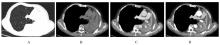
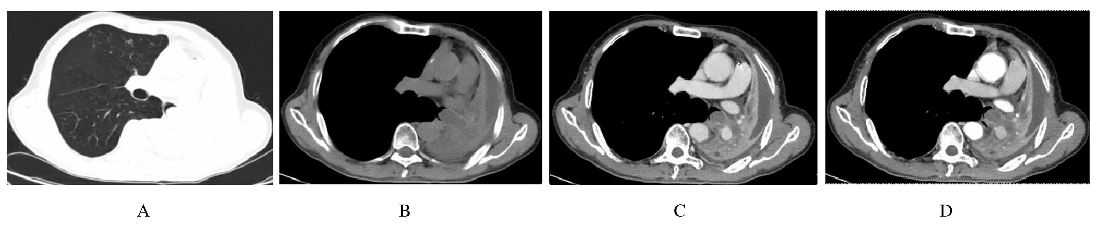
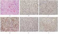
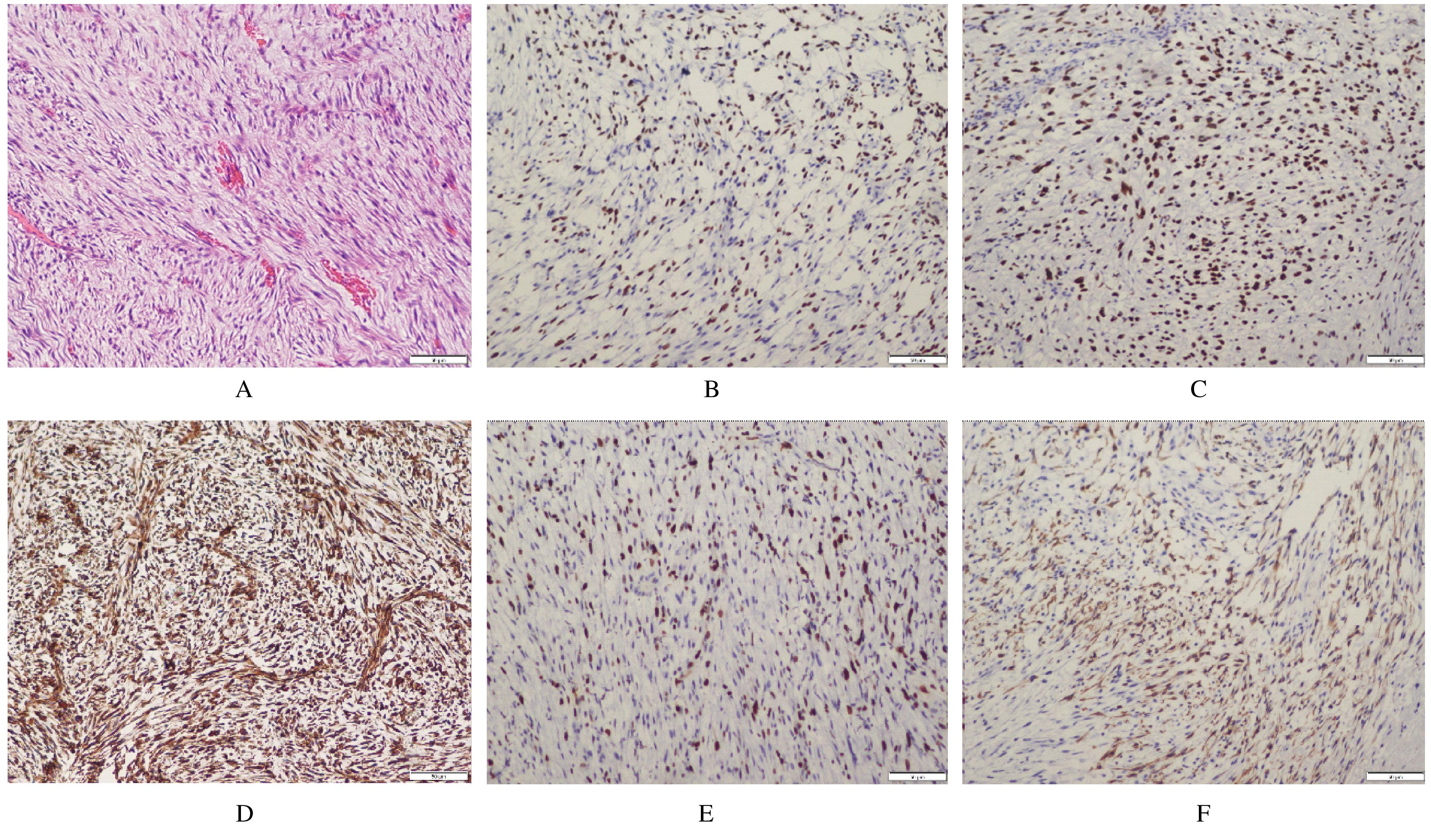
  • Received:2021-04-27 Online:2021-11-28 Published:2021-12-14
  • Contact: Ke WANG E-mail:kewangm1@hotmail.com

摘要: 目的

探讨肺梭形细胞癌(SpCC)患者的临床特征、诊断过程和治疗方法,旨在提高临床医生对该病的认识。

方法

收集1例SpCC患者的临床资料、影像学表现、支气管镜和病理检查结果,分析上述资料,并进行相关文献复习。

结果

患者,男性,71岁,因咳嗽和气短1个月入院。查体见左侧胸廓凹陷,肋间隙变窄,气管向左侧移位,叩诊左肺浊音,左肺呼吸音消失,无其他明显阳性体征。胸部CT显示左侧气道内高密度影,左肺体积减小。纤维支气管镜显示左主支气管内见带蒂新生物阻塞气道。病理检测结果显示支气管腺性乳头状瘤形成伴鳞化。入院后行胸部增强CT检查显示左主支气管条片状软组织密度影,增强扫描呈轻度强化,左肺体积减小,呈条片状软组织密度影,增强扫描呈不均匀强化。综合全面的实验室和影像学检查考虑支气管良性肿瘤可能性大,行2次纤维支气管镜下电圈套器治疗。支气管镜肺活检见大量梭形细胞,结合免疫组织化学染色结果确诊为 SpCC。

结论

SpCC是一种特殊类型的上皮来源肿瘤,形态与肉瘤相似,极易误诊。以主支气管占位性病变伴肺不张为主要影像学表现的SpCC患者较为少见,应与支气管良性肿瘤相鉴别。

关键词: 肉瘤样癌, 梭形细胞癌, 影像学表现, 纤维支气管镜, 病例报告

Abstract: Objective

To investigate the clinical features, diagnosis and treatment of the patients with spindle cell carcinoma of lung (SpCC), in order to improve the clinicians’understanding of this disease.

Methods

The clinical data, imaging manifestations, bronchoscope findings,and pathological findings of 1 patient with SpCC were collected, the above data were analyzed, and the relevant literatures were reviewed.

Results

A 71-year-old male patient was hospitalized due to cough and shortness of breath for 1 month.The physical examination results showed that the left thoracic cavity was depressed, the intercostal space was narrow, and the trachea shifted to the left;the dullness of left lung was detected by percussion, and the breath sounds of left lung disappeared without other obvious positive signs. The chest CT results showed the high-density shadow in the left airway and the reduced volume of left lung.The fiberoptic bronchoscope results showed that there were pedicled neoorganisms blocking the airway in the left main bronchus. The pathological examination results showed that the adenopapilloma of the bronchus was formed with scalification. After admission,the chest enhanced CT examination results showed the strip-shaped soft tissue density shadow of the left main bronchus,which showed mild enhancement in enhanced scan;the volume of left lung was reduced with strip-like soft tissue density shadow, which showed uneven enhancement in enhanced scan. After comprehensive laboratory and imaging examination, the possibility of bronchial benign tumor was considered, and two times of bronchoscopic electrical traps were performed. The final bronchoscopic lung biopsy showed a large number of spindle cells.Combined with the immunohistochemical staining results, SpCC was diagnosed.

Conclusion

SpCC is a special type of epithelial-derived tumor and it is easily misdiagnosed due to its similar morphology to sarcoma.The SpCC patients with main bronchial space occupying lesions accompanying with atelectasis as the main imaging manifestations are relatively rare, and SpCC should be differentiated from the bronchial benign tumor.

Key words: sarcomatoid carcinoma, spindle cell carcinoma, imaging manifestation, fiber bronchoscope, case report

中图分类号: 

  • R563.9