吉林大学学报(医学版) ›› 2022, Vol. 48 ›› Issue (1): 216-221.doi: 10.13481/j.1671-587X.20220127

• 临床医学 • 上一篇    下一篇

尾骨原发小细胞型骨肉瘤患者 18F-FDG PET/CT 显像特点分析1例报告及文献复习

胡萍,姜楠,李英华,赵红光()   

  1. 吉林大学第一医院核医学科,吉林 长春 130021
  • 收稿日期:2021-06-11 出版日期:2022-01-28 发布日期:2022-01-17
  • 通讯作者: 赵红光 E-mail:zhaohg@jlu.edu.cn
  • 作者简介:胡 萍 (1988-),女,吉林省长春市人,主治医师,主要从事核医学肿瘤诊断方面的研究。
  • 基金资助:
    吉林省卫健委卫生健康青年科技骨干培养计划项目(2019Q001)

Analysis on characteristics of 18F-FDG PET/CT imaging of patient with primary small cell osteosarcoma of coccyx:A case report and literature review

Ping HU,Nan JIANG,Yinghua LI,Hongguang ZHAO()   

  1. Department of Nuclear Medicine,First Hospital,Jilin University,Changchun 130021,China
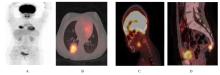
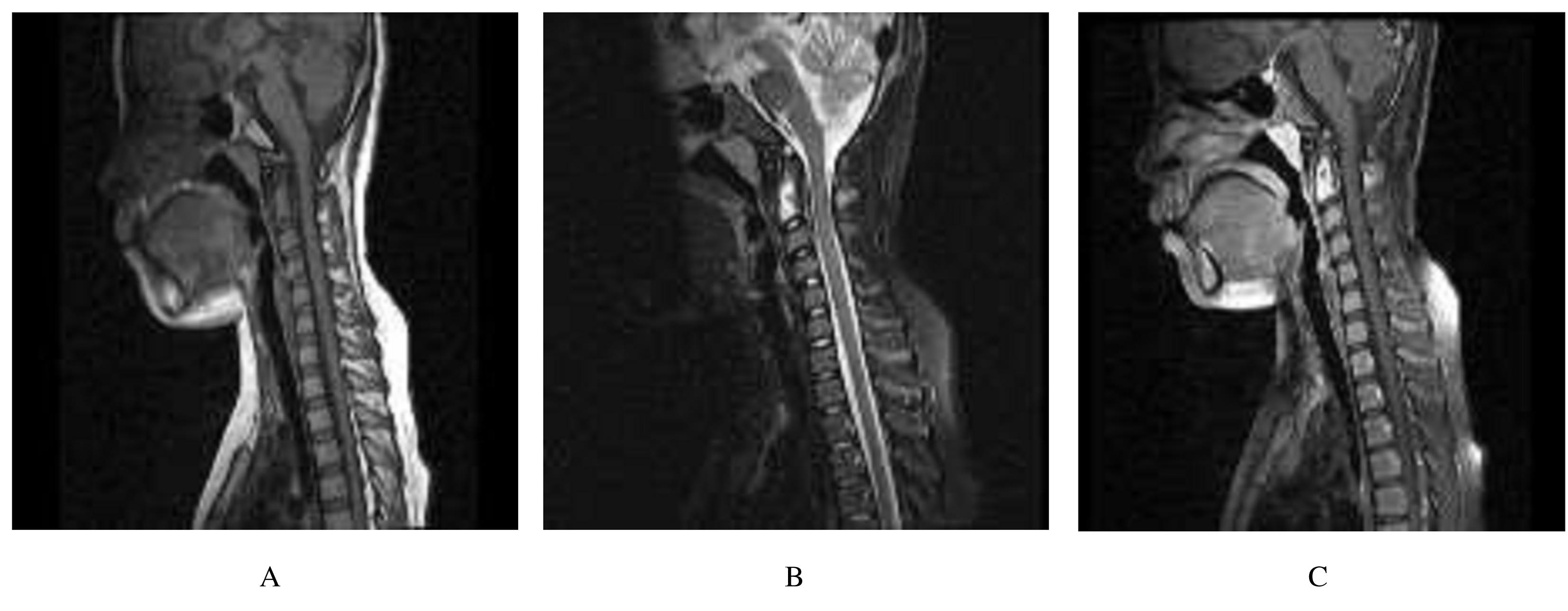
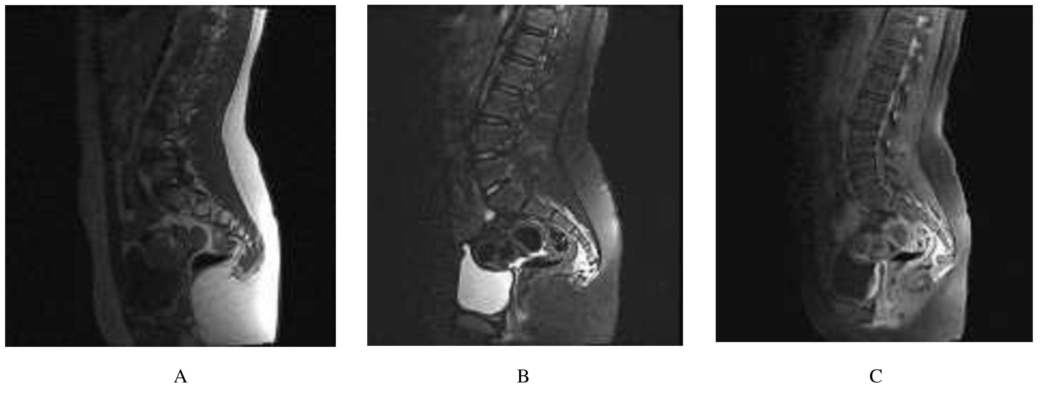
  • Received:2021-06-11 Online:2022-01-28 Published:2022-01-17
  • Contact: Hongguang ZHAO E-mail:zhaohg@jlu.edu.cn

摘要: 目的

分析 1 例尾骨原发小细胞型骨肉瘤并发多处转移患儿的临床症状、实验室检查和氟代脱氧葡萄糖(18F-FDG)正电子发射断层显像/X线计算机断层显像(PET/CT)的特点,以提高影像科医生对尾骨原发病变的诊断水平。

方法

收集 1 例尾骨原发小细胞型骨肉瘤并发多处转移患儿的临床资料,并通过相关文献复习,探讨其临床特点、影像学表现、诊断和治疗方法。

结果

患儿,女性,10 岁,因颈部疼痛 2 周、发热 1 周行肺 CT 检查发现右肺下叶占位性病变入院。 查体,体温最高达37.5 ℃,无其他不适。实验室检查, 碱性磷酸酶 221.3 U·L-1,肌酐 38.1 μmol·L-118F-FDG PET/CT 检查显示右肺下叶高代谢团块,考虑为恶性肿物;颈2椎体左侧代谢增高,不除外转移瘤;尾骨密度不均,局部软组织密度结节突向盆腔伴代谢增高,考虑恶性。核磁共振成像(MRI)检查显示,枢椎形态不规整,不规则长T1长T2信号,增强病灶明显强化;骶 5 水平椎管内、骶 5 椎体与尾椎间及周围囊片状稍长 T1和稍长T2信号,增强未见明显强化, 骶椎前方软组织条片样强化,考虑占位性病变。右肺下叶占位术后病理,考虑为肺的转移性小细胞型骨肉瘤。综合上述表现及病理学分析决定对尾骨软组织病灶穿刺活检,病理提示为小细胞型骨肉瘤。本例患儿最终诊断为尾骨原发小细胞型骨肉瘤,肺转移,骨转移。

结论

尾骨原发小细胞型骨肉瘤诊断难度大,18F-FDG PET/CT 分子影像的应用对明确病变受累部位、肿瘤代谢程度和定位活检组织部位起关键性作用。

关键词: 小细胞型, 骨肉瘤, 转移瘤, 尾骨, 核磁共振成像, 正电子发射断层显像/X线计算机断层显像

Abstract: Objective

To analyze the clinical symptoms, laboratory examination and 18F-FDG PET/CT imaging of a patient with primary small cell osteosarcoma of coccyx with concurrent metastases, and to improve the physician’s diagnosis of rare manifestation for this common disease.

Methods

The patient’s clinical data of the patient with primary small cell osteosarcoma of coccyx with concurrent metastases were collected, and its clinical characteristics, imaging performance, diagnosis and treatment methods were analyzed through the review of relevant literatures.

Results

A 10-year old girl admitted to hospital due to neck pain for two weeks and fever for one week, and a mass in lower right lung lobe found in lung CT examination. Body examination showed body temperature up to 37.5 ℃, no other discomfort. The laboratory results indicated alkaline phosphatase 221.3 U·L-1, creatinine 38.1 μmol·L-1.The 18F-FDG PET/CT results showed high metabolic mass in the lower lobe of the right lung, considering malignancy; the metabolism of the left side of the cervical 2 vertebrae was increased, maybe metastatic tumors; the tail bone density was not equal, a high metabolism soft tissue nodule protruding into the pelvis, considering malignant neoplasm.The MRI results showed that the axoid morphology was irregular, with long T1 and T2 signals, and the lesion was markedly enhanced; slightly longer T1 and slightly longer T2 signals lesions were found in sacral 5 level spinal canal, sacral 5 vertebral body and caudal vertebrae and around the capsule, and no obvious enhancement; anterior soft tissue around the sacral vertebrae anterior soft tissue was enhanced, space occupying lesion was considered. The postoperative pathology of the lower lobe of the right lung was considered metastatic small cell-type osteosarcoma for the lungs.Based on the above manifestations and pathological analysis, puncture biopsy of soft tissue lesions of the coccyx was performed, and the pathological result indicated small cell osteosarcoma. Finally the patient was diagnosed as primary small cell osteosarcoma of coccyx combined with lung and bone metastasis.

Conclusion

Primary tail bone small cell osteosarcoma is difficult to be diagnosed, but 18F-FDG PET/CT molecular imaging plays a key role in clarifying the lesion scope,tumor metabolic degree, and guiding the biopsy site.

Key words: Small cell, osteosarcoma, metastatic tumor, coccyx, magnetic resonance imaging, positron emission tomography/computed tomography

中图分类号: 

  • R734.2