吉林大学学报(医学版) ›› 2022, Vol. 48 ›› Issue (6): 1462-1473.doi: 10.13481/j.1671-587X.20220612

• 基础研究 • 上一篇    下一篇

lncRNA-MIAT对肿瘤相关巨噬细胞M2型极化的作用及其机制

许静1(),郭健1,蒲兴魏2,李大星1   

  1. 1.贵州省骨科医院骨内科,贵州 贵阳 550000
    2.贵州省骨科医院脊柱科,贵州 贵阳 550000
  • 收稿日期:2022-01-31 出版日期:2022-11-28 发布日期:2022-12-07
  • 通讯作者: 许静 E-mail:xuj1101@163.com
  • 作者简介:许 静(1981-),女,贵州省毕节市人,副主任医师,医学硕士,主要从事腰椎管狭窄症的保守治疗、骨质疏松和骨肉瘤治疗方面的研究。(E-mail:xuj1101@163.com
  • 基金资助:
    贵州省卫健委科学技术基金项目(gzwkj2021-438)

Effect of lncRNA-MIAT on M2-type polarization of tumor-associated macrophages and its mechanism

Jing XU1(),Jian GUO1,Xingwei PU2,Daxing LI1   

  1. 1.Department of Orthopedics, Guizhou Provincial Orthopedic Hospital, Guiyang 550000, China
    2.Department of Spine, Guizhou Orthopedic Hospital, Guiyang 550000, China
  • Received:2022-01-31 Online:2022-11-28 Published:2022-12-07
  • Contact: Jing XU E-mail:xuj1101@163.com

摘要:

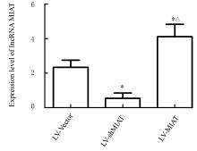
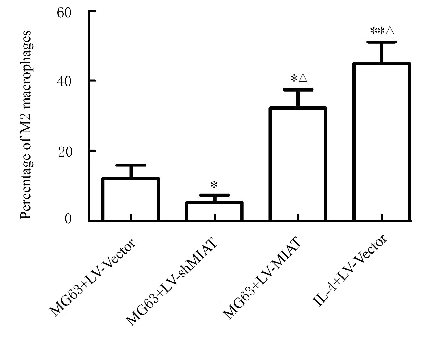
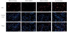
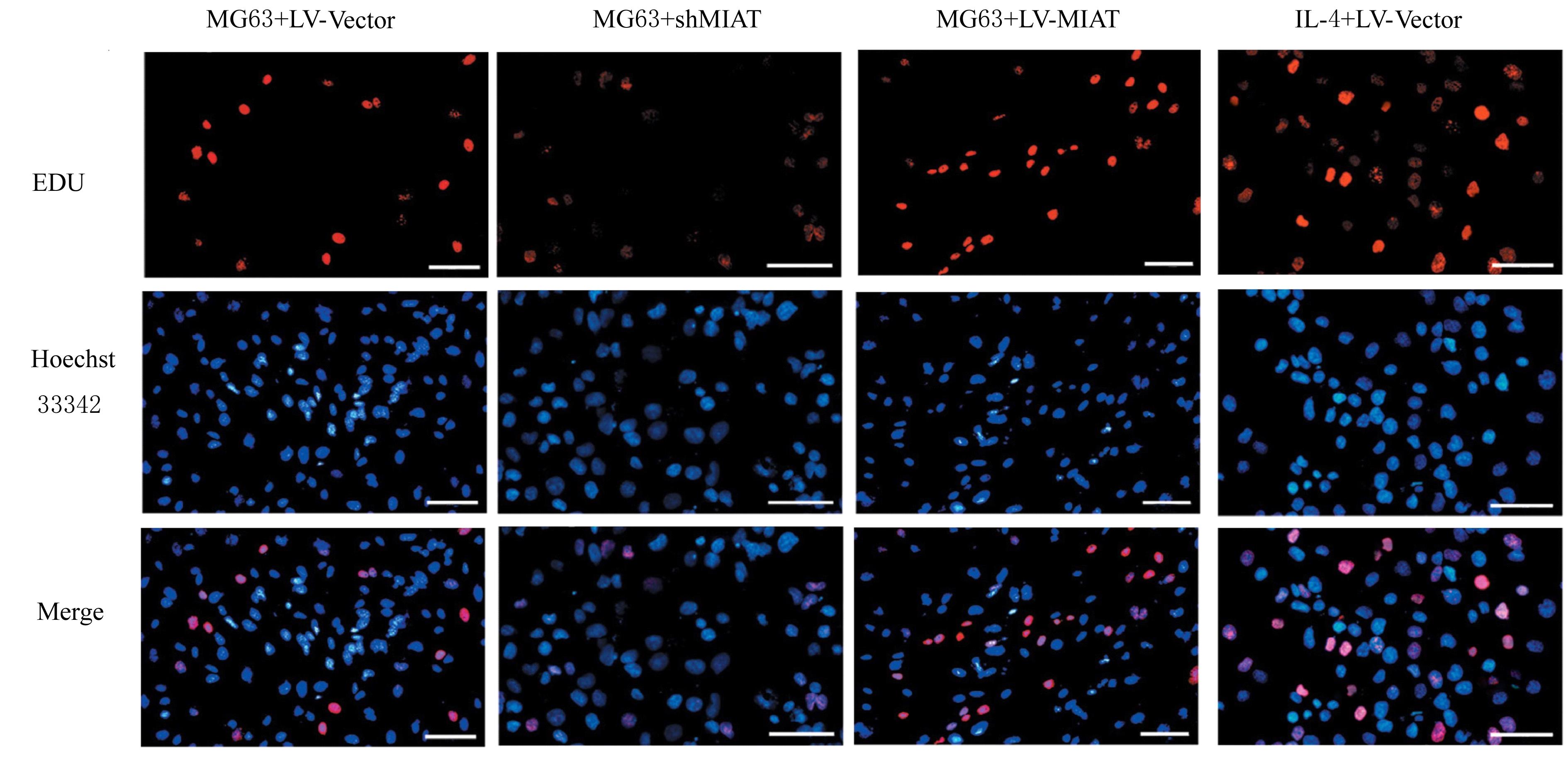
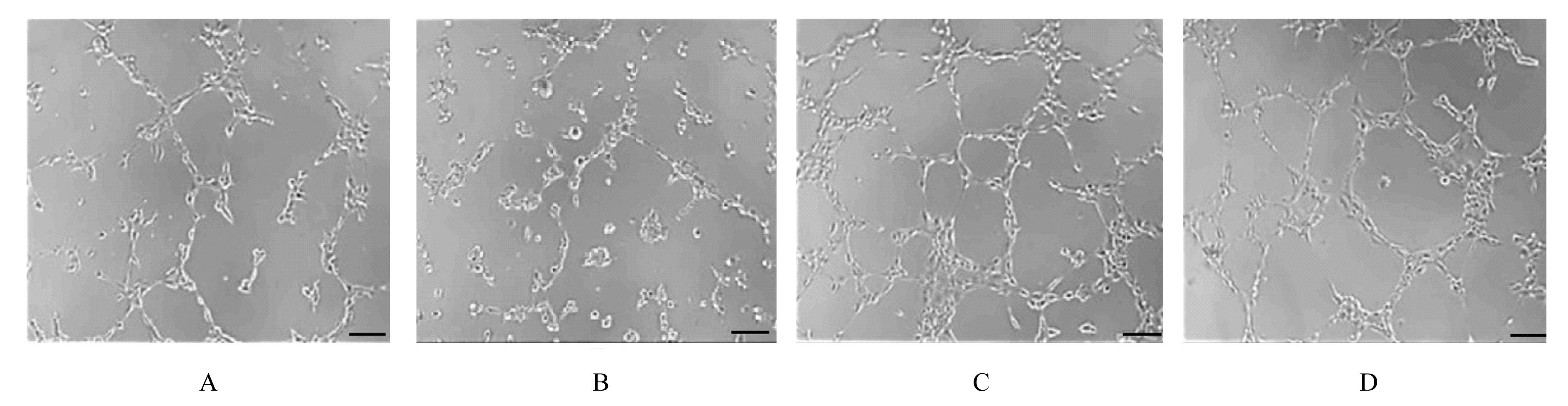
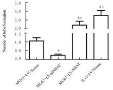
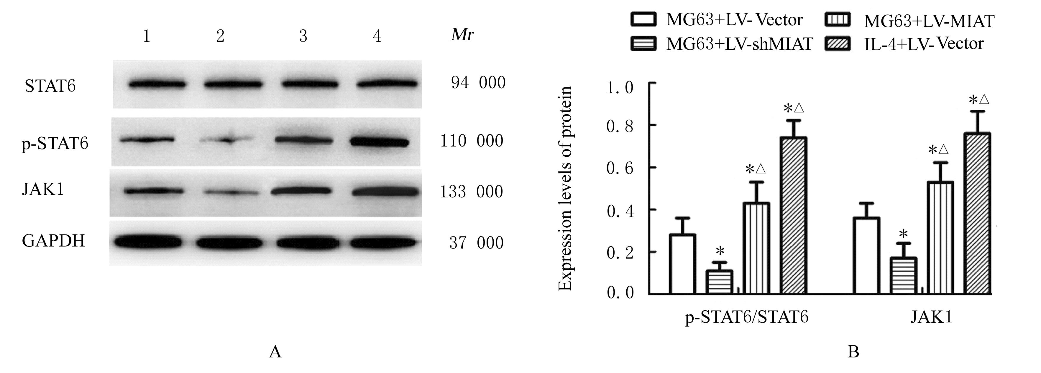
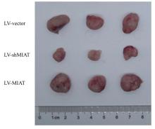
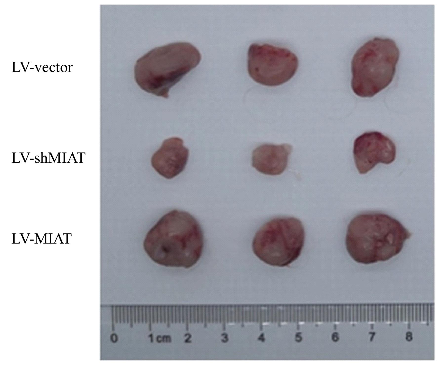
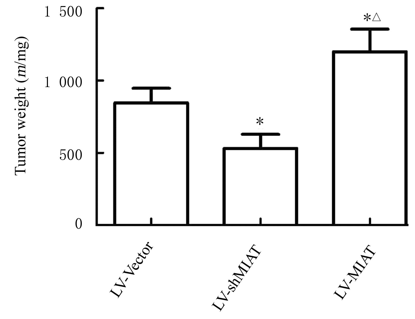
目的 探讨lncRNA-MIAT对肿瘤相关巨噬细胞M2型极化的作用,并阐明其可能的作用机制。 方法 体外培养THP-1细胞,将过表达(LV-MIAT)与下调lncRNA-MIAT表达(LV-shMIAT)的慢病毒颗粒及空载体(LV-Vector)转染入THP-1细胞中,将THP-1细胞诱导活化为巨噬细胞,并采用Transwell共培养体系将活化后的巨噬细胞与骨肉瘤(OS)MG63细胞共培养,将上述共培养体系分为MG63+LV-Vector组(阳性对照组)、MG63+LV-shMIAT组、MG63+LV-MIAT组和IL-4+LV-Vector组。检测各组THP-1细胞中lncRNA-MIAT表达水平,流式细胞术检测各组细胞中M2型巨噬细胞百分率,ELISA法检测各组THP-1细胞上清液中血管内皮生长因子(VEGF)、白细胞介素4(IL-10)和转化生长因子β1(TGF-β1)水平,检测各组HUVEC中血管内皮细胞生长因子受体2(VEGFR2)、Notch1和δ样蛋白4(DLL4)表达水平。取各培养体系中的巨噬细胞与人脐静脉内皮血管细胞(HUVEC)共培养,EDU染色法检测各组HUVEC增殖活力,成管实验检测HUVEC血管形成数。Western blotting法检测各组THP1中Janus激酶1(JAK1)、信号传导及转录激活蛋白6(STAT6)和磷酸化STAT6(p-STAT6)蛋白表达水平,并计算p-STAT6/STAT6比值。构建OS荷瘤小鼠模型,并将36只荷瘤小鼠分为LV-Vector组、LV-shMIAT组和LV-MIAT组(n=12)。检测干扰lncRNA-MIAT表达后各组小鼠肿瘤体积和质量及肿瘤组织中CD163和CD31阳性表达率。 结果 采用慢病毒感染成功建立稳定过表达或下调lncRNA-MIAT的THP-1细胞。与LV-Vector组比较,LV-shMIAT组细胞中lncRNA-MIAT表达水平明显降低(P<0.05),LV-MIAT组细胞中lncRNA-MIAT表达水平明显升高(P<0.05)。与MG63+LV-Vector组比较,MG63+LV-shMIAT组THP-1细胞中VEGF、IL-10和TGF-β1水平明显降低(P<0.05),HUVEC中VEGFR2、Notch1和DLL4蛋白表达水平明显降低(P<0.05),MG63+LV-MIAT组和IL-4+LV-Vector组THP-1细胞中VEGF、IL-10和TGF-β1水平明显升高(P<0.05或P<0.01), HUVEC中VEGFR2、Notch1和DLL4蛋白表达水平明显升高(P<0.05),M2型巨噬细胞百分率、THP-1细胞中p-STAT6/STAT6比值和JAK1蛋白表达水平升高(P<0.05);与MG63+sh-MIAT组比较,MG63+LV-MIAT组和IL-4+LV-Vector组THP-1细胞中VEGF水平明显降低(P<0.05),IL-10和TGF-β1水平明显升高(P<0.05),HUVEC中VEGFR2、Notch1和DLL4蛋白表达水平均明显升高(P<0.05);与MG63+LV-Vector组比较,MG63+LV-shMIAT组HUVEC的增殖活力与血管形成数均明显降低(P<0.05),MG63+LV-MIAT组和IL-4+LV-Vector组HUVEC的增殖活力与血管形成数均明显升高(P<0.05)。裸鼠体内成瘤实验,与LV-Vector组比较,LV-MIAT组小鼠移植瘤体积和质量明显升高(P<0.05),肿瘤组织中CD163和CD31阳性表达率明显升高(P<0.05);LV-shMIAT组小鼠移植瘤体积和质量、肿瘤组织中CD163和CD31阳性表达率均明显降低(P<0.05)。与LV-shMIAT组比较,LV-MIAT组小鼠移植瘤体积和质量明显升高(P<0.05),肿瘤组织中CD163与CD31阳性表达率明显升高(P<0.05)。 结论 lncRNA MIAT可能通过促进巨噬细胞的M2极化和上调肿瘤组织血管生成,参与调控OS进展。

关键词: 骨肉瘤, lncRNA-MIAT, M2型巨噬细胞, 肿瘤血管, 人脐静脉内皮血管细胞

Abstract:

Objective To investigate the effect of lncRNA-MIAT on M2-type polarization of tumor-associated macrophages, and to elucidate its possible mechanism. Methods The THP-1 cells were cultured in vitro. The lentivirus particles, which over expressed (LV-MIAT) and down regulated expression of lncRNA-MIAT(LV-shMIAT),and empty vector(LV-Vector) were transfected into the THP-1 cells. The THP-1 cells were activated into the macrophages, and the activated macrophages were co-cultured with the osteosarcoma (OS) MG63 cells by using Transwell co-culture system.The above co-culture systems were divided into MG63+LV-Vector group (positive control group),MG63+LV-shMIAT group, MG63+LV-MIAT group,and IL-4+LV-Vector group. The expression levels of lncRNA-MIAT in the THP-1 cells in various groups were detected;the percentages of M2-type macrophages in various groups were detected by flow cytometry; the levels of vascular endothelial growth factor (VEGF), interleukin-10(IL-10) ,and transforming growth factor-β1(TGF-β1) in supernatant of the THP-1 cells in various groups were detected by ELISA method;the macrophages in each culture system were co-cultured with the human umbilical vein endothelial cell (HUVEC); EDU staining was used to detect the proliferation activities of the HUVEC in various groups;the numbers of angiogenesis in the HUVEC in various groups were detected by tube formation assay;the expression levels of Janus kinase 1 (JAK1),signal transducer and activator of transcription 6 (STAT6),and phosphorylated STAT6 proteins in the THP1 cells and the expression levels of vascular endothelial growth factor receptor 2(VEGFR2),Notch1,and delta like protein 4(DLL4)in the HUVEC in various groups were detected by Western blotting method;the p-STAT6/STAT6 ratio was calculated.The OS tumor-bearing mouse model was constructed, and 36 nude mice were divided into LV-Vector group, LV-shMIAT group,and LV-MIAT group(n=12).The volumes and weights of the tumor and the positive expression rates of CD163 and CD31 in tumor tissue of the mice in various groups were detected after interfering lncRNA-MIAT expression. Results The THP-1 cells with stable over-expression or down-regulation of lncRNA-MIAT were successfully established by lentivirus infection. Compared with LV-Vector group, the expression level of lncRNA MIAT in the cells in LV-shMIAT group was significantly decreased (P<0.05), and the expression level of lncRNA MIAT in the cells in LV-MIAT group was significantly increased (P<0.05). Compared with MG63+LV-Vector group, the levels of VEGF, IL-10,and TGF- β1 in the THP-1 cells in MG63+LV-shMIAT group were significantly decreased (P<0.05),and the expression levels of VEGFR2, Notch1, and DLL4 proteins in the HUVEC were significantly decreased (P<0.05),the levels of VEGF,IL-10,and TNF-β1 in the THP-1 cells in MG63+LV-MIAT and IL-4+LV-Vector groups were increased significantly (P<0.05 or P<0.01);the expression levels of VEGFR2, Notch1,and DLL4 proteins in HUVEC were increased(P<0.05), the percentage of M2-type macrophages,and p-STAT6/STAT6 ratio and expression level of JAK1 protein in the THP-1 cells were increased(P<0.05);compared with MG63+sh-MIAT group, the VEGF level in the THP-1 cells in MG63+LV MIAT group was decreased significantly (P<0.05), the levels of IL-10 and TGF-β were decreased significantly (P<0.05),the expression levels of VEGFR2, Notch1,and DLL4 proteins in the HUVEC were increased significantly (P<0.05);compared with MG63+LV-Vector group, the proliferation activity and number of angiogenesis in the HUVEC in MG63+LV shMIAT group were significantly decreased(P<0.05),while the proliferation activities and number of angiogenesis in the HUVEC in MG63+LV-MIAT group and IL-4+LV Vector group were significantly increased (P<0.05).The results of in vivo tumorigenesis experiment of the nude mice showed that compared with LV-Vector group, the volume and weight of transplanted tumor of the mice in LV-MIAT group were significantly increased (P<0.05),and the positive expression rates of CD163 and CD31 in tumor tissue were significantly increased (P<0.05); the volume and mass of transplanted tumor and the positive expression rates of CD163 and CD31 in tumor tissue of the mice in LV-shMIAT group were significantly decreased (P<0.05). Compared with LV-shMIAT group, the volume and weight of transplanted tumor of the mice in LV-MIAT group were significantly increased (P<0.05), and the positive expression rates of CD163 and CD31 in tumor tissue were significantly increased (P<0.05). Conclusion LncRNA MIAT may regulate the progression of OS by promoting the M2 polarization of macrophages and up-regulating the angiogenesis in tumor tissue.

Key words: Oteosarcoma, LncRNA-MIAT, M2-type macrophage, Tumor vessel, Human umbilical vein endothelial cell

中图分类号: 

  • R33