吉林大学学报(医学版) ›› 2023, Vol. 49 ›› Issue (3): 697-705.doi: 10.13481/j.1671-587X.20230319

• 基础研究 • 上一篇    下一篇

lncRNA MALAT1对肝星状细胞活化的调节作用及其机制

徐菱遥,魏书堂,董勇,孙正路,赵俊波,韩大正()   

  1. 河南大学第一附属医院消化病科,河南 开封 475100
  • 收稿日期:2022-06-23 出版日期:2023-05-28 发布日期:2023-06-20
  • 通讯作者: 韩大正 E-mail:13569519096@163.com
  • 作者简介:徐菱遥(1991-),女,河南省驻马店市人,医学硕士,主治医师,主要从事消化系统疾病的基础和临床方面的研究。
  • 基金资助:
    河南省卫健委医学科技攻关计划项目(LHGJ20190532)

Regulatory effect of lncRNA MALAT1 on activation of hepatic stellate cell and its mechanism

Lingyao XU,Shutang WEI,Yong DONG,Zhenglu SUN,Junbo ZHAO,Dazheng HAN()   

  1. Department of Gastroenterology,First Affiliated Hospital,Henan University,Kaifeng 475100,China
  • Received:2022-06-23 Online:2023-05-28 Published:2023-06-20
  • Contact: Dazheng HAN E-mail:13569519096@163.com

摘要:

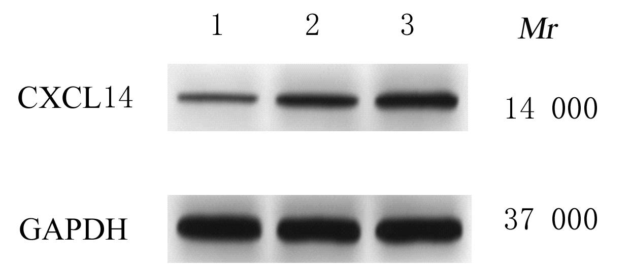
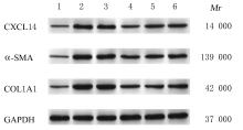
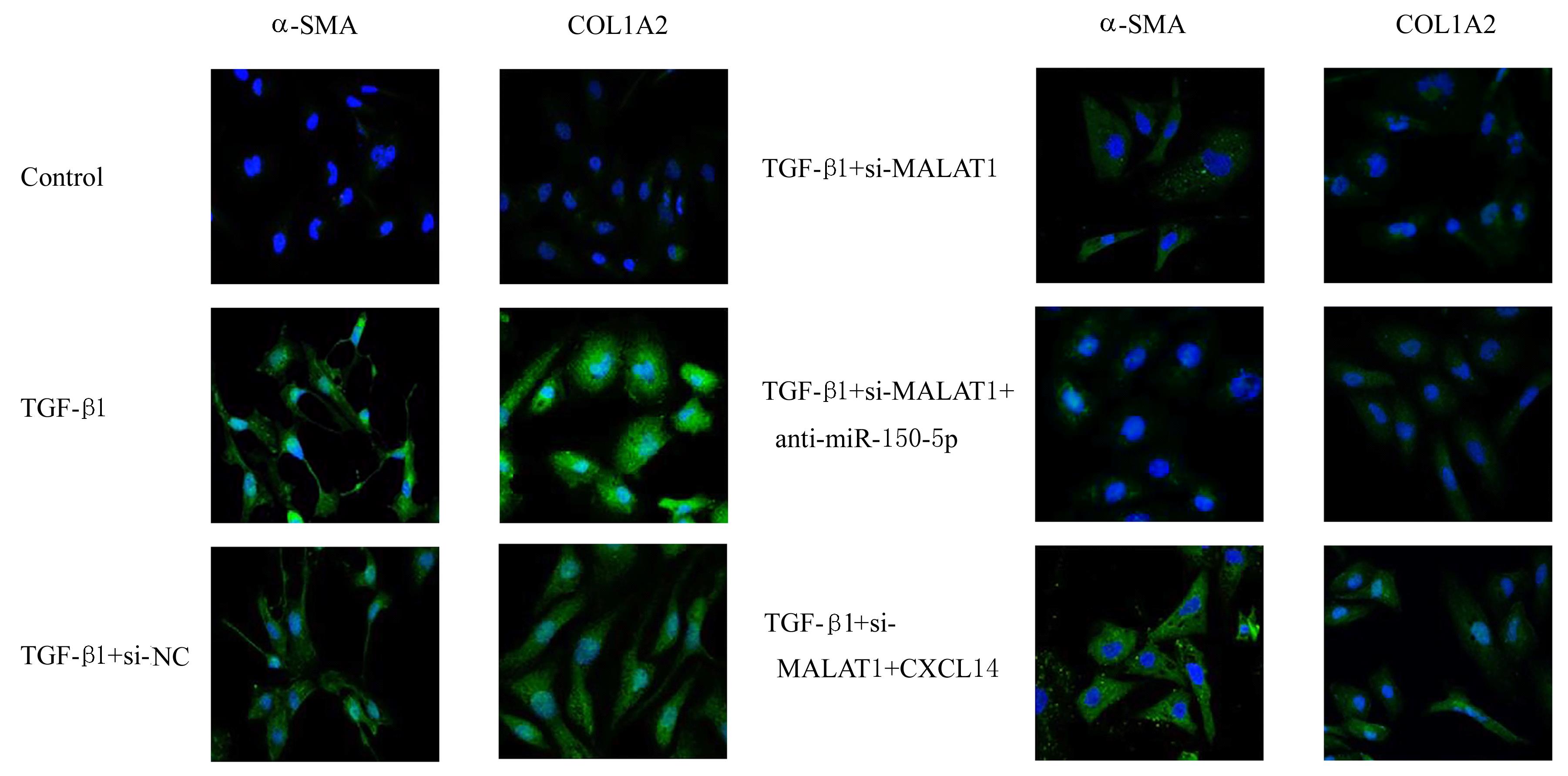
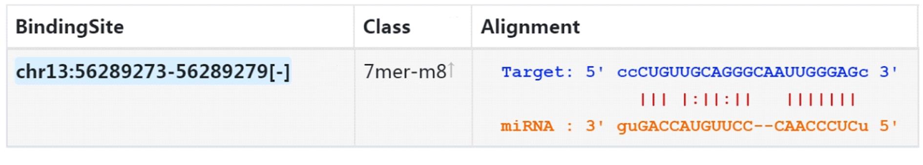
目的 探讨长链非编码RNA(lncRNA)肺腺癌转移相关转录本1(MALAT1)在肝纤维化进展过程中对肝星状细胞(HSC)活化的调节作用,并阐明其作用机制。 方法 收集25例健康志愿者(健康组,n=25)和25例肝纤维化患者[轻度肝纤维化组(n=12)和重度肝纤维化组(n=13)]血清样本。小鼠HSC分为对照组、转化生长因子β1(TGF-β1)组、TGF-β1+si-NC组、TGF-β1+si-MALAT1组、TGF-β1+si-MALAT1+anti-miR-150-5p组和TGF-β1+si-MALAT1+CXCL14组。采用实时荧光定量PCR(RT-qPCR)法检测各组研究对象血清和各组HSC中MALAT1 mRNA、miR-150-5p和CXC趋化因子配体14(CXCL14)mRNA表达水平,Western blotting法检测各组研究对象血清中CXCL14蛋白表达水平和各组HSC中CXCL14、α-平滑肌肌动蛋白(α-SMA)和Ⅰ型胶原蛋白α1(COL1A1)蛋白表达水平,CCK-8法检测各组HSC增殖活性,免疫荧光法检测各组HSC中α-SMA和COL1A1蛋白表达量,双荧光素酶报告系统检测miR-150-5p与MALAT1和CXCL14 3′-UTR基因的靶向关系。 结果 RT-qPCR法检测,与健康组比较,轻度和重度肝纤维化组患者血清中MALAT1 mRNA和CXCL14 mRNA表达水平升高(P<0.05),miR-150-5p表达水平降低(P<0.05);与轻度肝纤维化组比较,重度肝纤维化组患者血清中MALAT1 mRNA和CXCL14 mRNA表达水平升高(P<0.05),miR-150-5p表达水平降低(P<0.05);与对照组比较,TGF-β1组HSC中MALAT1 mRNA和CXCL14 mRNA表达水平均升高(P<0.05),miR-150-5p表达水平降低(P<0.05);与TGF-β1+si-NC组比较,TGF-β1+si-MALAT1组HSC中MALAT1 mRNA和CXCL14 mRNA表达水平均降低(P<0.05),miR-150-5p表达水平升高(P<0.05);与TGF-β1+si-MALAT1组比较,TGF-β1+si-MALAT1+anti-miR-150-5p组HSC中miR-150-5p表达水平降低(P<0.05),CXCL14 mRNA表达水平升高(P<0.05);与TGF-β1+si-MALAT1组比较,TGF-β1+si-MALAT1+CXCL14组HSC中CXCL14 mRNA表达水平升高(P<0.05)。Western blotting法检测,与健康组比较,轻度和重度肝纤维化组患者血清中CXCL14蛋白表达水平升高(P<0.05);与轻度肝纤维化组比较,重度肝纤维化组患者血清中CXCL14蛋白表达水平升高(P<0.05);与对照组比较,TGF-β1组HSC中CXCL14、α-SMA和COL1A1蛋白表达水平升高(P<0.05);与TGF-β1+si-NC组比较,TGF-β1+si-MALAT1组HSC中CXCL14、α-SMA和COL1A1蛋白表达水平降低(P<0.05);与TGF-β1+si-MALAT1组比较,TGF-β1+si-MALAT1+anti-miR-150-5p组和TGF-β1+si-MALAT1+CXCL14组HSC中CXCL14、α-SMA和COL1A1蛋白表达水平升高(P<0.05)。CCK-8法检测,与对照组比较,TGF-β1组HSC增殖活性升高(P<0.05);与TGF-β1+si-NC组比较,TGF-β1+si-MALAT1组HSC增殖活性降低(P<0.05);与TGF-β1+si-MALAT1组比较,TGF-β1+si-MALAT1+anti-miR-150-5p组和TGF-β1+si-MALAT1+CXCL14组HSC增殖活性升高(P<0.05)。免疫荧光检测,各组HSC中α-SMA和COL1A1蛋白表达与Western blotting法检测结果一致。MALAT1和CXCL14 3'-UTR与miR-150-5p存在靶向关系。双荧光素酶报告基因测定,与miR-NC组比较,与MALAT1 WT或CXCL14 WT共转染的miR-150-5p组HSC中荧光素酶活性降低(P<0.05)。 结论 敲低MALAT1可抑制TGF-β1诱导HSC活化,其机制可能与miR-150-5p/CXCL14信号通路有关。

关键词: 肝纤维化, 肝星状细胞, 长链非编码RNA肺腺癌转移相关转录本1, 微小RNA-150-5p, CXC趋化因子配体14

Abstract:

Objective To discuss the regulatory effect of long chain non-coding RNA (lncRNA) metastasis associated transcript 1 (MALAT1) on the activation of the hepatic stellate cells (HSC) during the progression of liver fibrosis, and to clarify its mechanism. Methods The serum samples were collected from 25 healthy volunteers (n=25,healthy group) and 25 liver fibrosis patients[ mild liver fibrosis group (n=12)and severe liver fibrosis group (n=13)]. The mouse HSC were divided into control group,transforming growth factor-β1(TGF-β1) group, TGF-β1+si-NC group, TGF-β1+si-MALAT1 group,TGF- β 1+si-MALAT1+anti-miR-150-5p group,and TGF-β1 group+si-MALAT1+CXCL14 group. Real-time fluorescence quantitative PCR (RT-qPCR) method was used to detect the expression levels of MALAT1 mRNA, microRNA-150-5p (miR-150-5p), and CXC chemokine ligand 14 (CXCL14) mRNA in serum and HSC of the subjects in various groups;Western blotting method was used to detect the expression levels of CXCL14, α-smooth muscle actin(α-SMA),and type Ⅰ collagen α 1 (COL1A1) proteins in serum of the subjects in various groups;CCK-8 method was used to detect the proliferation activities of the HSC in various groups; the expression levels of α- SMA,COL1A1 proteins in the HSC in various groups were detected by immunofluorescence;the targeting relationship between miR-150-5p and MALAT1 and CXCL14 3′-UTR genes was detected by dual luciferase reporting system. Results .The RT-qPCR results showed that compared with healthy group, the serum expression levels of MALAT1 mRNA and CXCL14 mRNA of the subjects in mild and severe liver fibrosis groups were increased(P<0.05), while the expression levels of miR-150-5p were decreased (P<0.05); compared with mild liver fibrosis group, the serum expression levels of MALAT1 mRNA and CXCL14 mRNA of the subjects in severe liver fibrosis group were increased (P<0.05),while the expression level of miR-150-5p was decreased (P<0.05); compared with control group, the expression levels of MALAT1 mRNA and CXCL14 mRNA in the HSC in TGF- β1 group were increased (P<0.05), while the expression level of miR-150-5p was decreased (P<0.05); compared with TGF-β1+si-NC group, the expression levels of MALAT1 mRNA and CXCL14 mRNA in the HSC in TGF-β1+si MALAT1 group were decreased (P<0.05), while the expression level of miR-150-5p was increased (P<0.05); compared with TGF-β1+si MALAT1 group, the expression levels of miR-150-5p in the HSC in TGF- β1+si-MALAT1 and anti-miR-150-5p groups were decreased (P<0.05), while the expression levels of CXCL14 mRNA were increased (P<0.05); compared with TGF-β1+si-MALAT1 group, the expression level of CXCL14 mRNA in the HSC in TGF-β1+si-MALAT1+CXCL14 group was increased (P<0.05).The Western blotting results showed that compared with healthy group, the serum expression levels of CXCL14 protein of the subjects in mild and severe liver fibrosis groups were increased (P<0.05); compared with mild liver fibrosis group, the serum expression level of CXCL14 protein of the subjects in severe liver fibrosis group was increased (P<0.05); compared with control group, the expression levels of α-SMA and COL1A1 proteins in the HSC in TGF-β1 group were increased (P<0.05); compared with TGF-β1+si-NC group, the expression levels of α- SMA and COL1A1 proteins in the HSC in TGF-β1+si-MALAT1 group were decreased (P<0.05); compared with TGF-β1+si-MALAT1 group, the expression levels of CXCL14,α-SMA and COL1A1 proteins of the HSC in TGF-β1+si-MALAT1+anti-miR-150-5p group and TGF-β1+siMALAT1+CXCL14 group were increased (P<0.05).The CCK-8 method results showed that compared with control group, the proliferation activity of the HSC in TGF-β1 group was increased (P<0.05); compared with TGF-β1+si-NC group, the proliferation activity of the HSC in TGF-β1+si-MALAT1 group was decreased (P<0.05); compared with TGF-β1+si-MALAT1 group,the proliferation activities of the HSC in TGF-β1+si-MALAT1+anti-miR-150-5p group and TGF-β1+si-MALAT1+CXCL14 group were increased (P<0.05). The immunofluorescence results showed that the expressions of α-SMA and COL1A1 proteins in the HSC in various groups was consistent with the results detected by Western blotting method. There was a targeting relationship between MALAT1 and CXCL14 3 '-UTR and miR-150-5p. The double luciferase reporter gene assay results showed that compared with miR-NC group, the luciferase activity of the HSC in miR-150-5p group co-transfected with MALAT1 WT or CXCL14 WT was decreased (P<0.05). Conclusion Knocking down of MALAT1 can inhibit the activation of the HSC induced by TGF- β 1,and the mechanism may be related to the miR-150-5p/CXCL14 signaling pathway.

Key words: Liver fibrosis, Hepatic stellate cell, Long non-coding RNA metastasis-associated lung adenocarcinoma transcript 1, MicroRNA-150-5p, CXC chemokine ligand 14

中图分类号: 

  • R575