吉林大学学报(医学版) ›› 2024, Vol. 50 ›› Issue (3): 718-727.doi: 10.13481/j.1671-587X.20240316

• 基础研究 • 上一篇    

脂肪干细胞源性外泌体对体外巨噬细胞迁移能力的影响

袁博1,2,3,谢佳忆4,江思瑜1,5,孟亚军1,朱清华1,李晓飞1,付秀美1,3(),谢利德1()   

  1. 1.承德医学院基础医学院人体解剖学教研室,河北 承德 067000
    2.承德护理职业学院病原生物与免疫教研室,河北 承德 067000
    3.河北省神经损伤与修复重点实验室,河北 承德 067000
    4.清华大学信息科学技术学院自动化系,北京 100062
    5.河北医科大学第一医院中心实验室,河北 石家庄 050000
  • 收稿日期:2023-06-26 出版日期:2024-05-28 发布日期:2024-07-01
  • 通讯作者: 付秀美,谢利德 E-mail:fuxiumei2012@163.com;xielide65@163.com
  • 作者简介:袁 博(1988-),女,河北省承德市人,讲师,医学硕士,主要从事神经损伤与修复方面的研究。
  • 基金资助:
    河北省科技厅自然科学基金项目(H2021406056);河北省高等学校科学研究计划项目(ZD2020178);承德医学院人体解剖与组织胚胎学优势学科资助项目(〔2023〕22号);河北省神经损伤与修复重点实验室开放项目(NJKF-202403)

Effect of adipose-derived stem cell-derived exosomes on migration ability of macrophages in vitro

Bo YUAN1,2,3,Jiayi XIE4,Siyu JIANG1,5,Yajun MENG1,Qinghua ZHU1,Xiaofei LI1,Xiumei FU1,3(),Lide XIE1()   

  1. 1.Department of Human Anatomy,School of Basic Medical Sciences,Chengde Medical University,Chengde 067000,China
    2.Department of Pathogens and Immunology,Chengde Nursing Vocational College,Chengde 067000,China
    3.Key Laboratory of Nerve Injury and Repair of Hebei Province,Chengde 067000,China
    4.Department of Automation,School of Information Science and Technology,Tsinghua University,Beijing 100062,China
    5.Central Laboratory,First Hospital,Hebei Medical University,Shijiazhuang 050000,China
  • Received:2023-06-26 Online:2024-05-28 Published:2024-07-01
  • Contact: Xiumei FU,Lide XIE E-mail:fuxiumei2012@163.com;xielide65@163.com

摘要:

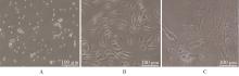
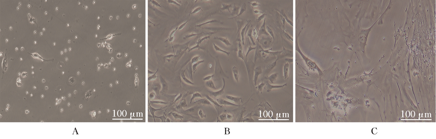
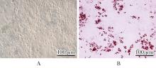
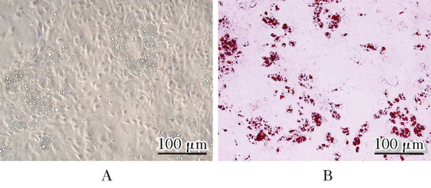
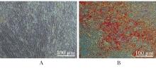
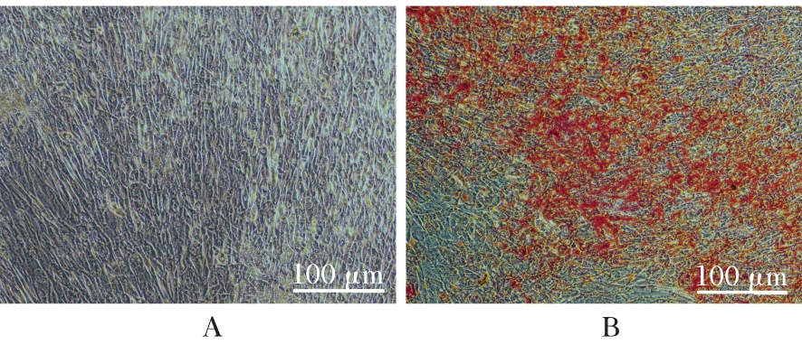
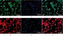
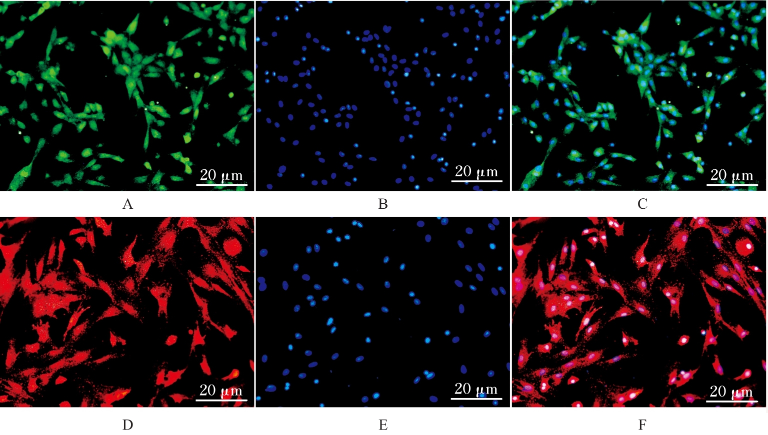
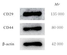
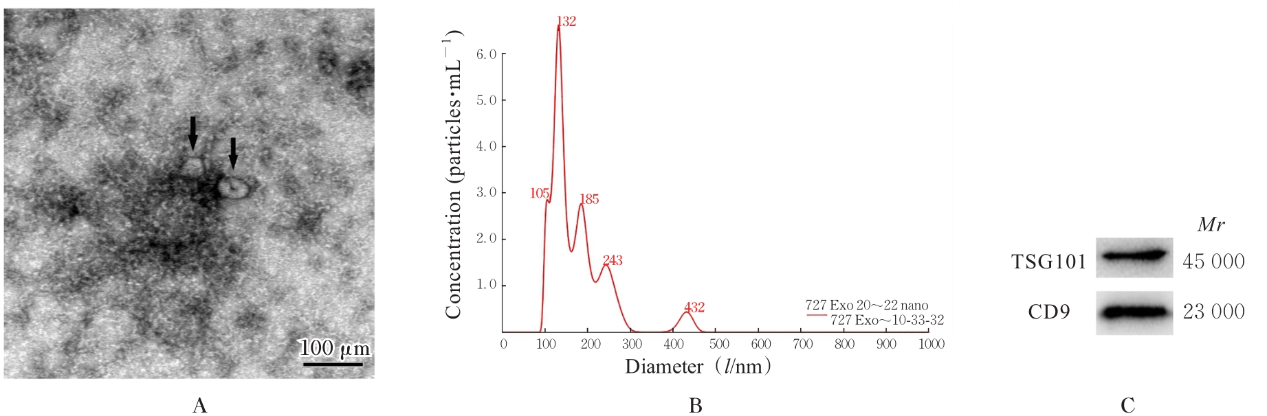
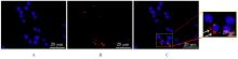
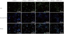
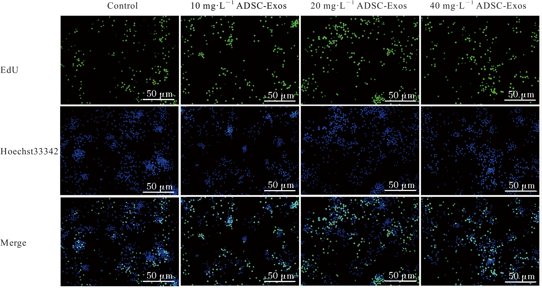
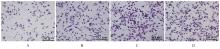
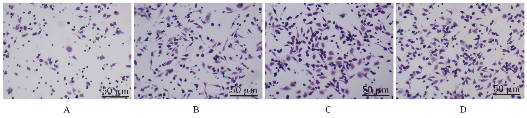
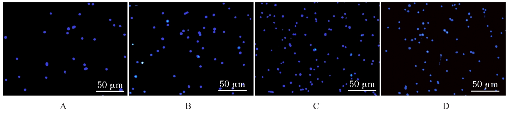
目的 探讨脂肪干细胞源性外泌体(ADSC-Exos)对巨噬细胞 RAW264.7 迁移能力的影响,阐明其促进巨噬细胞功能的作用。 方法 分离SD大鼠附睾旁脂肪组织,进行脂肪源性干细胞(ADSCs)原代培养,通过成脂和成骨分化诱导,结合油红O和茜素红染色检测ADSCs的多向分化潜能。采用Western blotting法和免疫荧光法检测ADSCs标记物CD29及CD44阳性表达情况,采用外泌体(Exos)分离试剂盒提取ADSC-Exos,透射电子显微镜和纳米颗粒跟踪分析仪检测ADSC-Exos的形态大小和粒径分布范围,Western blotting法检测Exos特异性标记物CD9和TSG101蛋白表达情况,示踪法观察巨噬细胞摄取ADSC-Exos情况。将巨噬细胞RAW264.7分为对照组、10 mg·L-1 ADSC-Exos组、20 mg·L-1 ADSC-Exos组和40 mg·L-1 ADSC-Exos组。5-乙基-2'-脱氧尿苷(EdU)染色检测各组巨噬细胞活性,Transwell小室实验检测各组巨噬细胞迁移细胞数,荧光显微镜观察各组巨噬细胞黏附情况。 结果 原代ADSCs培养24 h后细胞贴壁生长,呈散在、长梭形;培养7 d,贴壁细胞呈齿梳状、旋涡状有序排列,呈成纤维细胞样外观;培养10代后ADSCs形态不规则,增殖速度减慢。分离提取的ADSCs具有成骨和成脂分化潜能,CD29和CD44蛋白表达阳性。透射电子显微镜下ADSC-Exos呈茶碟状,ADSC-Exos的粒径主峰分布于132 nm附近,ADSC-Exos中CD9和TSG101蛋白表达阳性,即ADSC-Exos提取成功。荧光显微镜下RAW264.7细胞核周围可检测到红色荧光信号,即ADSC-Exos可以被巨噬细胞摄取。与对照组比较,10、20和40 mg·L-1 ADSC-Exos组EdU阳性细胞率均明显升高(P<0.05);与10 mg·L-1 ADSC-Exos组比较,20 mg·L-1 ADSC-Exos组EdU阳性细胞率均明显升高(P<0.05)。与对照组比较,10、20和40 mg·L-1 ADSC-Exos组巨噬细胞迁移细胞数均明显增加(P<0.05);与10 mg·L-1 ADSC-Exos组比较,20和40 mg·L-1 ADSC-Exos组巨噬细胞迁移细胞数均明显增加(P<0.05)。与对照组比较,10、20和40 mg·L-1 ADSC-Exos组巨噬细胞黏附细胞数均明显增加(P<0.05);与10 mg·L-1 ADSC-Exos组比较,20 mg·L-1 ADSC-Exos组巨噬细胞黏附细胞数明显增加(P<0.05)。 结论 ADSC-Exos可被巨噬细胞摄取,其通过影响细胞黏附提高巨噬细胞的迁移能力。

关键词: 脂肪干细胞, 外泌体, 巨噬细胞, 细胞迁移, 细胞黏附

Abstract:

Objective To discuss the effect of adipose-derived stem cell-derived exosomes (ADSC-Exos) on the migration ability of the macrophages RAW264.7, and to clarify its role in promoting function of the macrophages. Methods The adipose tissue adjacent to epididymis of the SD rats was isolated to perform primary culture of the adipose-derived stem cells (ADSCs). The adipogenic and osteogenic differentiation induction was conducted, and the multidirectional differentiation potential of the ADSCs was detected by oil Red O and Alizarin red staining. Western blotting and immunofluorescence methods were used to detect the positive expressions of the ADSCs markers CD29 and CD44; the ADSC-Exos were extracted by Exos isolation kit, and the morphology, size, and distribution of particle size of the ADSC-Exos were examined by transmission electron microscope and nanoparticle tracking analyzer; the expression levels of exosome-specific markers CD9 and TSG101 proteins in the ADSC-Exos were detected by Western blotting method; the uptake of ADSC-Exos by the macrophages was observed by tracing method. The macrophages RAW264.7 were divided into control group, 10 mg·L-1 ADSC-Exos group, 20 mg·L-1 ADSC-Exos group, and 40 mg·L-1 ADSC-Exos group. The activities of the macrophages in various groups were detected by 5-ethynyl-2'-deoxyuridine (EdU) staining; the number of migration macrophages in various groups was detected by Transwell chamber assay; the adhesion of macrophages in various groups was observed by fluorescence microscope. Results After 24 h of primary culture, the ADSCs adhered to the wall and exhibited scattered, elongated shapes; after 7 d of culture, the adherent cells showed a comb-like, vortex-like orderly arrangement, resembling fibroblasts; after 10 passages, the irregular morphology of the ADSCs and decreased proliferation rate were found. The isolated ADSCs showed potential for the osteogenic and adipogenic differentiation, and the expressions of CD29 and CD44 proteins were positive.The transmission electron microscope observation resuls showed that the ADSC-Exos appeared disc-shaped, and the main peak of particle size distribution was around 132 nm. The CD9 and TSG101 proteins were positively expressed in the ADSC-Exos, indicating successful extraction. The fluorescence microscope results showed red fluorescence signals around the nuclei of the RAW264.7 cells, indicating the uptake of ADSC-Exos by the macrophages. Compared with control group, the rates of EdU positive cells in 10,20, and 40 mg·L-1 ADSC-Exos groups were significantly increased(P<0.05); compared with 10 mg·L-1 ADSC-Exos group, the rate of EdU positive cells in 20 mg·L-1 ADSC-Exos group was significantly increased (P<0.05). Compared with control group, the numbers of migration cells in 10, 20, and 40 mg·L-1 ADSC-Exos groups were significantly increased (P<0.05); compared with 10 mg·L-1 ADSC-Exos group, the numbers of migration cells in 20 and 40 mg·L-1 ADSC-Exos groups were significantly increased (P<0.05). Compared with control group, the numbers of the adherent macrophages in 10, 20, and 40 mg·L-1 ADSC-Exos groups were significantly increased (P<0.05); compared with 10 mg·L-1 ADSC-Exos group, the number of adherent macrophages in 20 mg·L-1 ADSC-Exos group was significantly increased (P<0.05). Conclusion The ADSC-Exos can be internalized by the macrophages and they can enhance the migration ability of the macrophages by affecting the cell adhesion.

Key words: Adipogenic stem cells, Exosomes, Macrophages, Cell migration, Cell adhesion

中图分类号: 

  • Q25