吉林大学学报(医学版) ›› 2023, Vol. 49 ›› Issue (6): 1528-1538.doi: 10.13481/j.1671-587X.20230616

• 临床研究 • 上一篇    下一篇

Klotho蛋白在子痫前期患者胎盘外泌体中表达及其对血管内皮细胞氧化应激的影响

薛筱蕾,胥保梅()   

  1. 新疆医科大学第五附属医院产科,新疆 乌鲁木齐 830011
  • 收稿日期:2022-09-28 出版日期:2023-11-28 发布日期:2023-12-22
  • 通讯作者: 胥保梅 E-mail:xubaomei198@163.com
  • 作者简介:薛筱蕾(1982-),女,新疆维吾尔自治区乌鲁木齐市人,主治医师,医学学士,主要从事高危妊娠基础和临床方面的研究。
  • 基金资助:
    新疆维吾尔自治区科技厅自然科学基金项目(2020D01C224)

Expression of Klotho protein in placenta exosomes in patients with pre-eclampsia and its effect on oxidative stress in vascular endothelial cells

Xiaolei XUE,Baomei XU()   

  1. Department of Obstetrics,Fifth Affiliated Hospital,Xinjiang Medical University,Urumqi 830011,China
  • Received:2022-09-28 Online:2023-11-28 Published:2023-12-22
  • Contact: Baomei XU E-mail:xubaomei198@163.com

摘要:

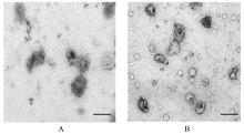
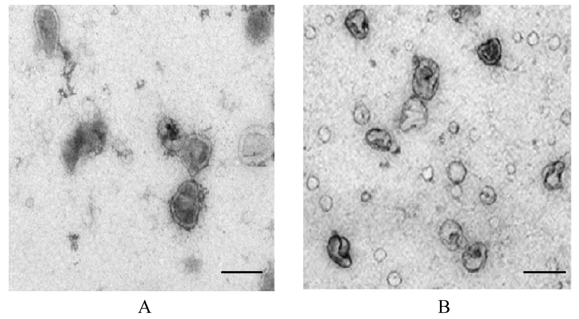
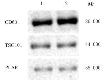
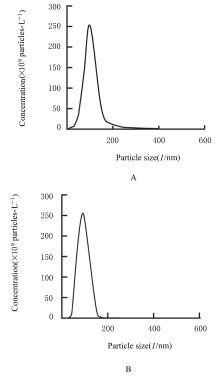
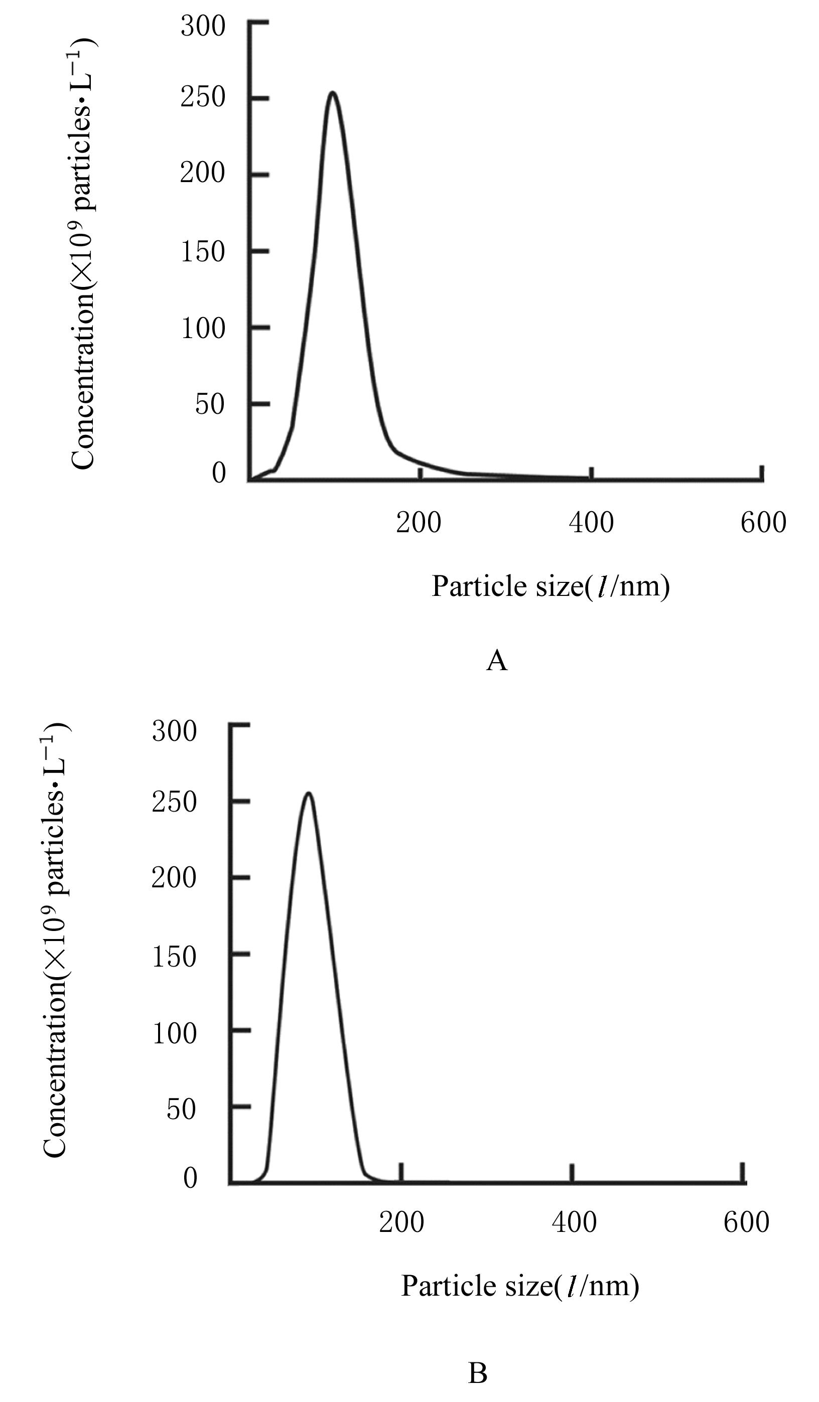
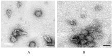
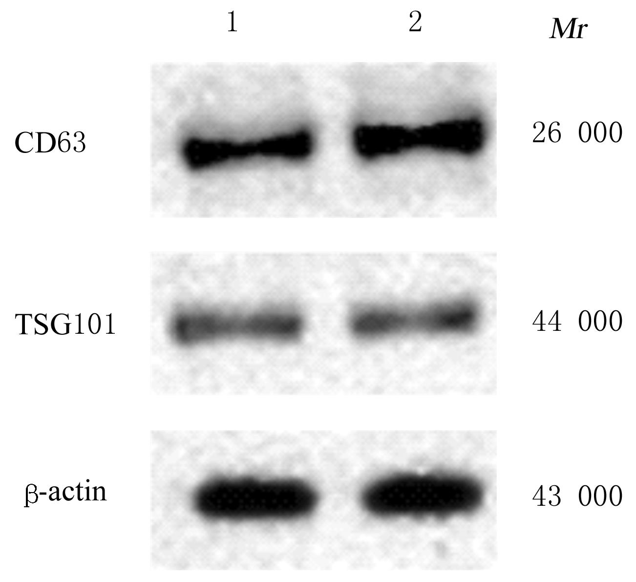
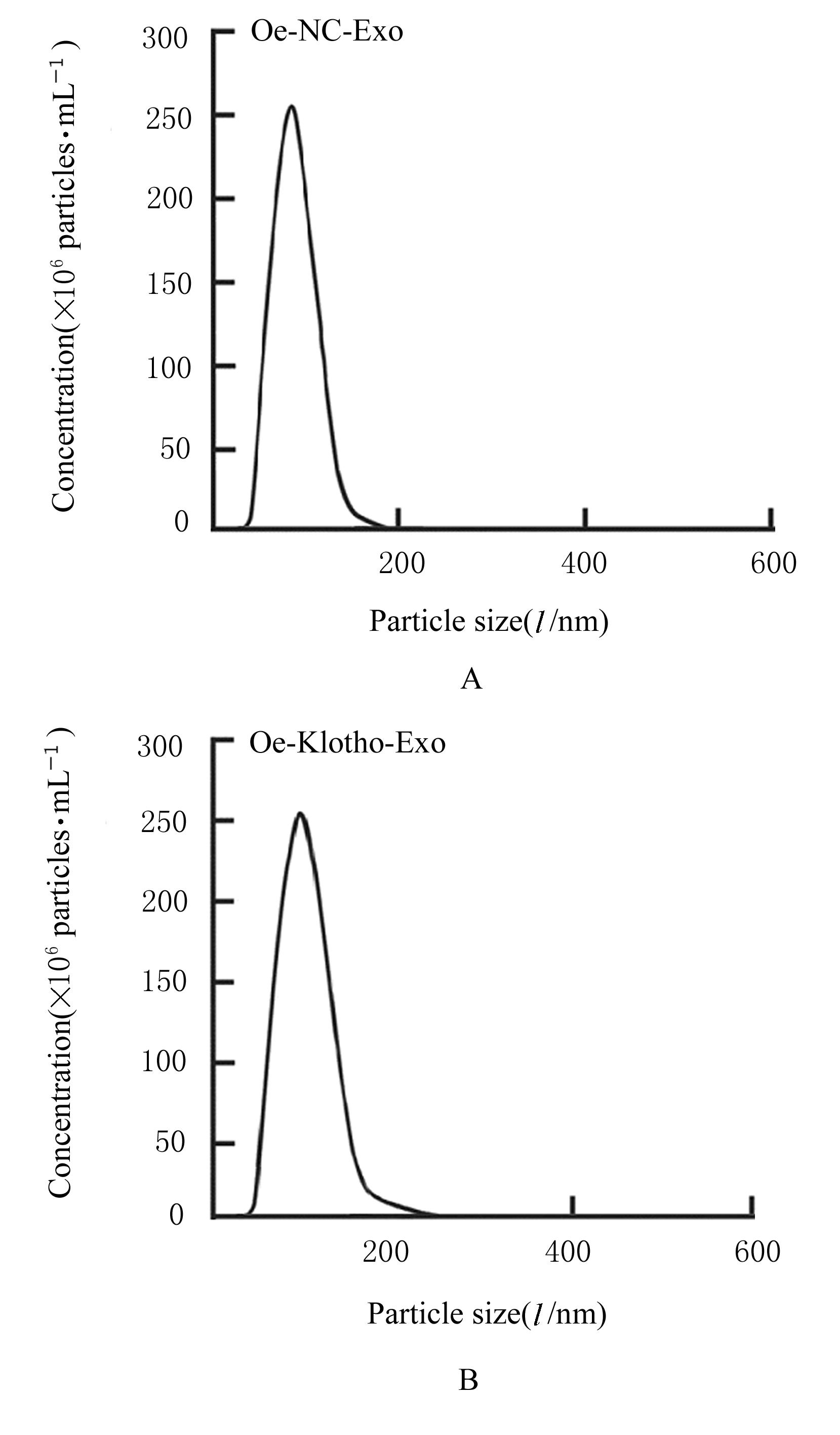
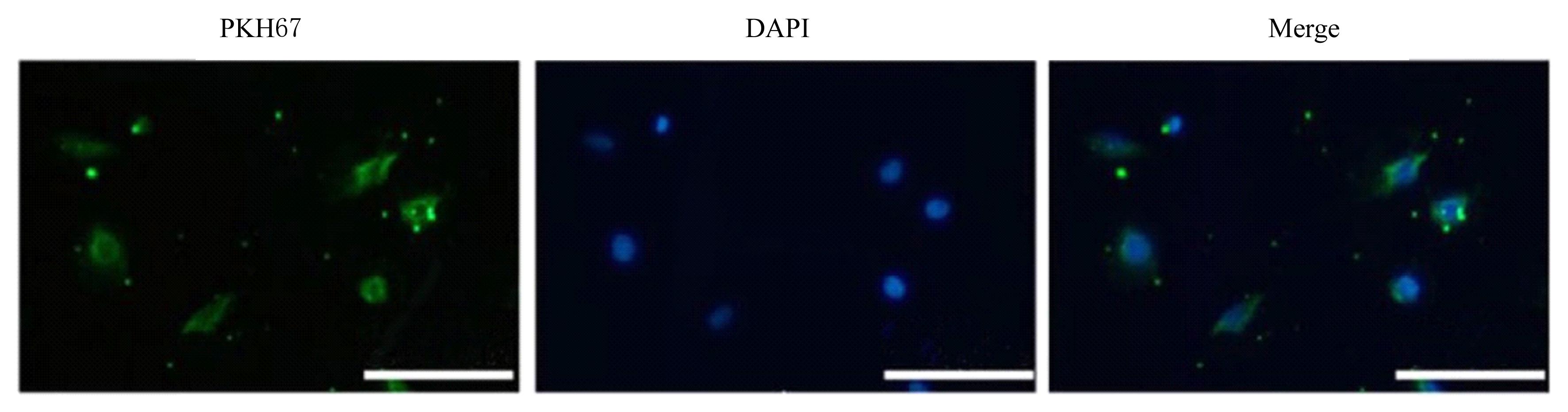
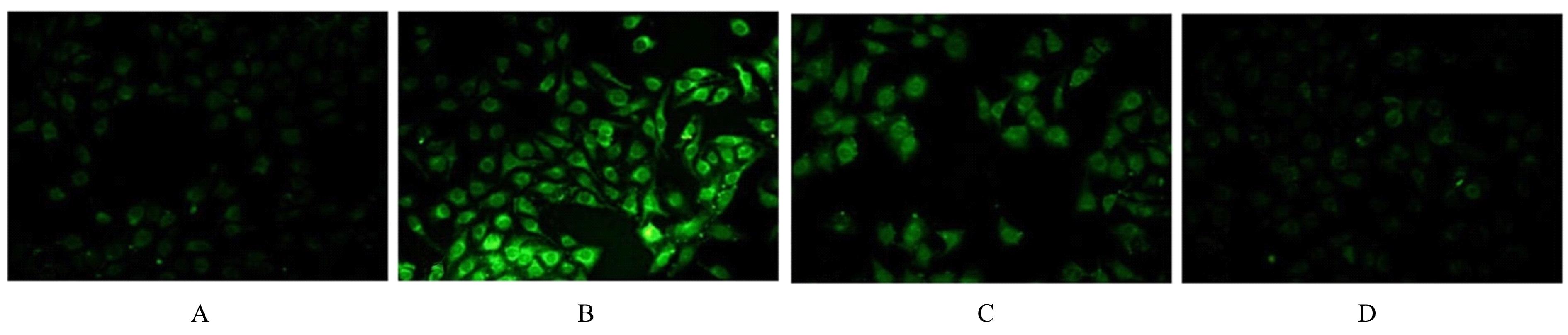
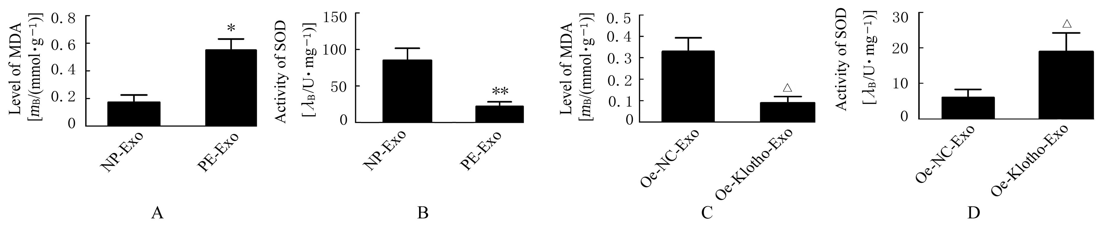
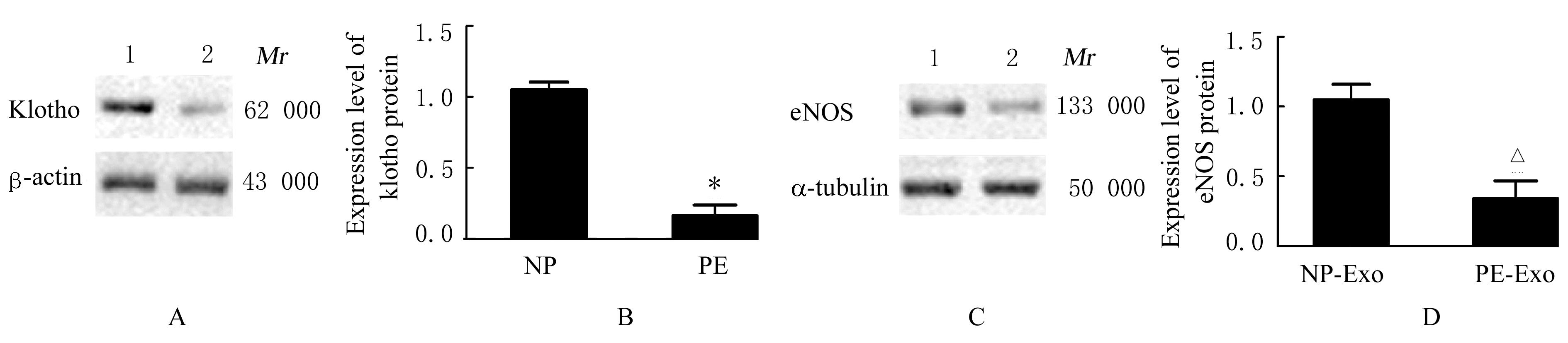
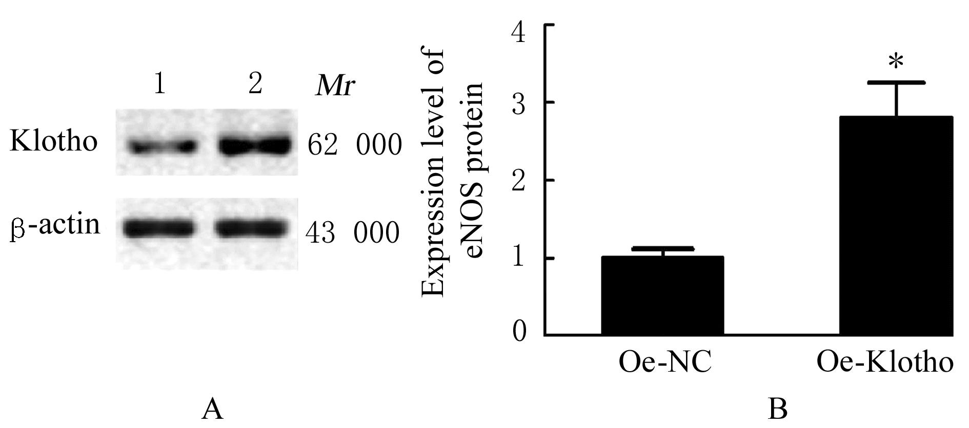
目的 探讨Klotho在子痫前期(PE)患者来源的胎盘外泌体(Exo)中的表达,阐明其对血管内皮细胞氧化应激的影响。 方法 收集40例孕产妇的临床资料,其中正常妊娠(NP)者20名,PE患者20例,设为NP组和PE组,分离2组研究对象外周血中胎盘Exo。将oe-Klotho和oe-NC质粒转染入人绒毛膜滋养层细胞HTR-8/SVneo中,作为oe-Klotho组和oe-NC组,收集HTR-8/SVneo细胞Exo。采用实时荧光定量PCR(RT-PCR)法和Western blotting法检测2组研究对象胎盘Exo和2组HTR-8/SVneo细胞Exo中Klotho mRNA及蛋白表达水平。取生长状态良好的人脐静脉内皮细胞(HUVECs),按Exo来源不同将HUVECs分为PE-Exo组(与PE患者胎盘Exo共培养)、NP-Exo组(与NP胎盘Exo共培养)、oe-Klotho-Exo组(与转染oe-Klotho的HTR-8/SVneo细胞Exo共培养)和oe-NC-Exo组(与转染oe-NC的HTR-8/SVneo细胞Exo共培养)。采用透射电子显微镜(TEM)观察Exo形态表现,Western blotting法检测Exo标记分子CD63、TSG101和胎盘Exo标记分子PLAP蛋白表达水平以鉴定Exo,荧光显微镜观察HUVECs对Exo的摄取情况。酶联免疫吸附试验(ELISA)法检测各组HUVECs中一氧化氮(NO)、活性氧(ROS)和丙二醛(MDA)水平及超氧化物歧化酶(SOD)活性,RT-qPCR法检测各组HUVECs中内皮型一氧化氮合酶(eNOS)mRNA表达水平,Western blotting法检测各组HUVECs中eNOS蛋白表达水平。 结果 与NP组比较,PE组患者收缩压和舒张压升高(P<0.05),新生儿体质量和胎盘质量均明显降低(P<0.05或P<0.01)。TEM观察,来源于NP组和PE组研究对象的胎盘Exo均呈圆形和椭圆形的囊泡盘状结构,包膜完整,形状相似,直径为50~100 nm;2组研究对象胎盘Exo均高表达Exo标记蛋白CD63和TSG101及胎盘Exo特异性蛋白PLAP。纳米示踪分析(NTA)检测,胎盘Exo的粒径为50~200 nm,提示由外周血分离的囊泡均是胎盘来源Exo。TEM、Western blotting和NTA检测,来源于oe-NC组和oe-Klotho组滋养层细胞的囊泡具有Exo的典型结构、分子标志物和粒径大小特征。HUVECs中均可见到明显的Exo绿色荧光表达,提示Exo可被HUVECs摄取。ELISA法检测,与NP-Exo组比较,PE-Exo组HUVECs中NO水平和SOD活性均明显降低(P<0.05或P<0.01),ROS和MDA水平明显升高(P<0.05或P<0.01);与oe-NC-Exo组比较,oe-Klotho-Exo组HUVECs中NO水平和SOD活性升高(P<0.05),ROS和MDA水平明显降低(P<0.05或P<0.01)。RT-qPCR法检测,与NP组比较,PE组研究对象胎盘Exo中Klotho mRNA表达水平明显降低(P<0.01);与NP-Exo组比较,PE-Exo组HUVECs中eNOS mRNA表达水平明显降低(P<0.01);与oe-NC组比较,oe-Klotho组滋养层细胞Exo中Klotho mRNA表达水平明显升高(P<0.01);与oe-NC-Exo组比较,oe-klotho-Exo组HUVECs中eNOS mRNA表达水平明显升高(P<0.01)。Western blotting法检测,与NP组比较,PE组研究对象胎盘Exo中Klotho蛋白表达水平明显降低(P<0.01);与NP-Exo组比较,PE-Exo组HUVECs中eNOS蛋白表达水平明显降低(P<0.01);与oe-NC组比较,oe-Klotho组滋养层细胞Exo中Klotho蛋白表达水平明显升高(P<0.01);与oe-NC-Exo组比较,oe-Klotho-Exo组HUVECs中eNOS蛋白表达水平均明显升高(P<0.01)。 结论 来源于PE患者的胎盘Exo可能通过抑制HUVECs中NO的产生和促进氧化应激反应以损伤内皮细胞功能,过表达Klotho的滋养层细胞Exo可减少HUVECs中NO的产生和氧化应激水平。

关键词: Klotho蛋白, 子痫前期, 外泌体, 血管内皮细胞, 氧化应激

Abstract:

Objective To discuss the expression of Klotho in placenta exosomes (Exo)of the patients with pre-eclampsia (PE),and to clarify its effect on the oxidative stress in the vascular endothelial cells. Methods The clinical data of 40 pregnant women including 20 with normal pregnancy(NP) women (NP group) and 20 PE patients (PE group) were collected. The placenta Exo in peripheral blood of the patients in two groups were isolated, and the oe-Klotho and oe-NC plasmids were transfected into the human chorionic trophoblast cells(HTR-8/SVneo), respectively, and were regarded as oe-Klotho group and oe-NC group.The expression levels of Klotho mRNA and protein in placenta Exo and the HTR8/SVneo cells in two groups were detected by real-time fluorescence quantitative PCR (RT-qPCR) method and Western blotting method. The human umbilical vein endothelial cells (HUVECs) with good growth status were taken and divided into PE-Exo group (co-cultured with placenta Exo from the patients with PE),NP-Exo group (co-cultured with placenta Exo from the NP subjects), oe-Klotho-Exo group (co-cultured with Exo from the HTR-8/SVneo cells transfected with oe-Klotho), and oe-NC-Exo group (co-cultured with Exo from the HTR-8/SVneo cells transfected with oe-NC) according to the sources of Exo.The expression levels of exosomal marker proteins CD63, TSG101, and placenta Exo-specific marker PLAP protein were identified by transmission electron microscope (TEM) and Western blotting method; the levels of nitric oxide (NO), reactive oxygen species (ROS), and malondialdehyde (MDA) and the activities of superoxide dismutase (SOD) in the HUVECs in various groups were detected by enzyme-linked immunosorbent assay (ELISA) method;the expression levels of endothelial nitric oxide synthase (eNOS) mRNA in the HUVECs in various groups was detected by RT-qPCR method;the expression levels of eNOS protein in the HUVECs in various groups were detected by Western blotting method. Results Compared with NP group, the systolic and diastolic blood pressures of the patients in PE group were increased (P<0.05), the body weight of newborn and placenta weight were significantly decreased (P<0.05 or P<0.01). The TEM observation results showed that the placenta Exo derived from the subjects in both NP and PE groups being round or oval vesicular discoid structures with complete membrane and similar shape, with a diameter of 50—100 nm. The Exo from the subjects in both groups highly expressed Exo marker proteins CD63 and TSG101, and placental Exo-specific protein PLAP. The nanoparticle tracking analysis (NTA) results showed that the particle size of Exo was 50—200 nm, suggesting that the vesicles isolated from the peripheral blood were derived from placental Exo. The TEM, Western blotting, and NTA results confirmed that the vesicles from oe-NC group and oe-klotho group had typical Exo structures, molecular markers, and particle size characteristics. The Exo with green fluorescence expression could be observed in the HUVECs, suggesting that the placenta Exo could be taken up by the HUVECs. The ELISA results showed that compared with NP-Exo group, the level of NO and activity of SOD in PE-Exo group were significantly decreasd(P<0.05 or P<0.01), and the levels of ROS and MDA were significantly increased(P<0.05 or P<0.01); compared with oe-NC-Exo group, the level of NO and activity of SOD in the HUVECs in oe-Klotho-Exo group were increased (P<0.05), and the levels of ROS and MDA were significantly decreased (P<0.05 or P<0.01). The RT-qPCR results showed that compared with NP group, the expression level of Klotho mRNA in placenta Exo of the patients in PE group was significantly decreased (P<0.01),and the expression level of eNOS mRNA in the HUVECs was significantly decreased (P<0.01); compared with oe-NC group, the expression level of Klotho mRNA in Exo in the trophoblast cells in oe-Klotho group was significantly increased (P<0.01); compared with oe-NC-Exo group, the expression level of eNOS mRNA in the HUVECs in oe-Klotho-Exo group was significantly increased (P<0.01). The Western blotting results showed that compared with NP group, the expression level of Klotho protein in placenta Exo of the patients in PE group was significantly decreased (P<0.01); compared with NP-Exo group, the expression level of eNOS protein in the HUVECs in PE-Exo group was significantly decreased (P<0.01); compared with oe-NC group, the expression level of Klotho protein in Exo in the trophoblast cells in oe-Klotho group was significantly increased (P<0.01); compared with oe-NC-Exo group, the expression level of eNOS protein in the HUVECs in oe-Klotho-Exo group was significantly increased (P<0.01). Conclusion The placenta Exo from the PE patients may inhibit the production of NO in the HUVECs and promote the oxidative stress to impair the endothelial cell function. The over-expression of Klotho in Exo in the trophoblast cells can reduce the production of NO and level of oxidative stress in the HUVECs.

Key words: Klotho protein, Pre-eclampsia, Exosomes, Vascular endothelial cell, Oxidative stress

中图分类号: 

  • R394