| 1 |
王 稳, 王兴国, 刘淑娟, 等. 交界性卵巢肿瘤诊治中国专家共识(2022年版)[J]. 中国实用妇科与产科杂志, 2022, 38(12): 1185-1194.
|
| 2 |
贾艳艳. PDLIM4在卵巢癌中的临床意义及其调控肿瘤生长和转移的机制研究[D].郑州:郑州大学,2020.
|
| 3 |
DHALIWAL D, SHEPHERD T G. Molecular and cellular mechanisms controlling integrin-mediated cell adhesion and tumor progression in ovarian cancer metastasis: a review[J]. Clin Exp Metastasis, 2022, 39(2): 291-301.
|
| 4 |
VALIPOUR M, ZARGHI A, EBRAHIMZADEH M A,et al. Therapeutic potential of chelerythrine as a multi-purpose adjuvant for the treatment of COVID-19[J]. Cell Cycle, 2021, 20(22): 2321-2336.
|
| 5 |
LIN Y L, ZHANG Q Z, XIE B F, et al. Chelerythrine-induced apoptotic cell death in HepG2 cells involves the inhibition of Akt pathway and the activation of oxidative stress and mitochondrial apoptotic pathway[J]. Antioxidants, 2022, 11(9): 1837.
|
| 6 |
GONG Y, YIN S L, SUN S J, et al. Chelerythrine reverses the drug resistance of resistant Candida albicans and the biofilm to fluconazole[J]. Future Microbiol, 2022, 17: 1325-1333.
|
| 7 |
QIAN W D, HUANG J, ZHANG J N, et al. Antimicrobial and antibiofilm activities and mechanism of action of chelerythrine against carbapenem-resistant Serratia marcescens in vitro [J]. Microb Drug Resist, 2021, 27(8): 1105-1116.
|
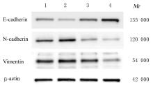
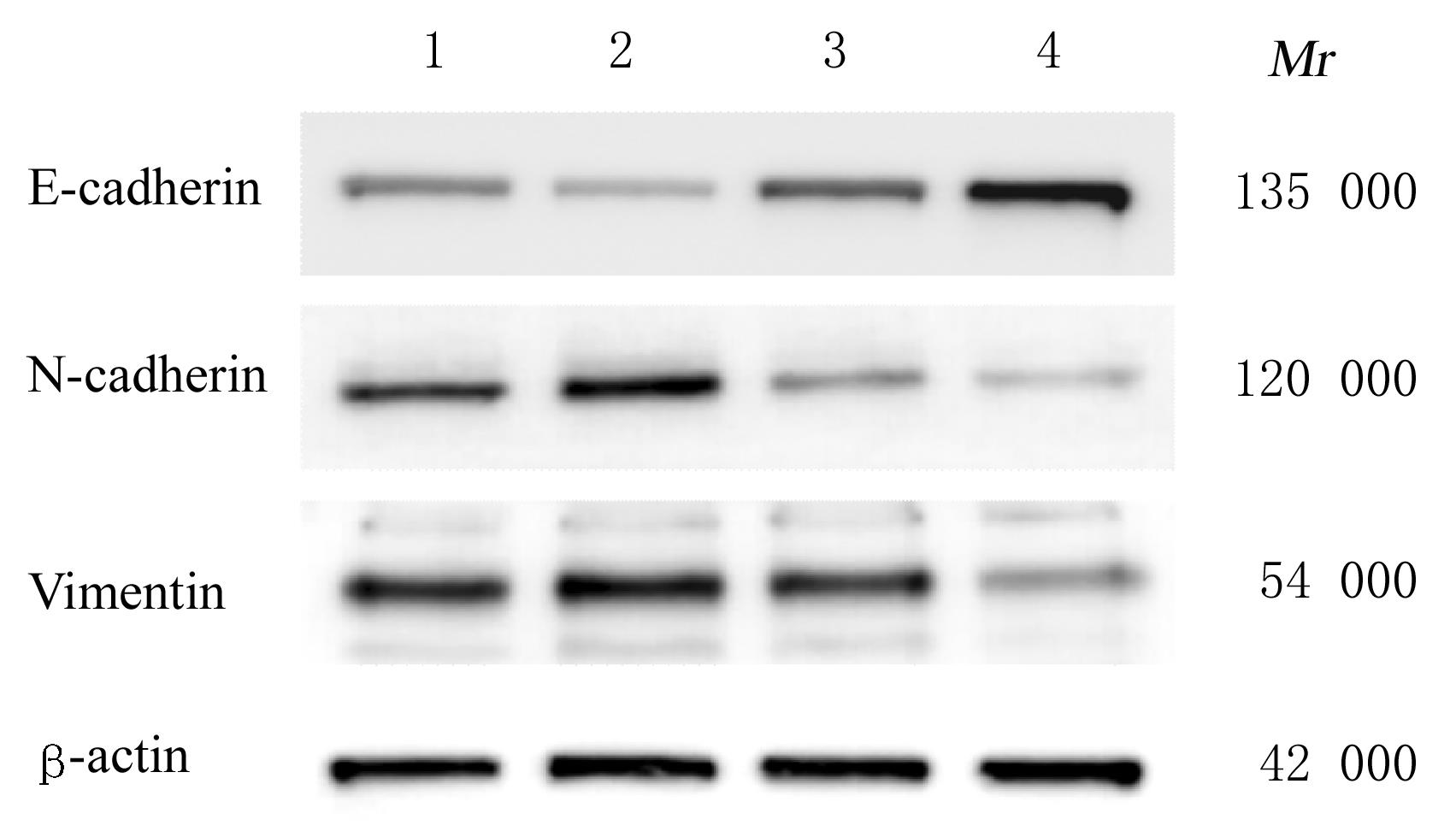
| 8 |
CHO O, LEE J W, KIM H S, et al. Chelerythrine, a novel small molecule targeting IL-2, inhibits melanoma progression by blocking the interaction between IL-2 and its receptor[J]. Life Sci, 2023, 320: 121559.
|
| 9 |
WANG M Z, MA B, NI Y F, et al. Restoration of the antibiotic susceptibility of methicillin-resistant Staphylococcus aureus and extended-spectrum β-lactamases Escherichia coli through combination with chelerythrine[J].Microb Drug Resist,2021,27(3):337-341.
|
| 10 |
贾茗博, 孙 莹, 王 莹, 等. 氯化两面针碱通过JAK2/STAT3信号通路对胶质瘤细胞上皮-间质转化的抑制作用[J]. 吉林大学学报(医学版), 2021, 47(1): 73-81.
|
| 11 |
王培卿, 尹震花, 康文艺. 白屈菜红碱药理活性研究进展[J]. 中国中药杂志, 2013, 38(17): 2745-2749.
|
| 12 |
YANG T F, XU R, SU Q, et al. Chelerythrine hydrochloride inhibits proliferation and induces mitochondrial apoptosis in cervical cancer cells via PI3K/BAD signaling pathway[J]. Toxicol In Vitro, 2020, 68: 104965.
|
| 13 |
WANG J H, SONG Y J, ZHANG N, et al. Using liposomes to alleviate the toxicity of chelerythrine, a natural PKC inhibitor, in treating non-small cell lung cancer[J]. Front Oncol, 2021, 11: 658543.
|
| 14 |
CAO L L, LIANG Y B, ZHAO F J, et al. Chelerythrine and Fe3O4 loaded multi-walled carbon nanotubes for targeted cancer therapy[J]. J Biomed Nanotechnol, 2016, 12(6): 1312-1322.
|
| 15 |
CHEN Z X, YANG H, ZHANG Q L, et al. Chelerythrine inhibits stemness of cancer stem-like cells of osteosarcoma and PI3K/AKT/mTOR signal[J]. J Oncol, 2022, 2022: 6435431.
|
| 16 |
CHEN N Z, QI Y L, MA X, et al. Rediscovery of traditional plant medicine: an underestimated anticancer drug of chelerythrine[J]. Front Pharmacol, 2022, 13: 906301.
|
| 17 |
TANG Z H, CAO W X, WANG Z Y, et al. Induction of reactive oxygen species-stimulated distinctive autophagy by chelerythrine in non-small cell lung cancer cells[J]. Redox Biol, 2017, 12: 367-376.
|
| 18 |
PLAZAS E, AVILA M M C, MUÑOZ D R, et al. Natural isoquinoline alkaloids: Pharmacological features and multi-target potential for complex diseases[J]. Pharmacol Res, 2022, 177: 106126.
|
| 19 |
DOHERTY J A, PERES L C, WANG C, et al. Challenges and opportunities in studying the epidemiology of ovarian cancer subtypes[J]. Curr Epidemiol Rep, 2017, 4(3): 211-220.
|
| 20 |
MELAMED A, RAUH-HAIN J A, GOCKLEY A A, et al. Association between overall survival and the tendency for cancer programs to administer neoadjuvant chemotherapy for patients with advanced ovarian cancer[J]. JAMA Oncol, 2021, 7(12): 1782-1790.
|
| 21 |
陈立兰, 狄 文. 柠檬酸合成酶对卵巢癌SKOV3细胞上皮间质转化的影响[J].现代肿瘤医学,2022,30(17): 3065-3068.
|
| 22 |
HUANG Y, LI C Q, ZHANG X, et al. Nanotechnology-integrated ovarian cancer metastasis therapy: insights from the metastatic mechanisms into administration routes and therapy strategies[J]. Int J Pharm, 2023, 636: 122827.
|
| 23 |
YOUSEFI M, DEHGHANI S, NOSRATI R,et al. Current insights into the metastasis of epithelial ovarian cancer - hopes and hurdles[J]. Cell Oncol,2020,43(4):515-538.
|
| 24 |
YEUNG T L, LEUNG C S, YIP K P, et al. Cellular and molecular processes in ovarian cancer metastasis. A Review in the Theme: cell and Molecular Processes in Cancer Metastasis[J]. Am J Physiol Cell Physiol, 2015, 309(7): C444-C456.
|
| 25 |
ANTONY J, THIERY J P, HUANG R Y. Epithelial-to-mesenchymal transition: lessons from development, insights into cancer and the potential of EMT-subtype based therapeutic intervention[J].Phys Biol,2019,16(4): 041004.
|
| 26 |
ASHRAFIZADEH M, MIRZAEI S, HASHEMI F,et al.New insight towards development of paclitaxel and docetaxel resistance in cancer cells: EMT as a novel molecular mechanism and therapeutic possibilities[J]. Biomedecine Pharmacother, 2021, 141: 111824.
|
| 27 |
白丽娜,刘 颖,唐春晓,等.可利霉素对胰腺癌细胞生物学功能的影响[J].临床肝胆病杂志, 2022,38(12): 2793-2801.
|
| 28 |
李 峣, 孙 莹, 宋燕珂, 等. PDGF-D通过Notch1信号通路对肿瘤细胞上皮-间质转化调控作用的研究进展[J].吉林大学学报(医学版),2022,48(1):265-270.
|
| 29 |
PAL M, BHATTACHARYA S, KALYAN G, et al. Cadherin profiling for therapeutic interventions in epithelial mesenchymal transition (EMT) and tumorigenesis[J]. Exp Cell Res,2018,368(2): 137-146.
|
| 30 |
GALVÁN J A, ZLOBEC I, WARTENBERG M, et al. Expression of E-cadherin repressors SNAIL, ZEB1 and ZEB2 by tumour and stromal cells influences tumour-budding phenotype and suggests heterogeneity of stromal cells in pancreatic cancer[J].Br J Cancer,2015,112(12): 1944-1950.
|
 ),黄晓巍1,3(