吉林大学学报(医学版) ›› 2024, Vol. 50 ›› Issue (1): 18-24.doi: 10.13481/j.1671-587X.20240103

• 基础研究 • 上一篇    下一篇

PD-L1对人口腔鳞状细胞癌细胞增殖、迁移和侵袭的影响

曾洁1,俞雪燕2,罗婷3,徐江1()   

  1. 1.石河子大学第一附属医院口腔科,新疆 石河子 832000
    2.石河子大学第一附属医院病理科,新疆 石河子 832000
    3.新疆维吾尔自治区儿童医院检验科,新疆 乌鲁木齐 830000
  • 收稿日期:2023-03-06 出版日期:2024-01-28 发布日期:2024-01-31
  • 通讯作者: 徐江 E-mail:1437759520@qq.com
  • 作者简介:曾 洁(1996-),女,新疆维吾尔自治区博乐市人,在读硕士研究生,主要从事口腔颌面部肿瘤基础和临床方面的研究。
  • 基金资助:
    国家自然科学基金项目(82160572)

Effect of PD-L1 on proliferation, migration, and invasion of human oral squamous carcinoma cells

Jie ZENG1,Xueyan YU2,Ting LUO3,Jiang XU1()   

  1. 1.Department of Stomatology,First Affiliated Hospital,Shihezi University,Shihezi 832000,China
    2.Department of Pathology,First Affiliated Hospital,Shihezi University,Shihezi 832000,China
    3.Department of Laboratory,Children’s Hospital,Xinjiang Uygur Autonomous Region,Urumqi 830000,China
  • Received:2023-03-06 Online:2024-01-28 Published:2024-01-31
  • Contact: Jiang XU E-mail:1437759520@qq.com

摘要:

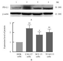
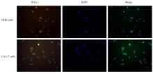
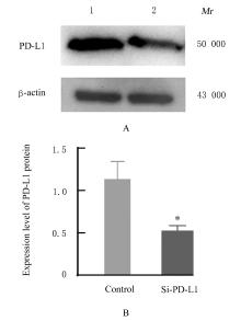
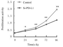
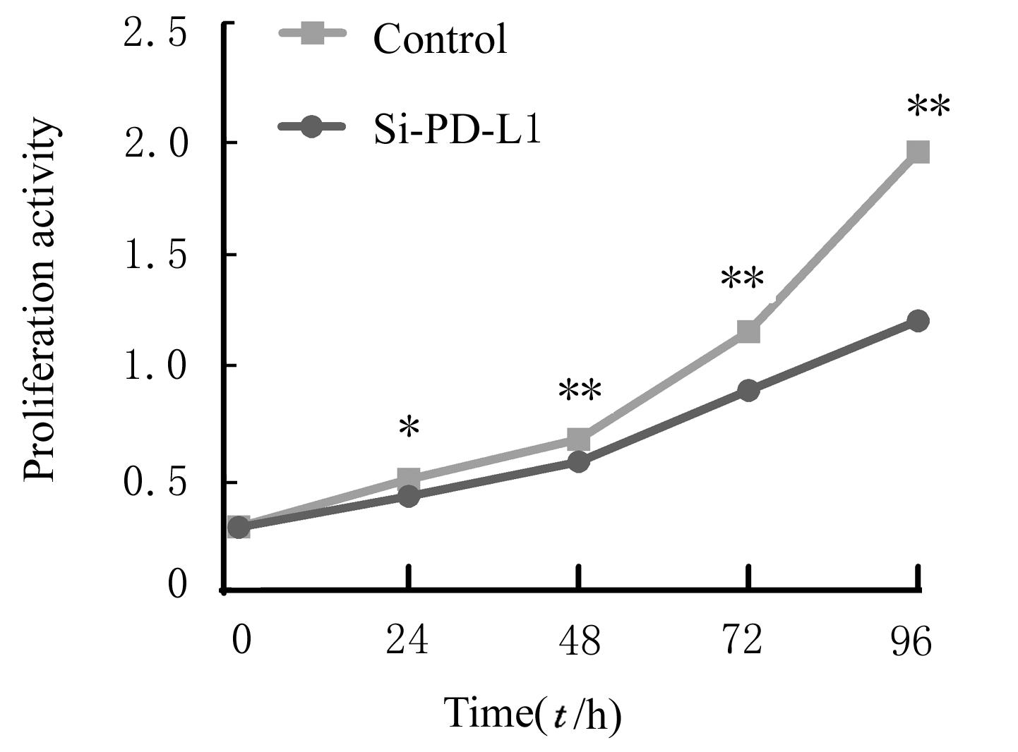
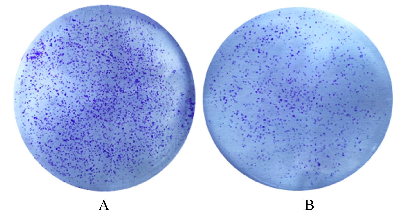
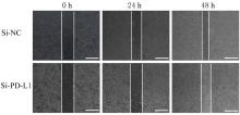
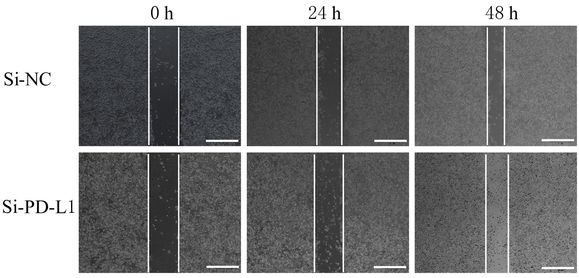
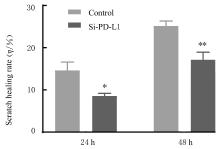
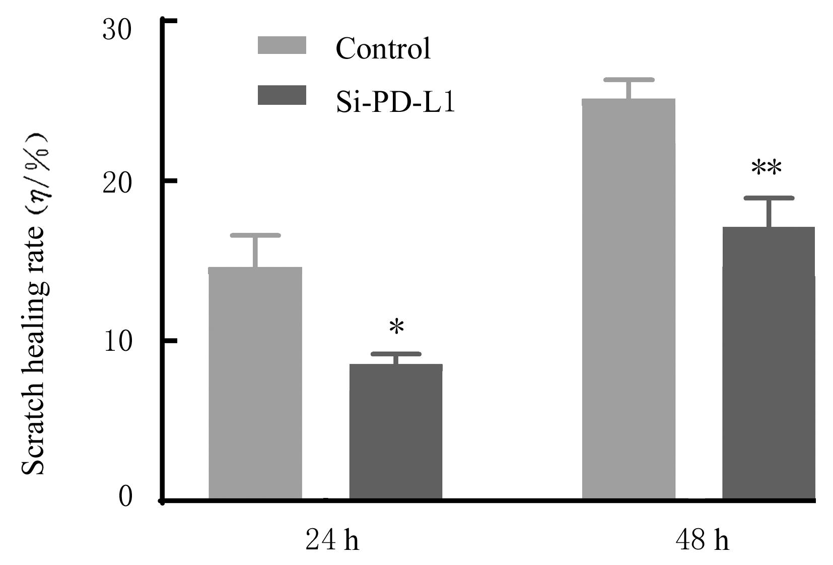
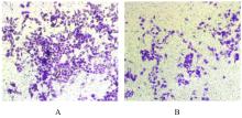
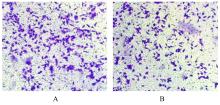
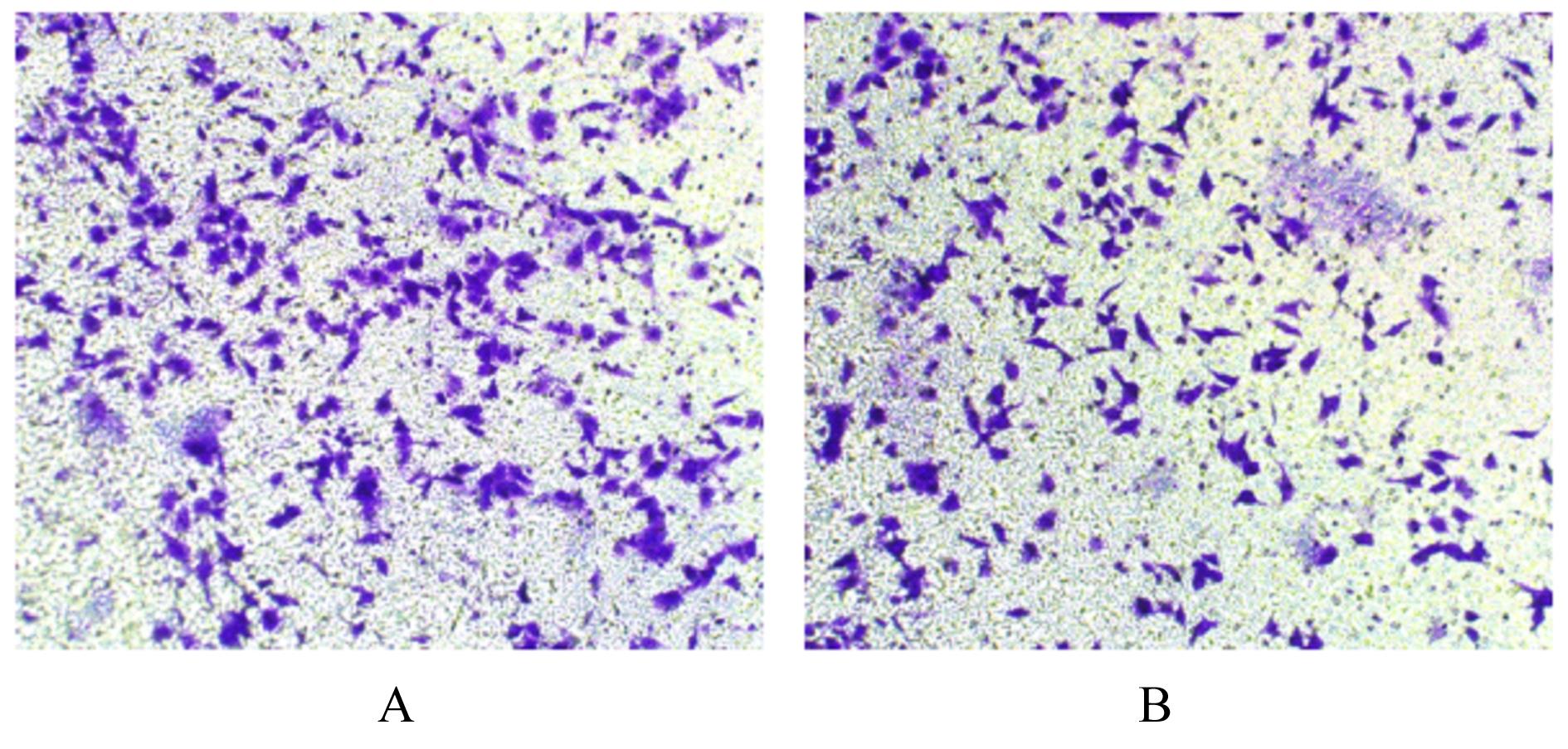
目的 探讨程序性细胞死亡配体1(PD-L1)在口腔鳞状细胞癌(OSCC)细胞中的表达及其对OSCC CAL27细胞生物学行为的影响,阐明其可能的作用机制。 方法 采用Western blotting法检测口腔上皮HOK细胞和OSCC CAL27、TCA8113和SCC15细胞中PD-L1蛋白表达水平。免疫荧光染色法检测CAL27细胞中PD-L1蛋白表达和定位情况。将CAL27细胞分为对照组(转染si-NC)和si-PD-L1组(转染si-PD-L1),Western blotting法检测2组细胞干扰效率,CCK-8法检测不同时间点2组细胞增殖活性,平板克隆实验检测2组细胞克隆形成数,细胞划痕愈合实验检测2组细胞划痕愈合率,Transwell小室实验检测2组细胞中迁移和侵袭细胞数。 结果 OSCC细胞中PD-L1蛋白表达水平均高于HOK细胞(P<0.05或P<0.01),PD-L1在CAL27细胞的细胞质和细胞核中均有表达。CCK-8法和平板克隆实验,与对照组比较,不同时间点si-PD-L1组CAL27细胞增殖活性明显降低(P<0.05或P<0.01),克隆形成数明显减少(P<0.01)。细胞划痕愈合实验,与对照组比较,si-PD-L1组CAL27细胞划痕愈合率明显降低(P<0.05或P<0.01)。Transwell小室实验,与对照组比较,si-PD-L1组CAL27细胞中迁移和侵袭细胞明显减少(P<0.01)。 结论 PD-L1在OSCC细胞中的表达高于口腔正常上皮细胞,敲低PD-L1表达可抑制OSCC细胞增殖、克隆形成及迁移和侵袭能力。

关键词: 口腔鳞状细胞癌, 程序性细胞死亡配体1, 细胞增殖, 细胞迁移, 细胞侵袭

Abstract:

Objective To discuss the expression of programmed cell death-ligand 1 (PD-L1) in the oral squamous cell carcinoma (OSCC) cells and its effect on biological behavior of the OSCC CAL27 cells, and to clarify the possible mechanism. Methods Western blotting method was used to detect the expression levels of PD-L1 protein in the oral epithelial HOK cells and OSCC CAL27, TCA8113, and SCC15 cells; immunofluorescence staining method was used to detect the expression and localization of PD-L1 protein in the CAL27 cells. The CAL27 cells were divided into control group (transfected with si-NC) and si-PD-L1 group (transfected with si-PD-L1). Western blotting method was used to detect the interference efficiency of the cells in two groups; CCK-8 assay was used to detect the proliferative activities of the cells in two groups at different time points; plate clone formation assay was used to detect the numbers of clone formation of the cells in two groups; cell scratch healing assay was used to detect the scratch healing rates of the cells in two groups; Transwell chamber assay was used to detect the numbers of migration and invasion cells in two groups. Results The expression level of PD-L1 protein in the OSCC cells was higher than that in the HOK cells (P<0.05 or P<0.01); PD-L1 expressed in the cytoplasm and nucleus of the CAL27 cells. The CCK-8 assay and plate clone formation assay results showed that compared with control group, the proliferative activities of the CAL27 cells in si-PD-L1 group at different time points were significantly decreased(P<0.05 or P<0.01), and the numbers of clone formation were significantly decreased (P<0.01). The cell scratch healing assay results showed that compared with control group, the scratch healing rates of the cells in si-PD-L1 group were significantly decreased (P<0.05 or P<0.01). The Transwell chamber assay results showed that compared with control group, the numbers of migration and invasion cells in si-PD-L1 group were significantly decreased (P<0.01). Conclusion The expression of PD-L1 in the OSCC cells is higher than that in normal oral epithelial cells, and knocking down PD-L1 expression can inhibit the proliferation, clone formation, migration and invasion capabilities of the OSCC cells.

Key words: Oral squamous cell carcinoma, Programmed cell death-ligand 1, Cell proliferation, Cell migration, Cell invasion

中图分类号: 

  • R739.8