吉林大学学报(医学版) ›› 2024, Vol. 50 ›› Issue (6): 1691-1702.doi: 10.13481/j.1671-587X.20240623

• 临床研究 • 上一篇    

宫颈病变和宫颈癌组织中嗜酸性粒细胞浸润及其临床意义

鲁艳艳1,2,许翔博1,3,吴亚梅2,刘雨齐1,王涵1,杨丽娟1,王振江1,肖梓屾1,刘艳波1()   

  1. 1.北华大学基础医学院病理生理学教研室,吉林 吉林 132013
    2.吉林省长春市妇产医院病理科,吉林 长春 130028
    3.吉林省吉林市人民医院妇产科,吉林 吉林 132011
  • 收稿日期:2024-01-08 出版日期:2024-11-28 发布日期:2024-12-10
  • 通讯作者: 刘艳波 E-mail:liuyanbobeihua@163.com
  • 作者简介:鲁艳艳(1989-),女,吉林省吉林市人,主治医师,医学硕士,主要从事女性生殖系统肿瘤发病机制方面的研究。
  • 基金资助:
    吉林省科技厅国际合作项目(20210402015GH);吉林省科技厅基础研究项目(YDZJ202201ZYTS194);北华大学研究生创新项目(研创合字〔2022〕056)

Eosinophil infiltration in cervical lesion and cervical cancer tissues and their clinical significances

Yanyan LU1,2,Xiangbo XU1,3,Yamei WU2,Yuqi LIU1,Han WANG1,Lijuan YANG1,Zhenjiang WANG1,Zishen XIAO1,Yanbo LIU1()   

  1. 1.Department of Pathophysiology,School of Basic Medical Sciences,Beihua University,Jilin 132013,China
    2.Department of Pathology,Gynaecology and Obstetrics Hospital,Changchun City,Jilin Province,Changchun 130028,China
    3.Department of Gynaecology and Obstetrics,People’s Hospital,Jilin City,Jilin Province,Jilin 132011,China
  • Received:2024-01-08 Online:2024-11-28 Published:2024-12-10
  • Contact: Yanbo LIU E-mail:liuyanbobeihua@163.com

摘要:

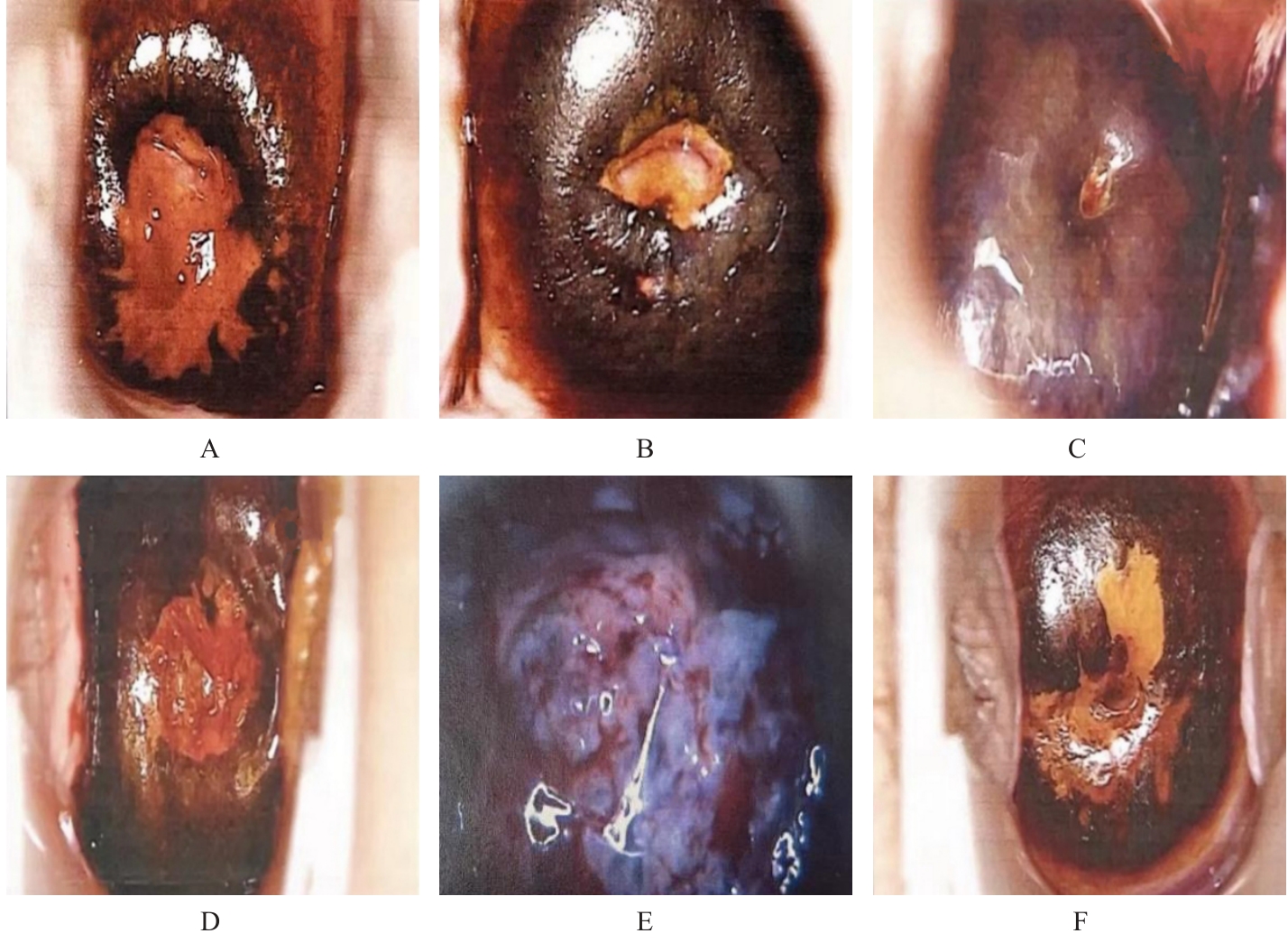
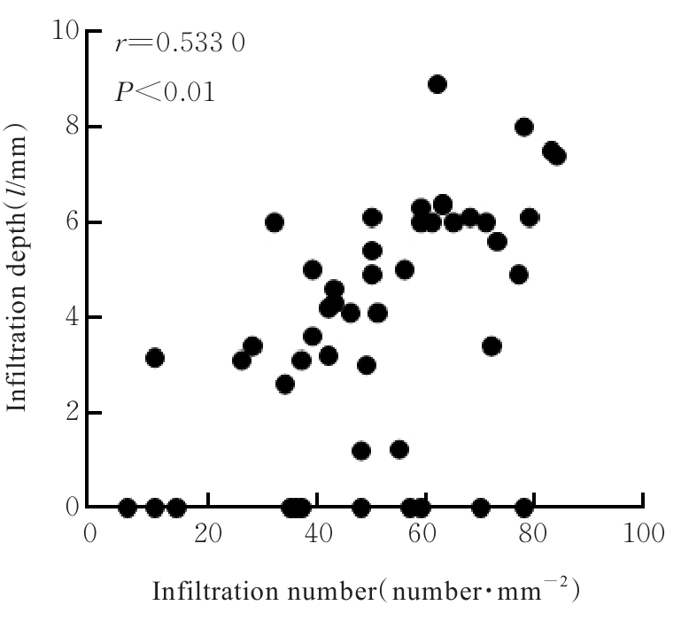
目的 探讨嗜酸性粒细胞(EOS)在宫颈组织中的浸润差异及其与宫颈相关疾病之间的关系,阐明EOS对宫颈上皮不典型增生(CIN)和宫颈癌发生发展的影响。 方法 收集256例宫颈疾病患者的临床资料,根据其发病情况分为宫颈癌组(n=46,其中宫颈鳞状细胞癌26例、宫颈腺癌15例和宫颈腺鳞癌5例)、慢性宫颈炎组(n=50)、CINⅠ期组(n=50)、CINⅡ期组(n=50)、CINⅢ期组(n=30)和正常组(癌旁正常宫颈组织,n=30)。阴道镜观察各组患者宫颈组织形态表现,薄层液基细胞学测试(TCT)法观察各组患者宫颈脱落细胞形态表现,杂交捕获-化学发光法检测各组患者宫颈组织人乳头瘤病毒(HPV)感染情况,HE染色观察各组患者宫颈组织病理形态表现,刚果红染色检测各组患者宫颈组织中EOS浸润数,Pearson相关性分析EOS浸润数与宫颈癌恶性程度的相关性。 结果 正常组患者宫颈表面光滑,呈粉红色,毛细血管均匀分布;慢性宫颈炎组患者宫颈表面呈红色炎性改变,部分伴有纳氏囊肿形成,可见不同程度的糜烂和溃疡等;CINⅠ期、CINⅡ期和CINⅢ期组患者宫颈可见上皮溃疡、增厚和形态不规则,细镶嵌及点状血管明显;宫颈癌组患者宫颈表面隆起,可见新生肿物及坏死性溃疡,质脆易出血。醋酸染色后,正常组患者宫颈无明显改变;慢性宫颈炎组患者宫颈呈少量白色改变,持续时间较短;CINⅠ期、CINⅡ期和CINⅢ期组患者宫颈薄醋白上皮不规则、呈地图样边界,其中CINⅠ期组患者宫颈部分组织呈醋白反应,CINⅡ期组患者宫颈出现明显醋白反应,CINⅢ期组患者宫颈醋白反应非常明显,面积较大,且持续时间较长;宫颈癌组患者宫颈醋白反应明显,白色上皮厚,持续时间久,轮廓硬直,边界清晰。正常组患者宫颈碘染色后呈棕褐色,着色均匀;慢性宫颈炎组患者宫颈炎性病变区着色差;CINⅠ期组患者宫颈上皮化生区碘着色不明显;CINⅢ期组患者宫颈病变区着色差,转化区周围面积较大;宫颈癌组患者宫颈表面不规则,呈菜花样生长,碘染色后不着色,呈现为橘黄或芥末黄色。TCT法观察,正常组患者宫颈脱落细胞中无异型性细胞,炎症细胞浸润少;慢性宫颈炎组患者宫颈脱落细胞中可见大量的中性粒细胞和EOS等炎症细胞,无异型性细胞;CINⅠ期和CINⅡ期组患者宫颈脱落细胞中可见双核异型性细胞,核质比较高,细胞核较深染,周围见空晕;CINⅢ期组患者脱落细胞中可见较多异型性细胞,核质比较高,核膜不规则;宫颈癌组患者宫颈脱落细胞中可见大而显著的核仁,聚集成片,合胞体样改变明显;与正常组比较,CINⅠ期组、CINⅡ期组、CINⅢ期组和宫颈癌组患者宫颈脱落细胞异型性均明显增加。杂交捕获-化学发光法检测,与正常组和慢性宫颈炎组比较,CINⅠ期、CINⅡ期和CINⅢ期组患者HPV感染数和TCT异型性细胞数均明显增加(P<0.05);与CINⅠ期、CINⅡ期和CINⅢ期组比较,宫颈癌组患者HPV感染数和TCT异型性细胞数均明显增加(P<0.05)。HE染色观察,正常组宫颈组织细胞形态正常,结构清晰,可见中性粒细胞、单核细胞、巨噬细胞、EOS和淋巴细胞等炎症细胞浸润;慢性宫颈炎组患者炎症细胞浸润增加;CIN组患者宫颈细胞核核仁稍大,可见异型性细胞,炎症细胞主要分布于异型性细胞周围;宫颈癌组患者宫颈细胞核仁大而深染,细胞异型性明显,癌细胞周围炎症细胞浸润增加。与正常组比较,慢性宫颈炎组患者宫颈组织中炎症细胞数和EOS浸润数均明显增加(P<0.05),CIN组患者炎症细胞数和EOS浸润数均明显增加(P<0.05);与慢性宫颈炎组比较,CIN组患者炎症细胞数和EOS浸润数均明显减少(P<0.05);与慢性宫颈炎组和CIN组比较,宫颈癌组患者炎症细胞数和EOS浸润数均明显增加(P<0.05)。宫颈癌组织中EOS主要分布于癌巢周围,与CINⅠ期组比较,CINⅡ期组和CINⅢ期组患者宫颈组织中EOS浸润数均明显增加(P<0.05);与CINⅡ期组比较,CINⅢ期组患者宫颈组织中EOS浸润数明显增加(P<0.05)。肿瘤恶性程度越高,EOS浸润越多,EOS浸润数与宫颈癌浸润深度呈正相关关系(r=0.533 0,P<0.01)。 结论 HPV感染和EOS浸润具有促进宫颈癌癌前病变及宫颈癌发生发展的作用。

关键词: 嗜酸性粒细胞, 宫颈肿瘤, 宫颈上皮不典型增生, 慢性宫颈炎, 细胞浸润

Abstract:

Objective To discuss the differences in eosinophil (EOS) infiltration in cervical tissue and its relationship with cervical-related diseases, and to clarify the effect of EOS on the occurrence and development of cervical intraepithelial neoplasia (CIN) and cervical cancer. Methods The clinical data of 256 patients with cervical diseases were collected and divided into cervical cancer group (n=46, including 26 cases of squamous cell carcinoma, 15 cases of adenocarcinoma, and 5 cases of adenosquamous carcinoma), chronic cervicitis group (n=50), CIN stageⅠ group (n=50), CIN stageⅡ group (n=50), CIN stageⅢ group (n=30), and normal group (adjacent normal cervical tissue, n=30) based on their conditions. Colposcopy was used to observe the morphology of cervical tissue of the patients in various groups; thin-layer liquid-based cytology test (TCT) was used to observe the morphology of the cervical exfoliated cells in various groups; hybrid capture-chemiluminescence method was used to detect the human papillomavirus (HPV) infection in cervical tissue of the patients in various groups; HE staining was used to observe the pathomorphology of cervical tissue of the patients in various groups; Congo red staining was used to detect the numbers of EOS infiltration in cervical tissue of the patients in various groups; Pearson correlation analysis was used to analyze the correlation between the number of EOS infiltration and the malignancy degree of cervical cancer. Results The cervical surface of the patients in normal group was smooth and pink, with uniformly distributed capillaries; the cervical surface of the patients in chronic cervicitis group showed red inflammatory changes, with some accompanied by Nabothian cysts and varying degrees of erosion and ulcers; the patients in CIN stageⅠ, CIN stageⅡ, and CIN stageⅢ groups showed epithelial ulcers, thickening, and irregular morphology, with mosaic and punctate vessels; the cervical surface of the patients in cervical cancer group showed raised areas with neoplasms and necrotic ulcers, and they were fragile and prone to bleeding. After acetic acid staining, no obvious changes of the patients in normal group were observed. The cervix of the patients in chronic cervicitis group showed slight white changes that lasted for a short time; in CIN stageⅠ, CIN stageⅡ, and CIN stageⅢ groups, irregular thin acetowhite epithelium with map-like borders was observed, with increasingly acetowhite reactions and larger areas as the stages advanced. The cervix of the patients in cervical cancer group showed thick acetowhite epithelium that lasted longer, with rigid and clear contours. After iodine staining, the cervix of the patients in normal group was brown, with uniform coloration; the cervix of the patients in chronic cervicitis group showed poor coloration in inflammatory lesion areas; the cervix of the patients in CIN stageⅠ group showed iodine coloration in metaplastic areas, while the cervix of the patients in CIN stageⅢ group showed poor coloration in larger lesion areas; the cervix of the patients in cervical cancer group showed irregular surfaces with cauliflower-like growth and no coloration after iodine staining, appearing orange-yellow or mustard yellow. The TCT observation results showed there were no heteromorphic cells and few inflammatory cells in cervical exfoliated cells of the patients in infiltration in normal group; there were numerous neutrophils and EOS in exfoliated cervical cells without heteromorphic cells in chronic cervicitis group. The heteromorphic binucleated cells with high nuclear-cytoplasmic ratios and deeply stained nuclei were observed in cervical exfoliated cells of the patients in CIN stageⅠ and CIN stageⅡ groups. More heteromorphic cells with high nuclear-cytoplasmic ratios and irregular nuclear membranes were showed in cervical exfoliated cells of the patients in CIN stageⅢ group. The cervical exfoliated cells of the patients in cervical cancer group showed large and prominent nucleoli, clustering into syncytial changes. Compared with normal group, the atypial of cervical exfoliated cells in CIN stageⅠ, CIN stageⅡ, CIN stageⅢ, and cervical cancer groups was increased. The hybrid capture-chemiluminescence results showed that compared with normal and chronic cervicitis groups, the numbers of HPV infection and TCT heteromorphic cells of the patients in CIN stageⅠ, CIN stageⅡ, and CIN stageⅢ groups were increased (P<0.05); compared with CIN stageⅠ, CIN stageⅡ, and CIN stageⅢ groups, the numbers of HPV infection and TCT heteromorphic cells of the patients in cervical cancer group were increased (P<0.05). The HE staining results showed normal cell morphology and structure in normal group, with infiltration of inflammation cells such as neutrophils, monocytes, macrophages, EOS, and lymphocytes; in chronic cervicitis group, the infiltration of inflammatory cells was increased; in CIN group, the cervical cells showed slightly larger nucleoli and heteromorphic cells, with inflammatory cells mainly distributing around the hetermomorphic cells; in cervical cancer group, the cervical cells showed large and deeply stained nucleoli with significant atypia, and the infiltration of inflammatory cells around the cancer cells was increased. Compared with normal group, the numbers of inflammatory cells and EOS infiltration in cervical tissue of the patients in chronic cervicitis group were increased (P<0.05), and the numbers of inflammatory cells and EOS infiltration of the patients in CIN group were increased (P<0.05); compared with chronic cervicitis group, the number of inflammatory cells and EOS infiltration of the patients in CIN group were decreased (P<0.05); compared with chronic cervicitis group and CIN group, the numbers of inflammatory cells and EOS infiltration of the patients in cervical cancer group were increased (P<0.05). The EOS in cervical cancer tissue was mainly distributed around the cancer nests; compared with CIN stageⅠ group, the numbers of EOS infiltration in CIN stageⅡ and CIN stageⅢ groups were increased (P<0.05); compared with CIN stageⅡ group, the number of EOS infiltration in CIN stageⅢ group was increased (P<0.05). The higher the malignancy degree of the tumor, the more EOS infiltration was observed, and the number of EOS infiltration was positively correlated with the invasion depth of cervical cancer (r=0.533 0, P<0.01). Conclusion HPV infection and EOS infiltration play a role in promoting the and occurrence development of cervical precancerous lesions and cervical cancer.

Key words: Eosinophils, Cervical tumors, Cervical intraepithelial neoplasia, Chronic cervicitis, Cell infiltration

中图分类号: 

  • R737.33