吉林大学学报(医学版) ›› 2021, Vol. 47 ›› Issue (5): 1187-1193.doi: 10.13481/j.1671-587X.20210515

• 基础研究 • 上一篇    下一篇

人脐带间充质干细胞对宫颈癌HeLa细胞增殖和凋亡的影响及其作用机制

王琛(),田君,程海玲   

  1. 河南大学淮河医院妇科, 河南 开封 475000
  • 收稿日期:2021-01-26 出版日期:2021-09-28 发布日期:2021-10-26
  • 通讯作者: 王琛 E-mail:qingtianxiayu_88@126.com
  • 作者简介:王 琛(1980-),女,河南省开封市人,主治医师,主要从事宫颈癌及癌前病变方面的研究。
  • 基金资助:
    河南省科技厅重点科技攻关项目(182102311175)

Effect of human umbilical cord mesenchymal stem cells on proliferation and apoptosis of cervical cancer HeLa cells and its mechanism

Chen WANG(),Jun TIAN,Hailing CHENG   

  1. Department of Gynecology,Huaihe Hospital,Henan University,Kaifeng 475000,China
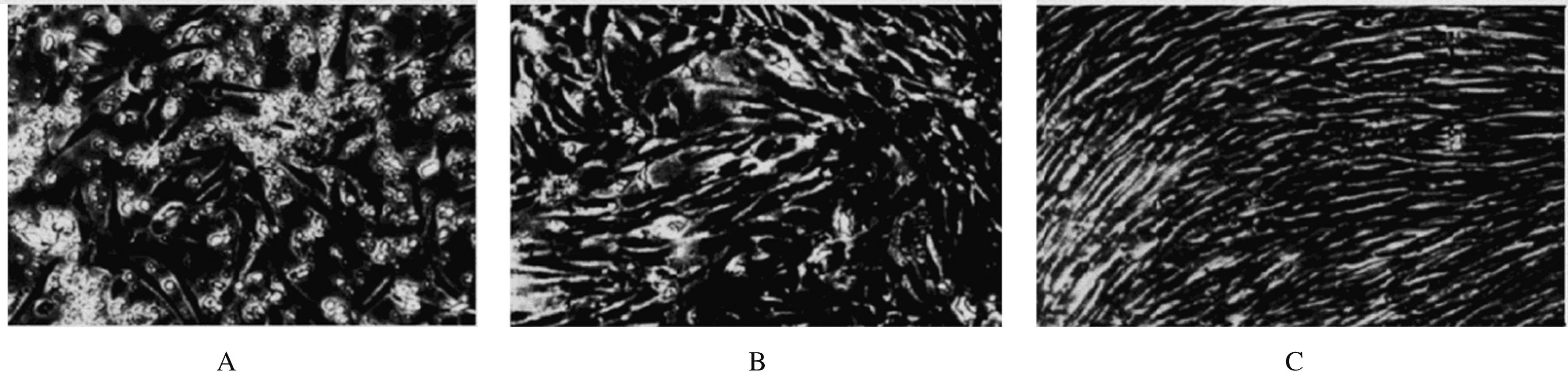
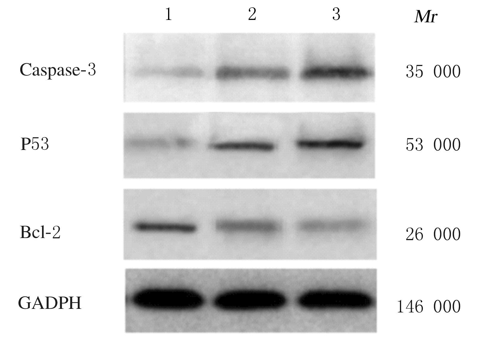
  • Received:2021-01-26 Online:2021-09-28 Published:2021-10-26
  • Contact: Chen WANG E-mail:qingtianxiayu_88@126.com

摘要: 目的

探讨人脐带间充质干细胞(MSCs)对宫颈癌HeLa细胞增殖和凋亡的影响,阐明其可能的作用机制。

方法

分离培养人脐带MSCs,采用光学显微镜观察细胞形态表现,并采用流式细胞仪分析鉴定其表面抗原标记物CD90、CD105、CD34和CD45的表达,以未加一抗仅加二抗的MSCs为平行对照组。将20%和60%浓度的MSCs条件培养基和宫颈癌HeLa细胞共培养72 h,集落形成实验观察MSCs条件培养基作用下HeLa细胞集落的形成能力;CCK-8实验检测HeLa细胞增殖抑制率;流式细胞术检测HeLa细胞凋亡率;蛋白质印迹法检测MSCs条件培养基作用下HeLa细胞增殖和凋亡相关基因蛋白表达水平。

结果

脐带分离细胞接种72 h后有少量细胞贴壁,1周后逐渐变成扁平单层细胞,呈簇状和旋涡状生长,伴随细胞密度增加,细胞体随之变细长,形态与成纤维细胞类似。MSCs细胞表面抗原标记物CD90和CD105呈阳性表达,CD34和CD45呈阴性表达,符合干细胞表型。集落形成实验,加入20%和60%的MSCs条件培养基,宫颈癌HeLa细胞集落形成的抑制率分别为18.56%和37.64%。与对照组比较,MSCs处理48 h后宫颈癌HeLa细胞早期凋亡率升高(P<0.05)。MSCs处理0、24和48 h后HeLa细胞中半胱天冬氨酸蛋白酶3(caspase-3)和P53蛋白表达水平持续升高(P<0.05),B细胞淋巴瘤2(Bcl-2)蛋白相对表达水平持续降低(P<0.05)。

结论

MSCs体外可抑制宫颈癌HeLa细胞增殖,并诱导HeLa细胞凋亡,其机制可能与上调促凋亡分子caspase-3与P53表达、下调抗凋亡因子Bcl-2表达有关。

关键词: 宫颈肿瘤, 人脐带间充质干细胞, 细胞增殖, 细胞凋亡

Abstract: Objective

To investigate the effect of human umbilical cord mesenchymal stem cells (MSCs) on the proliferation and apoptosis of cervical cancer HeLa cells, and to elucidate its possible mechanism.

Methods

The human umbilical cord MSCs were isolated and cultured. The morphology of cells were observed by light microscope. The expressions of surface antigen markers CD90, CD105, CD34 and CD45 were identified by flow cytometry. The MSCs were added with second antibody (without first antibody) were used as parallel control group.The conditioned medium containing 20% and 60% MSCs and cervical cancer HeLa cells were co-cultured for 72 h, colony formation experiment was used to observe the colony forming ability of HeLa cells after treated with of MSCs conditioned medium.CCK-8 experiment was used to detect the inhibitory rates of proliferation of the HeLa cells. Flow cytometry was used to detect the apoptotic rates of HeLa cells. Western blotting method was used to detect the expression levels of proliferation- and apoptosis-related gene proteins in the HeLa cellsn.

Results

After 72 h of inoculation, a small number of cells adhered to the wall, and gradually became flat monolayer cells, which grew in clusters and vortices after one week. With the increase of cell density, the cell body became slender, similar to that of fibroblasts.The MSCs showed positive expressions of CD90 and CD105, but negative expressions of CD34 and CD45, which conformed to the phenotype of stem cells.The results of colony formation test showed that the inhibitory rates of colony formation of HeLa cells were 18.56% and 37.64% respectively when the conditioned medium containing 20% and 60% MSCs were added.Compared with control group,the early apoptotic rate of HeLa cells after treated with MSCs for 48 h was increased (P<0.05). After the HeLa cells were treated with MSCs for 0, 24 and 48 h, the expression levels of cysteinyl aspartate-specific proteinase-3 (Caspase-3) and P53 proteins were increased continuously (P<0.05), and the expression levels of B-cell lymphoma-2 (Bcl-2) protein were decreased continuously (P<0.05).

Conclusion

The MSCs can inhibit the proliferation of HeLa cells and induce the apoptosis of HeLa cells in vitro,and its mechanism may be related to up-regulating the expression of Caspase-3 and P53 and down-regulating the expression of anti-apoptotic factor Bcl-2.

Key words: cervical cancer, human umbilical cord mesenchymal stem cells, cell proliferation, cell apoptosis

中图分类号: 

  • R285.5