吉林大学学报(医学版) ›› 2021, Vol. 47 ›› Issue (4): 951-957.doi: 10.13481/j.1671-587X.20210418

• 基础研究 • 上一篇    下一篇

氧化苦参碱对宫颈癌SiHa细胞增殖和凋亡的影响及其机制

张冬丽(),付瑞芳,孙桂霞   

  1. 河南大学淮河医院妇科,河南 开封 475200
  • 收稿日期:2020-10-26 出版日期:2021-07-28 发布日期:2021-07-22
  • 通讯作者: 张冬丽 E-mail:jingjingwang_19@163.com
  • 作者简介:张冬丽(1983-),女,河南省开封市人,副主任医师,医学硕士,主要从事妇科肿瘤临床方面的研究。
  • 基金资助:
    河南省科技厅科技攻关项目(182102311175);河南省教育厅重点科研项目(17A320023);河南省开封市科技局科技攻关项目(2003015)

Effect of oxymatrine on proliferation and apoptosis of cervical cancer SiHa cells and its mechanism

Dongli ZHANG(),Ruifang FU,Guixia SUN   

  1. Department of Gynecology,Huaihe Hospital,Henan University,Kaifeng 475200,China
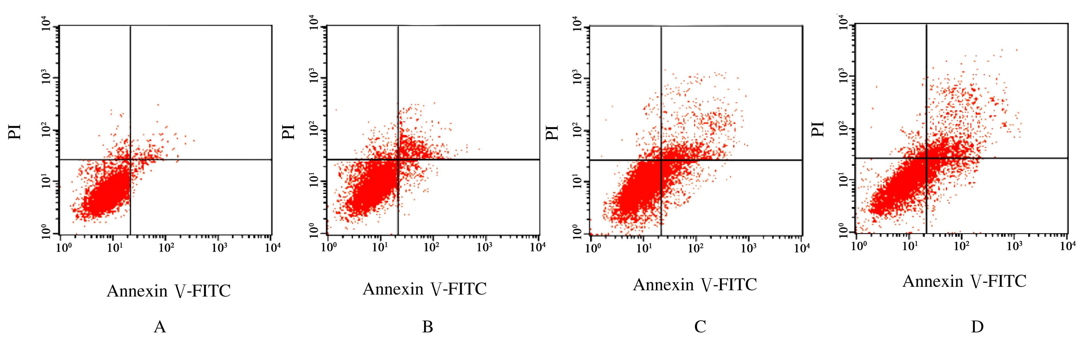
  • Received:2020-10-26 Online:2021-07-28 Published:2021-07-22
  • Contact: Dongli ZHANG E-mail:jingjingwang_19@163.com

摘要: 目的

探讨氧化苦参碱(OMT)对宫颈癌SiHa细胞增殖和凋亡的影响,并阐明其作用机制。

方法

SiHa细胞分为对照组、低剂量OMT组、中剂量OMT组和高剂量OMT组,分别以含二甲基亚砜(DMSO)及含0.4、0.8和1.6 g·L-1 OMT的RPMI 1640培养基培养。培养24 h后,Hoechst33258染色法观察细胞形态表现;分别培养24、48和72 h后,MTT法检测各组细胞增殖活性;培养48 h后,流式细胞术检测各组不同细胞周期细胞百分率和凋亡率;实时荧光定量PCR(RT-qPCR)和Western blotting法分别检测各组细胞中β-连环蛋白(β-catenin)、细胞周期蛋白D1(cyclinD1)、B细胞淋巴瘤2(Bcl-2)及Bcl-2相关X蛋白(Bax)mRNA和蛋白表达水平。

结果

荧光显微镜下观察,对照组细胞生长良好,蓝色荧光较弱;低剂量OMT组细胞可见少量凋亡小体;中剂量OMT组细胞数量减少,凋亡小体数量增多;高剂量OMT组蓝色荧光增多,细胞解体,核皱缩,凋亡小体数量明显增加。与对照组比较,培养24、48和72 h后,低、中和高剂量OMT组细胞增殖活性明显降低(P<0.05),G0/G1期细胞百分率明显升高(P<0.05),S期和G2/M期细胞百分率明显降低(P<0.05),细胞凋亡率明显升高(P<0.05),细胞中β-catenin、cyclinD1及Bcl-2 mRNA和蛋白表达水平明显降低(P<0.05),Bax mRNA和蛋白表达水平明显升高(P<0.05)。

结论

OMT可将SiHa细胞阻滞在G1期,抑制细胞增殖并诱导细胞凋亡,其机制可能与抑制Wnt/β-catenin信号通路有关。

关键词: 宫颈肿瘤, 氧化苦参碱, 细胞增殖, 细胞凋亡, β-连环蛋白

Abstract: Objective

To explore the effect of oxymatrine (OMT) on the proliferation and apoptosis of cervical cancer SiHa cells, and to clarify its mechanism.

Methods

The SiHa cells were randomly divided into control group, low dose of OMT group, medium dose of OMT group, and high dose of OMT group. The cells were cultured with RPMI 1640 medium containing dimethyl sulfoxide (DMSO), 0.4, 0.8 and 1.6 g·L-1 OMT. After 24 h of culture, the morphology of cells in various groups was detected by Hoechst33258 staining method; after 24, 48 and 72 h of culture, the proliferation activities of cells in various groups were detected by MTT method; after 48 h, the percentage of cells in different cell cycles and the apoptotic rates in various groups were detected by flow cytometry; RT-qPCR and Western blotting methods were used to detect the expression levels of β-catenin, cyclin D1, B-cell lymphoma-2 (Bcl-2), Bcl-2 assaciated X protein (Bax) mRNA and proteins.

Results

Under fluorescence microscope, the cells in control group grew well, and the blue fluorescence was weak; a small amount of apoptotic bodies were seen in low dose of OMT group; the number of cells in medium dose of OMT group was decreased, and the number of apoptotic bodies were increased; the blue fluorescence in high dose of OMT group was increased, cell disintegration and nuclear shrinkage were found, and the number of apoptotic bodies were increased significantly. Compared with control group, at 24, 48, and 72 h after culture,the proliferation activities of the cells in low dose of OMT group, medium dose of OMTgroup and high dose of OMT group were decreased significantly (P<0.05), the percentage of cells in G0/G1 phase were increased(P<0.05), the percentages of cells in S and G2/M phases were decreased (P<0.05), the apoptotic rates of cells were increased(P<0.05),the expression levels of β-catenin, cyclinD1, Bcl-2 mRNA and proteins were decreased(P<0.05),and the expression levels of Bax mRNA and protein were increased(P<0.05).

Conclusion

OMT can block the SiHa cells in the G1 phase,and inhibit the cell proliferation and induce the apoptosis;its machanism may be related to the inhibition of Wnt/β-catenin signaling pathway.

Key words: cervical neaplasms, oxymatrine, cell proliferation, apoptosis, β-catenin

中图分类号: 

  • R737.33