吉林大学学报(医学版) ›› 2024, Vol. 50 ›› Issue (6): 1499-1511.doi: 10.13481/j.1671-587X.20240603

• 基础研究 • 上一篇    

草苁蓉多糖对THP-1巨噬细胞炎症反应的抑制作用及其机制

马新月1,2,徐慧2,刁佳雯2,金爱花1(),全吉淑2()   

  1. 1.延边大学附属医院检验科,吉林 延吉 133000
    2.延边大学医学院生物化学与分子生物学教研室,吉林 延吉 133000
  • 收稿日期:2024-01-20 出版日期:2024-11-28 发布日期:2024-12-10
  • 通讯作者: 金爱花,全吉淑 E-mail:aihua1028@sina.com;quanjs@ybu.edu.cn
  • 作者简介:马新月(1998-),女,吉林省长春市人,在读硕士研究生,主要从事天然成分活性方面的研究。
  • 基金资助:
    国家自然科学基金项目(82060113)

Inhibitory effect of Boschnikia rossica polysaccharides on THP-1 macrophage inflammation and its mechanism

Xinyue MA1,2,Hui XU2,Jiawen DIAO2,Aihua JIN1(),Jishu QUAN2()   

  1. 1.Department of Clinical Laboratory,Affiliated Hospital,Yanbian University,Yanji 133000,China
    2.Department of Biochemistry and Molecular Biology,School of Medical Sciences,Yanbian University,Yanji 133000,China
  • Received:2024-01-20 Online:2024-11-28 Published:2024-12-10
  • Contact: Aihua JIN,Jishu QUAN E-mail:aihua1028@sina.com;quanjs@ybu.edu.cn

摘要:

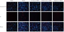
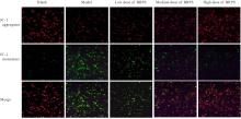
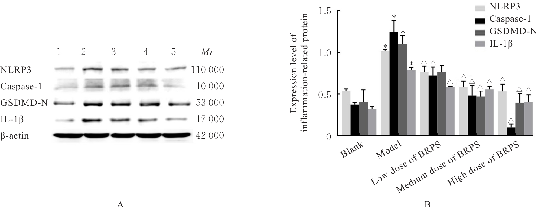
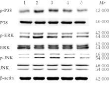
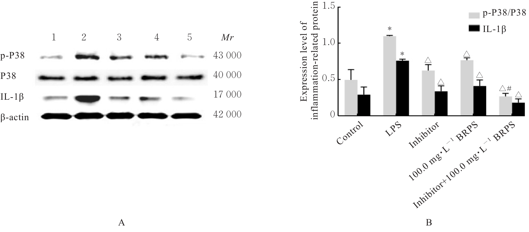
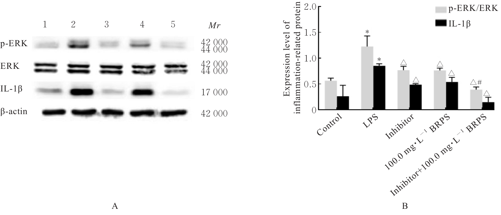
目的 探讨草苁蓉多糖(BRPS)对脂多糖(LPS)诱导的THP-1巨噬细胞炎症反应的影响,并阐明其作用机制。 方法 将THP-1单核细胞分化为巨噬细胞,采用LPS诱导THP-1巨噬细胞,建立炎症模型。CCK-8法检测不同浓度(0、100、200、500、1 000和2 000 μg·L-1)LPS及不同浓度(0、12.5、25.0、50.0、100.0和200.0 mg·L-1)BRPS处理后THP-1巨噬细胞存活率,选取后续实验药物浓度。将THP-1巨噬细胞分为空白组、模型组、低剂量BRPS组(25.0 mg·L-1 BRPS)、中剂量BRPS组(50.0 mg·L-1 BRPS)和高剂量BRPS组(100.0 mg·L-1 BRPS)。采用P38抑制剂SB203580、ERK抑制剂U0126、c-Jun氨基末端激酶(JNK)抑制剂SP600125和核因子κB(NF-κB)抑制剂BAY11-7082对THP-1细胞进行验证。另取THP-1细胞,分为对照组、LPS组、抑制剂组、100.0 mg·L-1 BRPS组和抑制剂+100.0 mg·L-1 BRPS组。酶联免疫吸附试验(ELISA)法检测各组THP-1巨噬细胞培养液中肿瘤坏死因子α(TNF-α)、白细胞介素(IL)-6和IL-1β水平,2,7-二氯荧光素二乙酸酯(DCFH-DA)荧光探针法检测各组THP-1巨噬细胞中活性氧(ROS)水平,Hoechst33342/PI荧光染色法观察各组THP-1巨噬细胞膜损伤情况,JC-1荧光染色法观察各组THP-1巨噬细胞线粒体膜电位,蛋白印迹法检测各组THP-1巨噬细胞中环氧合酶2(COX-2)、高迁移率族蛋白B1(HMGB1)、NOD样受体热蛋白结构域相关蛋白3(NLRP3)、含半胱氨酸的天冬氨酸蛋白酶(Caspase)-1、消皮素D(GSDMD)-N、IL-1β、丝裂原活化蛋白激酶(MAPK)和NF-κB相关蛋白表达水平。 结果 CCK-8法检测,LPS浓度为100~2 000 μg·L-1时,THP-1巨噬细胞存活率均>90%;与0 μg·L-1 LPS组比较,100、200、500、1 000和2 000 μg·L-1 LPS组THP-1巨噬细胞培养液中IL-6水平均明显升高(P<0.05),提示巨噬细胞炎症反应明显增强,因此选用100 μg·L-1 LPS构建炎症模型;12.5、25.0、50.0、100.0和200.0 mg·L-1 BRPS处理THP-1巨噬细胞,THP-1巨噬细胞存活率分别为91.2%、93.8%、91.4%、90.6%和91.8%,选取25.0、50.0和100.0 mg·L-1 BRPS作为后续实验中低、中和高剂量BRPS组药物浓度。ELISA法检测,与空白组比较,模型组THP-1巨噬细胞培养液中IL-6、TNF-α和IL-1β水平均明显升高(P<0.05);与模型组比较,低、中和高剂量BRPS组THP-1巨噬细胞培养液中IL-6、TNF-α和IL-1β水平均明显降低(P<0.05)。DCFH-DA荧光探针法检测,与空白组比较,模型组THP-1巨噬细胞中ROS水平明显升高(P<0.05);与模型组比较,低、中和高剂量BRPS组THP-1巨噬细胞中ROS水平均明显降低(P<0.05)。Hoechst33342/PI荧光染色法观察,与空白组比较,模型组THP-1巨噬细胞膜损伤程度明显增加;与模型组比较,低、中和高剂量BRPS组THP-1巨噬细胞膜损伤程度明显减少。JC-1荧光染色法观察,空白组THP-1巨噬细胞线粒体膜电位较高;与空白组比较,模型组THP-1巨噬细胞线粒体跨膜电位明显降低;与模型组比较,低、中和高剂量BRPS组THP-1巨噬细胞线粒体跨膜电位逐渐升高。蛋白印迹法检测,与空白组比较,模型组THP-1巨噬细胞中COX-2、HMGB1、NLRP3、Caspase-1、GSDMD-N和IL-1β蛋白表达水平及p-P38/P38、p-ERK/ERK、p-JNK/JNK和p-NF-κB/NF-κB比值均明显升高(P<0.05);与模型组比较,中和高剂量BRPS组THP-1巨噬细胞中HMGB1、NLRP3、Caspase-1、GSDMD-N和IL-1β蛋白表达水平及p-P38/P38、p-ERK/ERK、p-JNK/JNK和p-NF-κB/NF-κB比值均明显降低(P<0.05),低剂量BRPS组THP-1巨噬细胞中NLRP3、Caspase-1和IL-1β蛋白表达水平均明显降低(P<0.05),高剂量BRPS组THP-1巨噬细胞中COX-2蛋白表达水平明显降低(P<0.05);与对照组比较,LPS组THP-1巨噬细胞p-P38/P38、p-ERK/ERK、p-JNK/JNK和p-NF-κB/NF-κB比值及IL-1β蛋白表达水平均明显升高(P<0.05);与LPS组比较,抑制剂组、100 mg·L-1 BRPS组和抑制剂+100 mg·L-1 BRPS组THP-1巨噬细胞中p-P38/P38、p-ERK/ERK、p-JNK/JNK和p-NF-κB/NF-κB比值及IL-1β蛋白表达水平均明显降低(P<0.05);与抑制剂组比较,抑制剂+ 100 mg·L-1 BRPS组THP-1巨噬细胞p-P38/P38、p-ERK/ERK、p-JNK/JNK和p-NF-κB/NF-κB比值均明显降低(P<0.05)。 结论 BRPS抑制THP-1细胞巨噬细胞的炎症反应,其机制可能与BRPS调控MAPK和NF-κB信号通路有关。

关键词: 草苁蓉多糖, NOD样受体热蛋白结构域相关蛋白3, 丝裂原活化蛋白激酶, 核因子κB, 焦亡

Abstract:

Objective To discuss the effect of Boschnikia rossica polysaccharides rapa polysaccharides (BRPS) on lipopolysaccharide (LPS)-induced inflammatory responses in the THP-1 macrophages, and to clarify its mechanism. Methods The THP-1 monocytes were differentiated into the macrophages, and the inflammation model was established using LPS to induce the THP-1 macrophages. CCK-8 method was used to detect the survial rates of the THP-1 macrophages after treated with different concentrations (0, 100, 200, 500, 1 000, and 2 000 μg·L-1) of LPS and different concentrations (0, 12.5, 25.0, 50.0, 100.0, and 200.0 mg·L-1) of BRPS to select the concentrations for the subsequent experiments. The THP-1 macrophages were divided into blank group, model group, low dose of BRPS group (25.0 mg·L-1 BRPS), medium dose of BRPS group (50.0 mg·L-1 BRPS), and high dose of BRPS group (100.0 mg·L-1 BRPS). P38 inhibitor SB203580, ERK inhibitor U0126, c-Jun N-terminal kinase(JNK) inhibitor SP600125, and nuclear factor of kappa B(NF-κB) inhibitor BAY11-7082 were used to verify the effects on THP-1 cells. The THP-1 cells were divided into control group, LPS group, inhibitor group, 100.0 mg·L-1 BRPS group, and inhibitor+100.0 mg·L-1 BRPS group. ELISA method was used to detect the levels of tumor necrosis factor α (TNF-α), interleukin (IL)-6, and IL-1β in culture fluid of the THP-1 macrophages in various groups; DCFH-DA fluorescence probe method was used to detect the reactive oxygen species (ROS) levels in the THP-1 macrophages in various groups; Hoechst33342/PI fluorescence staining method was used to detect the membrane damage in the THP-1 macrophages in various groups; JC-1 fluorescence staining was used to observe mitochondrial membrane potential in the THP-1 macrophages in various groups; Western blotting method was used to detect the expression levels of cyclooxygenase-2 (COX-2), high mobility group protein B1 (HMGB1), NOD-like receptor thermal protein domain assciated protein 3 (NLRP3), cysteinyl aspartate specific protease (Caspase)-1, gasdermin D (GSDMD)-N, IL-1β, mitogen-activated protein kinase (MAPK), and nuclear factor-kappa B (NF-κB) related proteins in the THP-1 macrophages in various groups. Results The CCK-8 method results showed that when the LPS concentration was 100-2 000 μg·L-1, the survival rates of the THP-1 macrophages were over 90%. Compared with 0 μg·L-1 LPS group, the IL-6 levels in culture fluid of the THP-1 macrophages in 100, 200, 500, 1 000, and 2 000 μg·L-1 LPS group were increased (P<0.05), indicating a significant enhancement of the inflammatory response in the macrophages, so 100 μg·L-1 LPS was used to construct the inflammation model.After treated with 12.5, 25.0, 50.0, 100.0, and 200.0 mg·L-1 BRPS, the survival rates of the THP-1 macrophage were 91.2%, 93.8%, 91.4%, 90.6%, and 91.8%, respectively, so 25.0, 50.0, and 100.0 mg·L-1 BRPS were selected as the drug concentrations for low, medium, and high doses of BRPS groups in the subsequent experiments.The ELISA results showed that compared with blank group, the levels of IL-6, TNF-α, and IL-1β in culture fluid of the THP-1 macrophages in model group were increased (P<0.05); compared with model group, the levels of IL-6, TNF-α, and IL-1β in low, medium, and high doses of BRPS groups were decreased (P<0.05). The DCFH-DA fluorescence probe method results showed that compared with blank group, the ROS level in the THP-1 macrophages in model group was increased (P<0.05); compared with model group,the ROS levels in low, medium, and high doses of BRPS groups were decreased (P<0.05). The Hoechst33342/PI fluorescence staining results showed that compared with blank group, the degree of membrane damage in the THP-1 macrophages in model group was increased; compared with model group, the degrees of membrane damage in low, medium, and high doses of BRPS groups were decreased. The JC-1 fluorescence staining results showed that compared with blank group, the mitochondrial membrane potential in the THP-1 macrophages in model group was decreased significantly; compared with model group, the mitochondrial membrane potential in low, medium, and high doses of BRPS groups were increased gradually.The Western blotting results showed that compared with blank group, the expression levels of COX-2, HMGB1, NLRP3, Caspase 1, GSDMD-N, and IL-1β proteins and the ratios of p-P38/P38, p-ERK/ERK, p-JNK/JNK, and p-NF-κB/NF-κB in the THP-1 macrophages in model group were increased (P<0.05); compared with model group, the expression levels of HMGB1, NLRP3, Caspase-1, GSDMD-N, and IL-1β proteins and the ratios of p-P38/P38, p-ERK/ERK, p-JNK/JNK, and p-NF-κB/NF-κB in the THP-1 macrophages in medium and high doses of BRPS groups were decreased (P<0.05), the expression levels of NLRP3, Caspase-1, and IL-1β proteins in the cells in low dose of BRPS group were decreased (P<0.05), the expression level of COX-2 protein in the cells in high dose of BRPS group was decreased (P<0.05). Compared with control group, the ratios of p-P38/P38, p-ERK/ERK, p-JNK/JNK, and p-NF-κB/NF-κB, and the expression level of IL-1β protein in the THP-1 macrophages in LPS group were increased (P<0.05); compared with LPS group, the ratios of p-P38/P38, p-ERK/ERK, p-JNK/JNK, and p-NF-κB/NF-κB, and the expression level of IL-1β protein in the THP-1 macrophages in inhibitor group, 100 mg·L-1 BRPS group, and inhibitor+100 mg·L-1 BRPS group were decreased (P<0.05); compared with inhibitor group, the ratios of p-P38/P38, p-ERK/ERK, p-JNK/JNK, and p-NF-κB/NF-κB in the THP-1 macrophages in inhibitor+100 mg·L-1 BRPS group were decreased (P<0.05). Conclusion BRPS inhibits the inflammatory response of the THP-1 macrophages, which may be related to the MAPK and NF-κB signaling pathways regulated by BRPS.

Key words: Boschnikia rossica polysaccharides, NOD-like receptor family pyrin domain-containing protein 3, Mitogen-activated protein kinase, Nuclear factor-κB, Pyroptosis

中图分类号: 

  • R285.5