吉林大学学报(医学版) ›› 2021, Vol. 47 ›› Issue (1): 133-138.doi: 10.13481/j.1671-587x.20210118

• 基础研究 • 上一篇    下一篇

妊娠期糖尿病对新生子代大鼠膈肌功能的影响

张瑞丽1,杨筱青1,张晓鸽2,郭华峰1(),朱继红3()   

  1. 1.河南省郑州市妇幼保健院产科,河南 郑州 450012
    2.郑州大学第一附属医院产科,河南 郑州 450000
    3.吉林大学第一医院生殖中心,吉林 长春 130021
  • 收稿日期:2020-07-27 出版日期:2021-01-28 发布日期:2021-01-27
  • 通讯作者: 郭华峰,朱继红 E-mail:ghf0905@163.com;jhzhu@jlu.edu.cn
  • 作者简介:张瑞丽(1988-),女,河南省郑州市人,医学硕士,主要从事妊娠期糖尿病临床方面的研究。
  • 基金资助:
    河南省卫健委医学科技攻关计划联合共建项目(LHGJ20191132);河南省郑州市科技局普通科技公关计划项目(20150180)

Effect of gestational diabetes mellitus on diaphragmatic function of newborn offsprings of rats

Ruili ZHANG1,Xiaoqing YANG1,Xiaoge ZHANG2,Huafeng GUO1(),Jihong ZHU3()   

  1. 1.Department of Obstetrics,Women and Infants Hospital of Zhengzhou,Henan Province,Zhengzhou 450012,China
    2.Department of Obstetrics,First Affiliated Hospital,Zhengzhou University,Zhengzhou 450000,China
    3.Department of Reproductive Center,First Hospital,Jilin University,Changchun 130021,China
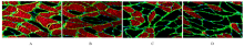
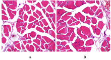
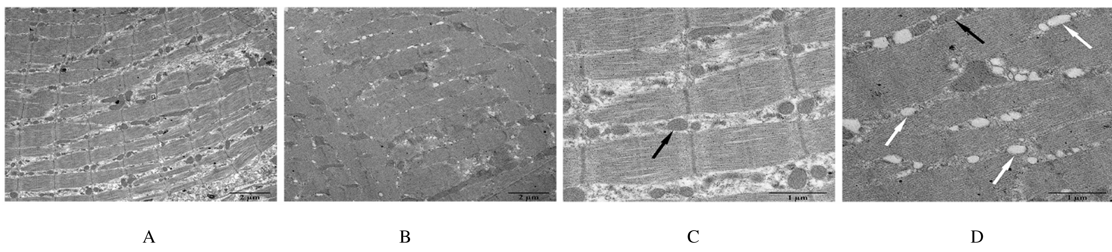
  • Received:2020-07-27 Online:2021-01-28 Published:2021-01-27
  • Contact: Huafeng GUO,Jihong ZHU E-mail:ghf0905@163.com;jhzhu@jlu.edu.cn

摘要: 目的

探讨妊娠期糖尿病(GDM)对新生子代大鼠膈肌功能和氧化应激水平的影响,阐明其可能作用机制。

方法

44只怀孕的8周龄SD雌性大鼠随机分为正常对照组和GDM模型组。妊娠第1天,GDM组大鼠单次腹腔注射35 mg·kg-1 链脲佐菌素(STZ),建立GDM大鼠模型(建模成功18只);正常对照组大鼠单次腹腔注射相同体积的柠檬酸-柠檬酸钠缓冲液。分娩后,体外检测2组新生子代大鼠的膈肌收缩力,免疫荧光法检测子代大鼠膈肌纤维横截面积,常规HE染色观察子代大鼠膈肌病理形态表现,透射电镜下观察子代大鼠膈肌超微结构,采用试剂盒检测子代大鼠膈肌组织中丙二醛(MDA)和羰基化蛋白水平及超氧化物歧化酶(SOD)和过氧化氢酶(CAT)活性。

结果

体外膈肌收缩力检测,与正常对照组比较,GDM组子代大鼠膈肌最大紧张收缩力、最大强直收缩力和不同刺激频率下的膈肌收缩力均明显降低(P<0.05),膈肌疲劳指数明显升高(P<0.05)。免疫荧光法检测,与正常对照组比较,GDM组子代大鼠膈肌组织中Ⅰ型和Ⅱ型膈肌纤维横截面积均明显缩小(P<0.05)。HE染色,2组子代大鼠膈肌纤维结构完整,排列整齐、致密,膈肌细胞无明显肿胀,细胞核染色清晰。透射电镜下观察,正常对照组子代大鼠膈肌纤维排列整齐,明暗带及“Z”线规整且清晰可见,线粒体结构完整,嵴成隔板状,清晰可见;GDM组子代大鼠膈肌纤维明暗带及“Z”线无规则排列或消失,线粒体数量减少,更多线粒体出现凋亡呈空泡化。与正常对照组比较,GDM组子代大鼠膈肌组织中MDA和羰基化蛋白水平均明显升高(P<0.05),SOD和CAT活性明显降低(P<0.05)。

结论

GDM可引起子代大鼠膈肌收缩力降低、纤维横截面积减小和超微结构改变,而氧化应激水平的升高是影响其膈肌功能的原因之一。

关键词: 妊娠期糖尿病, 大鼠, 新生子代, 膈肌, 氧化应激

Abstract: Objective

To investigate the effects of gestational diabetes mellitus (GDM) on the diaphragmatic function and the oxidative stress levels of the newborn offsprings of the SD rats, and to clarify the possible mechanisms,

Methods

Forty-four pregnant female SD rats(aged 8 weeks) were randomly divided into normal control group and GDM model group. On the first day after pregnancy, the rats in GDM group were intraperitoneally injected with 35 mg·kg-1 streptozotocin(STZ) to establish the GDM rat models. The rats in normal control group were intraperitoneally injected with the equal volume of citric acid-sodium citrate buffer. After delivery, the diaphragm contractile force of the newborn rats in two groups was measured in vitro. Meanwhile,the cross section area of the diaphragm fiber was detected by immunofluorescence method. Routine pathological staining and transmission electron microscope (TEM) were used to observe the phathomorphology and ultrastructure of diaphragm tissue of the newborn offsprings of the rats. The levels of malondialdehyde(MDA) and carbonylated protein and the activities of superoxidase dismutase(SOD) and catalase(CAT) in the diaphragm tissue were measured with commercial kits.

Results

The results of contractile force detection in vitro showed that compared with normal control group, the diaphragmatic maximum tetanic force, maximum tension force, and contractile force under different stimulation frequencies of the newborn offsprings in GDM group were all significantly decreased(P<0.05), and the diaphragmatic fatigue index was increased (P<0.05). The immunofluorescence detection results showed that the cross section areas of type Ⅰ and type Ⅱ fibers of the newborn offspring rats in GDM group were significantly smaller than those in normal control group (P<0.05). The HE staining results showed the neatly and densely arranged diaphragm fibers, intact nuclear without obvious cellular swelling, and clear nucleus staining in diaphragm tissue of the newborn offspvings in two groups. The TEM observation results showed that the diaphragm fibers in normal control group were neatly arranged with regular and visible light-dark bands or “Z” lines, and the mitochondrial structure was intact with partitioned and visible ridges; however, the diaphragm fibers in GDM group were arranged with irregularly or even the light-dark bands and “Z” lines were disappeared,the amount of mitochondria was decreased,and the signs of mitochondrial apoptosis or vacuolation were found in diaphragm tissue of the newborn offsprings GDM group. Compared with normal control group, the levels of MDA and carbonylated protein in diaphragm tissue of the newborn offspring rats in GDM group were significantly increased(P<0.05), and the activities of SOD and CAT in GDM group were significantly decreased(P<0.05).

Conclusion

GDM can cause the decrease of diaphragmatic contractile force, reduction of cross section area of diaphragmatic fibers and changes of diaphragmatic ultrastructures,and the increase in oxidative stress level is one of the reasons that affect its diaphragm function.

Key words: gestational diabetes mellitus, rats, newborn offspring, diaphragm, oxidative stress

中图分类号: 

  • R714.25