吉林大学学报(医学版) ›› 2024, Vol. 50 ›› Issue (5): 1432-1437.doi: 10.13481/j.1671-587X.20240530

• 临床医学 • 上一篇    

腹壁寄生性平滑肌瘤并发播散性腹膜平滑肌瘤病1例报告及文献复习

张金萍1,佟玲玲1,高璐1,程洪晶2,盛敏佳1()   

  1. 1.吉林大学中日联谊医院妇产科, 吉林 长春 130033
    2.吉林大学中日联谊医院病理科, 吉林 长春 130033
  • 收稿日期:2023-09-28 出版日期:2024-09-28 发布日期:2024-10-28
  • 通讯作者: 盛敏佳 E-mail:shengmj@jlu.edu.cn
  • 作者简介:张金萍(1997-),女,吉林省长春市人,住院医师,在读硕士研究生,主要从事妇产科基础和临床方面的研究。
  • 基金资助:
    吴阶平医学基金会项目(320.6750.2021-19-1)

Parasitic leiomyoma of abdominal wall complicated with disseminated peritoneal leiomyomatosis : A case report and literature review

Jinping ZHANG1,Lingling TONG1,Lu GAO1,Hongjing CHENG2,Minjia SHENG1()   

  1. 1.Department of Obstetrics and Gynecology,China-Japan Union Hospital,Jilin University,Changchun 130033,China
    2.Department of Pathology,China-Japan Union Hospital,Jilin University,Changchun 130033,China
  • Received:2023-09-28 Online:2024-09-28 Published:2024-10-28
  • Contact: Minjia SHENG E-mail:shengmj@jlu.edu.cn

摘要:

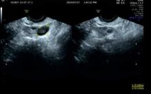
目的 探讨腹腔镜下子宫肌瘤核出术后出现腹壁寄生性平滑肌瘤(PM)并发播散性腹膜平滑肌瘤病(DPL)患者的诊疗经过,以提高对该病的临床认识水平和诊疗水平。 方法 收集1例腹腔镜下子宫肌瘤核出术后出现腹壁PM并发DPL患者的临床资料,分析其发病原因、临床特点、诊断、鉴别诊断和治疗经过,并回顾相关文献。 结果 患者,女性,49岁,因自觉腹部包块1年入院。专科查体,脐左下侧腹壁扪及大小约为6 cm×4 cm包块,活动性欠佳,边界尚清,无压痛;脐下方右侧腹部扪及大小约为7 cm×5 cm包块,活动性尚可,边界清晰,无压痛。妇科彩超,脐孔左下方皮下可探及大小约为6.6 cm×2.7 cm的低回声。脐孔下方腹腔内可探及大小约为7.6 cm×3.3 cm的低回声。浅表局部彩超,左下腹腹直肌内可见大小约为5.79 cm×2.55 cm×4.74 cm低回声,边缘光滑,较浅处距皮约1.97 cm,较深处距皮约4.73 cm,深方及浅方未穿破腹直肌外膜,深方紧邻腹膜。诊断为子宫肌瘤、腹部肿物和子宫肌瘤核出术后。择期在静吸复合全麻下行开腹子宫平滑肌瘤核出术、腹壁平滑肌瘤切除术和腹膜平滑肌瘤切除术,手术过程顺利,患者术后恢复良好,顺利出院。 结论 PM和DPL无典型临床特点,需借助影像学检查等辅助诊断,手术探查为主要治疗手段,多为良性,有恶性转化可能,患者术后需进一步随访。

关键词: 寄生性平滑肌瘤, 播散性腹膜平滑肌瘤病, 种植, 病例报告

Abstract:

Objective To discuss the diagnosis and treatment process of the patients with parasitic leiomyoma (PM) of the abdominal wall complicated with disseminated peritoneal leiomyomatosis (DPL) after laparoscopic myomectomy,and to improve the clinical understanding and management of this condition. Methods The clinical data of one patient with PM of the abdominal wall complicated with DPL after laparoscopic myomectomy were collected. The causes, clinical features, diagnosis, differential diagnosis and treatment process were analyzed, and the relevant literatures were reviewed. Results The patient, a 49-year-old woman, was admitted due to a self-discovered abdominal mass lasting for one year. The physical examination results showed a palpable mass, approximately 6 cm×4 cm, in the lower left abdominal wall with poor mobility, with clear borders, and without tenderness. Another palpable mass, approximately 7 cm×5 cm, was found in the lower right abdomen with fair mobility,with clear borders, and without tenderness. The gynecological ultrasonography results showed a hypoechoic area of approximately 6.6 cm×2.7 cm in the subcutaneous tissue below the left umbilicus and another hypoechoic area of approximately 7.6 cm×3.3 cm in the abdominal cavity below the umbilicus. The superficial ultrasonography of the local area showed a hypoechoic area of approximately 5.79 cm×2.55 cm×4.74 cm within the left lower abdominal rectus muscle, with smooth edges, located 1.97 cm from the skin at its shallowest point and 4.73 cm at its deepest point, without penetration of the rectus sheath but adjacent to the peritoneum. The patient was diagnosed as uterine leiomyoma, abdominal mass, and post-myomectomy status. The elective surgeries for uterine leiomyoma enucleation, abdominal wall leiomyoma excision, and peritoneal leiomyoma excision were performed under combined intravenous-inhalation anesthesia. The operation procedure was successful, and the patient recovered well and was discharged smoothly. Conclusion PM and DPL lack typical clinical features and require imaging examinations for diagnosis. Surgical exploration is the main treatment modality, and while PM and DPL are generally benign, there is a potential for malignant transformation, and the patients need further postoperative follow-up.

Key words: Parasitic leiomyoma, Disseminated peritoneal leiomyomatosis, Implantation, Case report

中图分类号: 

  • R711.74