吉林大学学报(医学版) ›› 2023, Vol. 49 ›› Issue (3): 777-781.doi: 10.13481/j.1671-587X.20230329

• 临床医学 • 上一篇    下一篇

颅内动脉瘤破裂并发急性心肌梗死1例报告及文献复习

罗云珂1,张剑1,张文文1,段宗生1,王虎山1(),王以恒2   

  1. 1.吉林大学第一医院麻醉科,吉林 长春 130021
    2.吉林大学第一医院神经血管外科,吉林 长春 130021
  • 收稿日期:2022-02-10 出版日期:2023-05-28 发布日期:2023-06-20
  • 通讯作者: 王虎山 E-mail:421058260@qq.com
  • 作者简介:罗云珂(1995-),女,四川省内江市人,在读硕士研究生,主要从事临床麻醉学方面的研究。
  • 基金资助:
    吉林省科技厅科技发展计划项目(20210101304JC)

Intracranial aneurysm rupture complicated with acute myocardial infarction: A case report and literature review

Yunke LUO1,Jian ZHANG1,Wenwen ZHANG1,Zongsheng DUAN1,Hushan WANG1(),Yiheng WANG2   

  1. 1.Department of Anesthesiology, First Hospital, Jiin University, Changchun 130021, China
    2.Department of Neurovascular Surgery, First Hospital, Jiin University, Changchun 130021, China
  • Received:2022-02-10 Online:2023-05-28 Published:2023-06-20
  • Contact: Hushan WANG E-mail:421058260@qq.com

摘要:

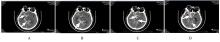
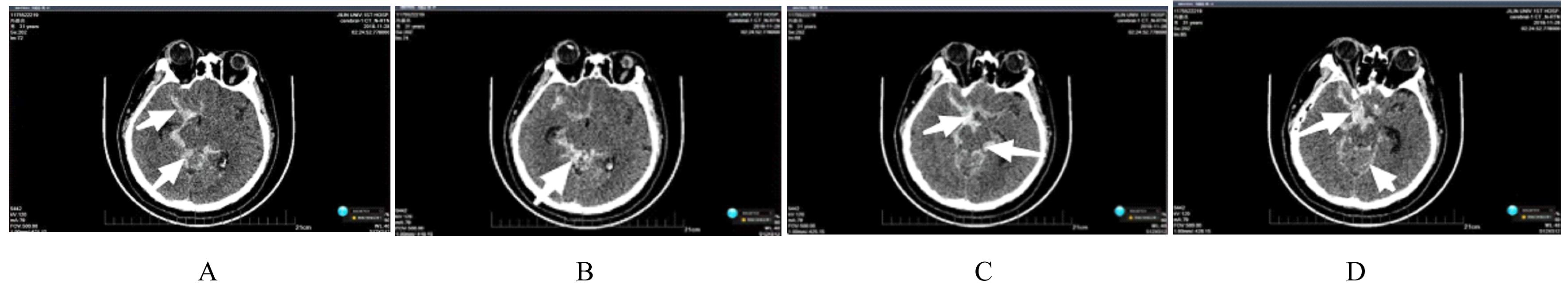
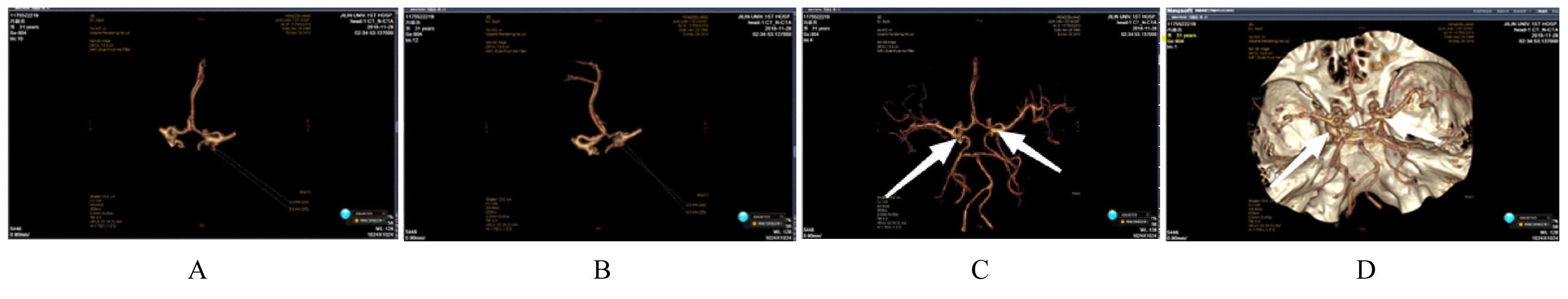
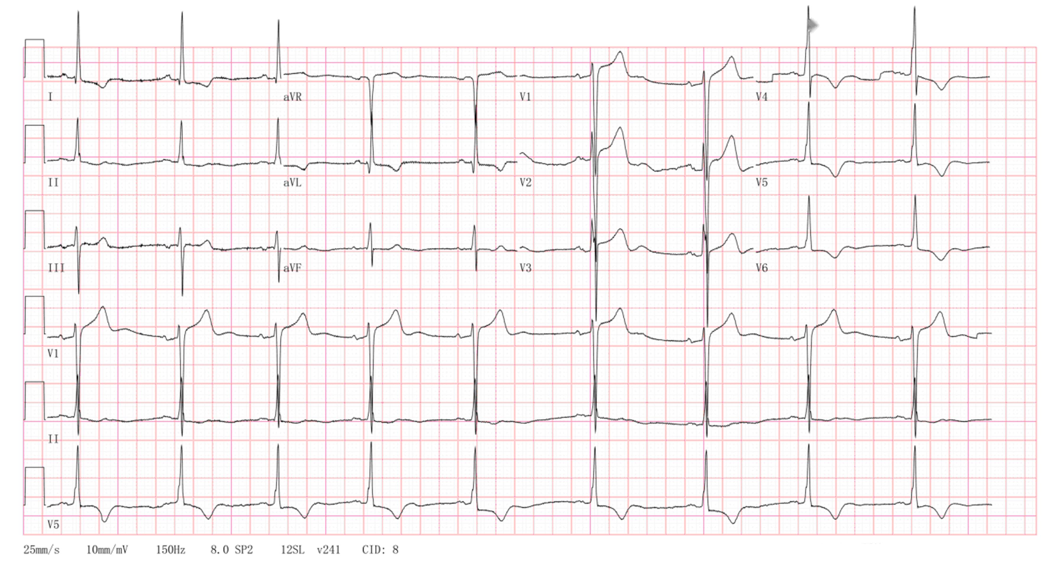
目的 探讨1例颅内动脉瘤破裂同时并发急性心肌梗死(AMI)患者的诊治过程,为该病的诊断、治疗和麻醉提供参考。 方法 回顾性分析1例颅内动脉瘤破裂同时并发AMI患者的临床资料、影像学表现和麻醉方法,结合相关文献进行分析。 结果 患者因突发剧烈头疼伴恶心呕吐4 h入院。入院1 h 10 min后颅脑多排CT显示蛛网膜下腔出血(SAH);双侧脑室少许积液,颅内血管造影显示右侧颈内动脉后交通段瘤。入院2 h 52 min后,肌红蛋白为483.6 μg·L-1,肌钙蛋白I为4.990 μg·L-1,肌酸激酶同工酶MB(CK-MB)为45.70 μg·L-1。入院16 h 31 min后心电图显示窦性心动过缓,左心室肥大,ST-T段改变。患者初诊为SAH、AMI和高血压病3级(极高危)。采取早期综合治疗手段,3 d后患者行急诊脑动脉瘤夹闭术。麻醉方式选择气管插管麻醉,慎重选择麻醉药物,以取得最好的血流和麻醉效果。术中生命体征平稳,7 d后病情好转出院。 结论 颅内动脉瘤破裂同时并发AMI的患者,CT、颅内动脉造影和心肌标志物均为诊断和鉴别诊断的重要检查,控制血压是治疗和麻醉的关键。

关键词: 颅内动脉瘤, 急性心肌梗死, 颅内动脉造影, 心肌标志物, 病例报告

Abstract:

Objective To discuss the diagnosis and treatment of one patient with ruptured intracranial aneurysm complicated with acute myocardial infarction(AMI), and to provide the reference for the clinical diagnosis, treatment, and anesthesia of the disease. Methods The clinical data, imaging findings, and anesthesia methods of one patient with ruptured intracranial aneurysm complicated with AMI were retrospectively analyzed,and the analysis was performed combined with the relevant literatures. Results The patient was admitted to hospital due to sudden severe headache with nausea and vomiting for 4 h. A total of 1 h and 10 min after admission,the multi-slice CT results showed subarachnoid hemorrhage(SAH).There was little bilateral ventricular effusion, and the intracranial angiography results showed a tumor in the posterior communicating segment of the right internal carotid artery.A total of 2 h 52 min after admission,the myoglobin was 483.6 μg·L-1,the troponin I was 4.990 μg·L-1,the creatine kinase isoenzyme-MB(CK-MB) was 45.70 μg·L-1.A total of 16 h 31 min after admission, the ECG results showed sinus bradycardia, left ventricular hypertrophy, and ST-T segment changes. The initial diagnosis of the patient was SAH, AMI, and hypertension grade 3 (very high risk). After early comprehensive treatment, the patient underwent emergency clipping of cerebral aneurysm after 3 d.The anesthesia method was tracheal intubation ansthesia,and the anesthetic drugs were carefully selected to achieve the best blood flow and anesthesia effect.The vital signs of the patient were stable during the operation, and the condition of the patient was improved and discharged after 7 d. Conclusion For the patients with intracranial aneurysm rupture complicated with AMI,CT, intracranial angiography, and myocardial markers are the important examinations for the diagnosis and differential diagnosis; controlling the blood pressure is the key point for the treatment and anesthesia.

Key words: Intracranial aneurysms, Acute myocardial infarction, Intracranial angiography, Cardiac markers, Case report

中图分类号: 

  • R743