吉林大学学报(医学版) ›› 2026, Vol. 52 ›› Issue (1): 116-124.doi: 10.13481/j.1671-587X.20260112

• 基础研究 • 上一篇    下一篇

丰富环境通过转录因子EB介导自噬对缺血性脑卒中大鼠神经功能损伤的影响

朱慧艳,陈敏,李金贤,李春丽()   

  1. 新疆维吾尔自治区人民医院康复医学科,新疆 乌鲁木齐 830001
  • 收稿日期:2025-02-28 接受日期:2025-04-27 出版日期:2026-01-28 发布日期:2026-02-24
  • 通讯作者: 李春丽 E-mail:874897249@qq.com
  • 作者简介:朱慧艳(1987-),女,河南省周口市人,主治医师,医学硕士,主要从事脑卒中、脊髓损伤和骨关节疾病康复治疗及相关方面的研究。
  • 基金资助:
    新疆维吾尔自治区科技厅自然科学基金项目(2022D01C125);新疆维吾尔自治区科技厅自然科学基金项目(2024D01C72)

Effect of enriched environment on neurofunctional damage in rats with ischemic stroke via transcription factor EB-mediated autophagy

Huiyan ZHU,Min CHEN,Jinxian LI,Chunli LI()   

  1. Department of Rehabilitation Medicine,People’s Hospital,Xinjiang Uygur Autonomous Region,Urumqi 830001,China
  • Received:2025-02-28 Accepted:2025-04-27 Online:2026-01-28 Published:2026-02-24
  • Contact: Chunli LI E-mail:874897249@qq.com

摘要:

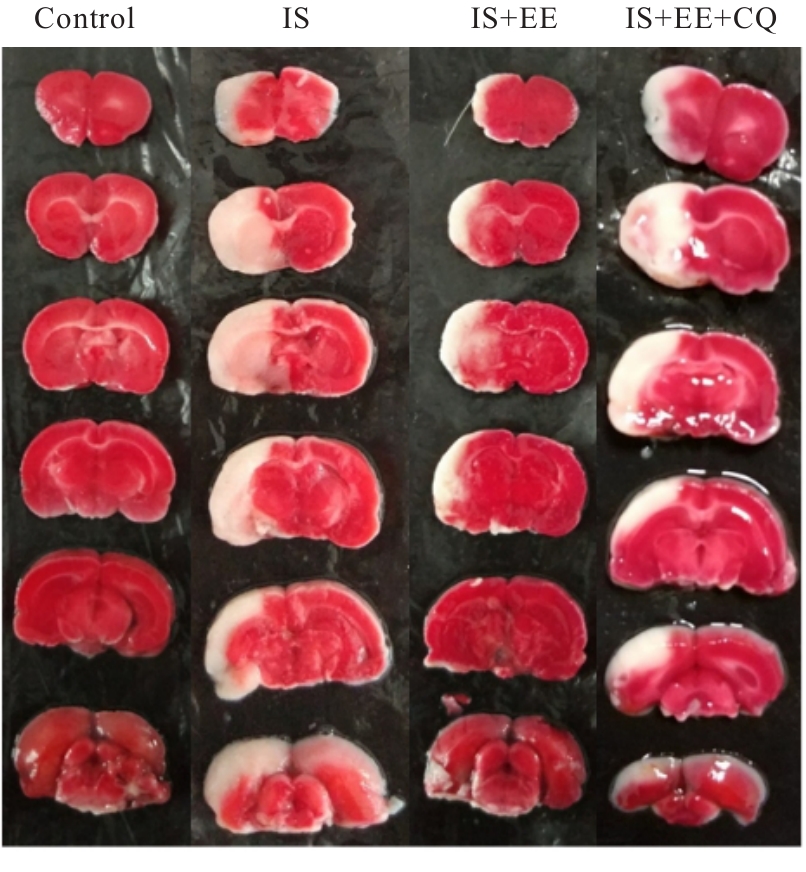
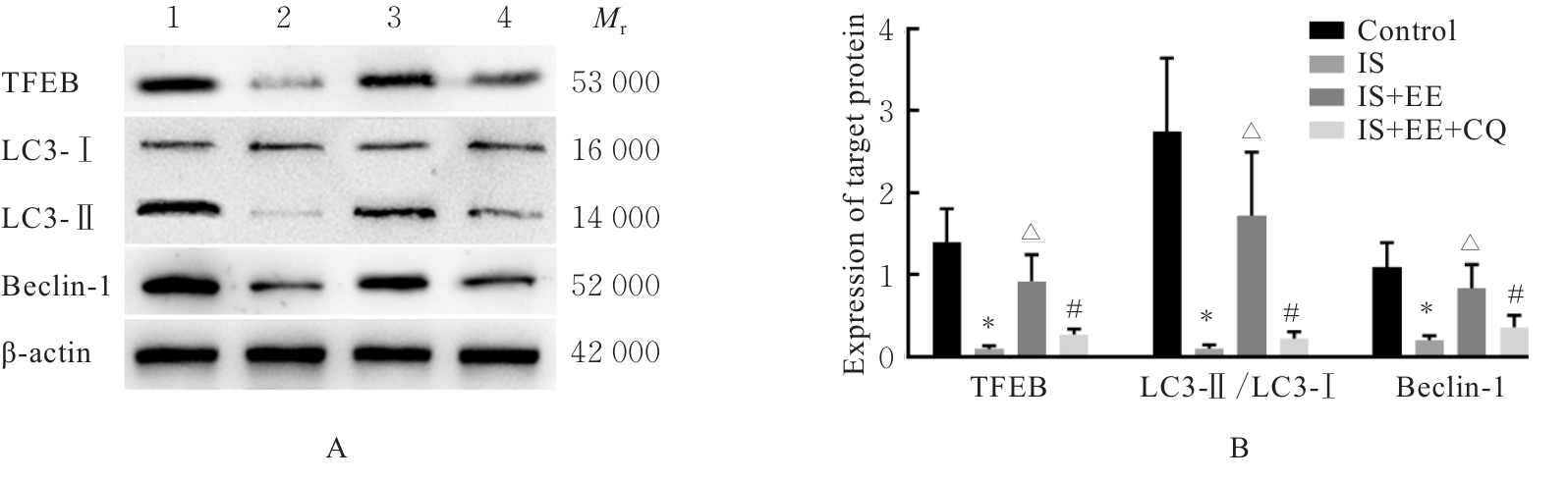
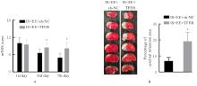
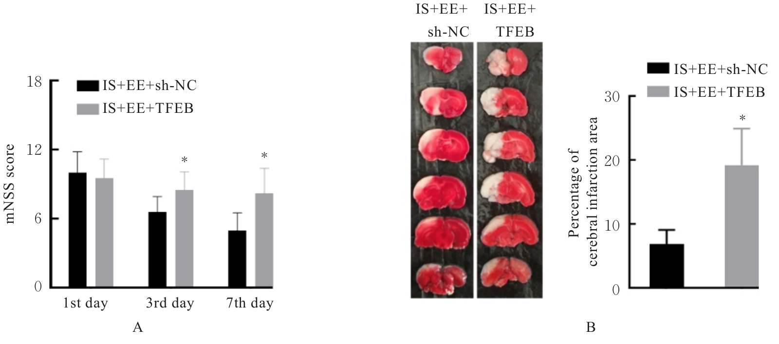
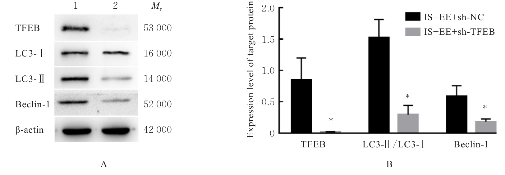
目的 探讨丰富环境(EE)对缺血性脑卒中(IS)损伤的影响,初步阐明转录因子EB(TFEB)蛋白在这一过程中发挥的作用以及EE与炎症反应和氧化应激反应的关系。 方法 实验Ⅰ,选取48只SD大鼠,随机分为对照组、IS组、IS+EE组和IS+EE+氯喹(CQ)组(IS+EE+CQ组),每组12只。实验Ⅱ,再取16只大鼠,随机分为IS+EE+sh-NC组和IS+EE+sh-TFEB组,每组8只。模型构建前,IS+EE+sh-TFEB组大鼠脑室注射TFEB shRNA沉默脑组织中TFEB基因表达。除对照组外,其余各组大鼠采用Longa线栓法构建IS模型。采用改良神经损伤严重程度评分(mNSS)法评估各组大鼠神经功能损伤程度,氯化三苯基四氮唑(TTC)染色法检测各组大鼠脑梗死区面积百分率,试剂盒检测各组大鼠炎性细胞因子和氧化应激因子水平,Western blotting法检测各组大鼠自噬相关蛋白表达水平。 结果 与对照组比较,IS组大鼠mNSS评分升高(P<0.05),缺血半暗带区脑组织中白细胞介素6(IL-6)、白细胞介素1β(IL-1β)、肿瘤坏死因子α(TNF-α)和丙二醛(MDA)水平明显升高(P<0.05),超氧化物歧化酶(SOD)活性明显降低(P<0.05),TFEB和Beclin-1蛋白表达水平以及微管相关蛋白1轻链3(LC3)-Ⅱ/LC3-Ⅰ比值明显降低(P<0.05);与IS组比较,IS+EE组大鼠mNSS评分和脑梗死面积百分率明显降低(P<0.05),缺血半暗带区脑组织中IL-6、IL-1β、TNF-α和MDA水平明显降低(P<0.05),SOD活性明显升高(P<0.05),TFEB和Beclin-1蛋白表达水平以及LC3-Ⅱ/LC3-Ⅰ比值明显升高(P<0.05)。与IS+EE组比较,IS+EE+CQ组大鼠mNSS评分和脑梗死面积百分率明显升高(P<0.05),缺血半暗带区脑组织中IL-6、IL-1β、TNF-α和MDA水平明显升高(P<0.05),SOD活性明显降低(P<0.05),TFEB和Beclin-1蛋白表达水平以及LC3-Ⅱ/LC3-Ⅰ比值明显降低(P<0.05)。与IS+EE+sh-NC组比较,IS+EE+sh-TFEB组大鼠mNSS评分降低(P<0.05),脑梗死区面积百分率明显升高(P<0.05),缺血半暗带区脑组织中TFEB和Beclin-1蛋白表达水平以及LC3-Ⅱ/LC3-Ⅰ比值明显降低(P<0.05)。 结论 EE对IS大鼠神经功能损伤具有明显的改善作用,其机制可能与EE通过升高TFEB蛋白表达诱导自噬、减轻脑缺血区神经炎症和氧化应激有关。

关键词: 丰富环境, 炎症, 自噬, 转录因子EB, 缺血性脑卒中

Abstract:

Objective To discuss the impact of an enriched environment (EE) on ischemic stroke (IS) injury, and to preliminarily clarify the role of transcription factor EB (TFEB) protein in this process as well as the relationship between EE and inflammatory response and oxidative stress response. Methods Forty?eight SD rats were randomly divided into control group, cerebral ischemia (IS) group, IS+EE group, and IS+EE+chloroquine (CQ) group (IS+EE+CQ group), with 12 rats in each group in experiment Ⅰ. Another 16 rats were randomly divided into IS+EE+sh-NC group and IS+EE+sh-TFEB group, with 8 rats in each group in experiment Ⅱ. Before model establishment, the rats in IS+EE+sh-TFEB group were injected with TFEB shRNA into the cerebral ventricle to silence TFEB expression in brain tissue. Except for control group, the IS model was established in the rats in the other groups using the Longa suture?occlusion method. The modified neurological severity score (mNSS) was used to evaluate the neurological function injury of the rats in various groups; triphenyltetrazolium chloride (TTC) staining was used to detect the percentages of cerebral infarction area of the rats in various groups; kits were used to detect the levels of inflammatory cytokines and oxidative stress factors of the rats in various groups; Western blotting method was used to detect the expression levels of autophagy-related proteins of the rats in various groups. Results Compared with control group, the mNSS score of the rats in IS group was increased (P<0.05), the levels of interleukin-6 (IL-6), interleukin-1β (IL-1β), tumor necrosis factor-α (TNF-α), and malondialdehyde (MDA) in the brain tissue in ischemic penumbra region were increased (P<0.05), the activity of superoxide dismutase (SOD) was decreased (P<0.05), and the expression levels of TFEB and Beclin-1 proteins and the ratio of microtubule-associated protein 1 light chain 3 (LC3)-Ⅱ/LC3-Ⅰ were decreased (P<0.05). Compared with IS group, the mNSS score and the percentage of cerebral infarction area of the rats in IS+EE group were decreased (P<0.05), the levels of IL-6, IL-1β, TNF-α, and MDA in the brain tissue in ischemic penumbra region were decreased (P<0.05), the activity of SOD was increased (P<0.05), and the expression levels of TFEB and Beclin-1 proteins and the ratio of LC3-Ⅱ/LC3-Ⅰ were increased (P<0.05). Compared with IS+EE group, the mNSS score and the percentage of cerebral infarction area of the rats in IS+EE+CQ group were increased (P<0.05), the levels of IL-6, IL-1β, TNF-α, and MDA in the brain tissue in ischemic penumbra region were increased (P<0.05), the activity of SOD was decreased (P<0.05), and the expression levels of TFEB and Beclin-1 proteins and the ratio of LC3-Ⅱ/LC3-Ⅰ were decreased (P<0.05). Compared with IS+EE+sh?NC group, the mNSS score of the rats in IS+EE+sh-TFEB group was decreased (P<0.05), the percentage of cerebral infarction area was increased (P<0.05), and the expression levels of TFEB and Beclin-1 proteins and the ratio of LC3-Ⅱ/LC3-Ⅰ in the brain tissue in ischemic penumbra region were decreased (P<0.05). Conclusion EE has a significant ameliorative effect on neurological function injury in the IS rats, and its mechanism may be related to EE inducing autophagy by increasing TFEB protein expression, thereby alleviating neuroinflammation and oxidative stress in the ischemic brain region.

Key words: Enriched environment, Inflammation, Autophagy, Transcription factor EB, Ischemic stroke

中图分类号: 

  • R743