吉林大学学报(医学版) ›› 2020, Vol. 46 ›› Issue (6): 1309-1314.doi: 10.13481/j.1671-587x.20200632

• 影像学 • 上一篇    

磁共振弥散加权成像ADC和rADC在腮腺良恶性肿瘤鉴别诊断中的应用

史灵雪1,刘硕1,郑雪微1,程晓亮1,陈昭辉2,马宝琳1,张红果1,丁军1()   

  1. 1.吉林大学中日联谊医院放射科,吉林 长春 130033
    2.吉林省长春铁北监狱医院放射科,吉林 长春 130052
  • 收稿日期:2020-03-25 出版日期:2020-11-28 发布日期:2021-01-27
  • 通讯作者: 丁军 E-mail:dingjun@jlu.edu.cn
  • 作者简介:史灵雪(1995-),女,河北省任丘市人,在读医学硕士,主要从事头颈部疾病影像诊断方面的研究。
  • 基金资助:
    吉林省教育厅科研基金资助课题(JJKH20201057KJ)

Application of magnetic resonance diffusion weighted imaging ADC and rADC in differential diagnosis of benign and malignant parotid gland tumors

Lingxue SHI1,Shuo LIU1,Xuewei ZHENG1,Xiaoliang CHENG1,Zhaohui CHEN2,Baolin MA1,Hongguo ZHANG1,Jun DING1()   

  1. 1.Department of Radiology,China-Japan Union Hospital,Jilin University,Changchun 130033,China
    2.Department of Radiology,Changchun Tiebei Prison Hospital,Jilin Province,Changchun 130052,China
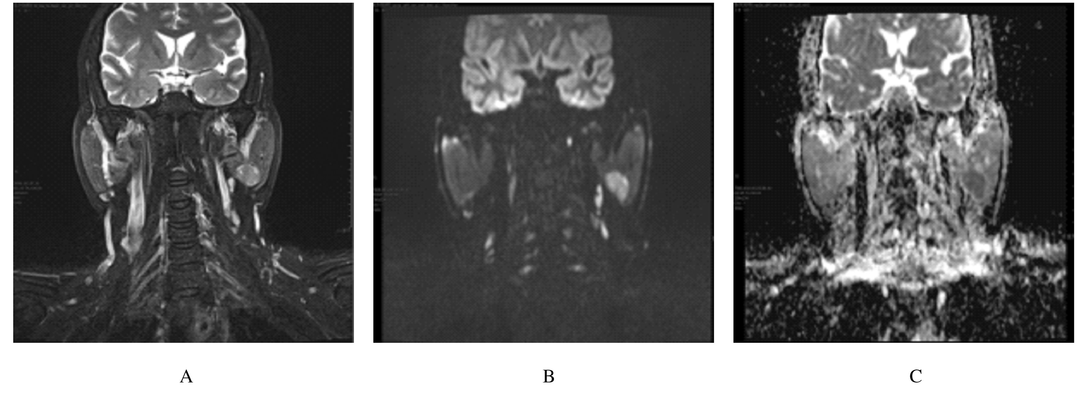
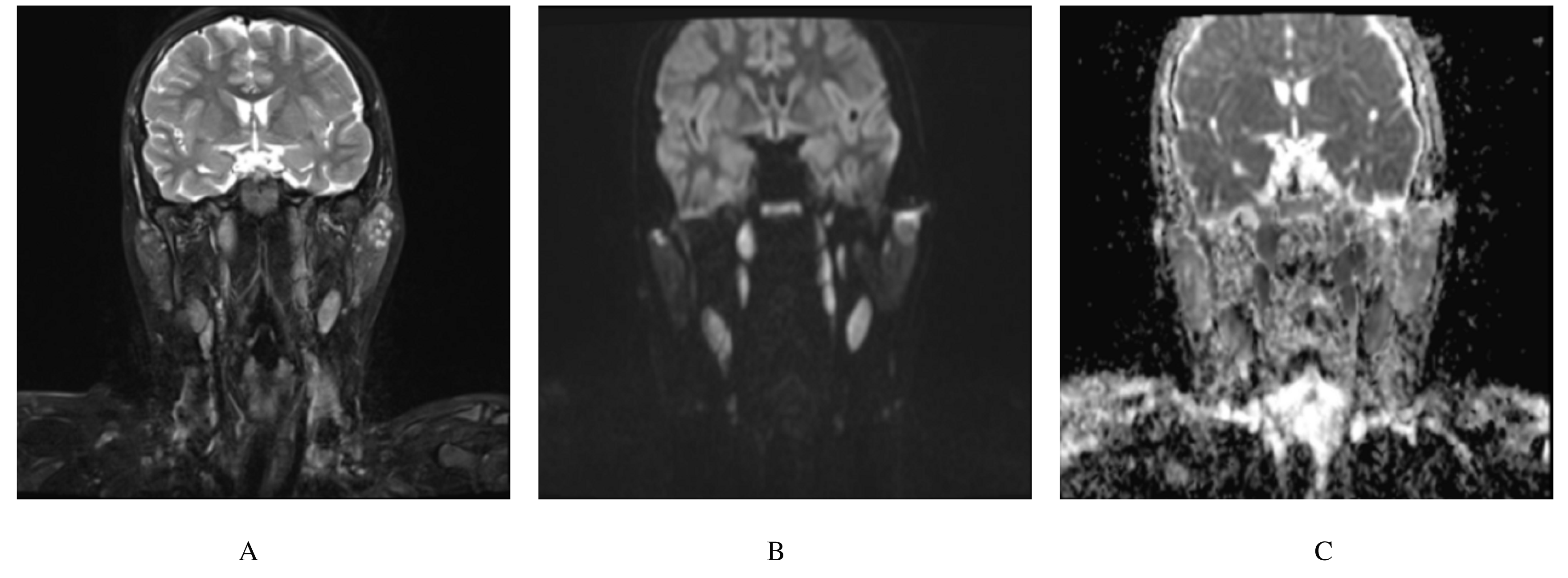
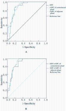
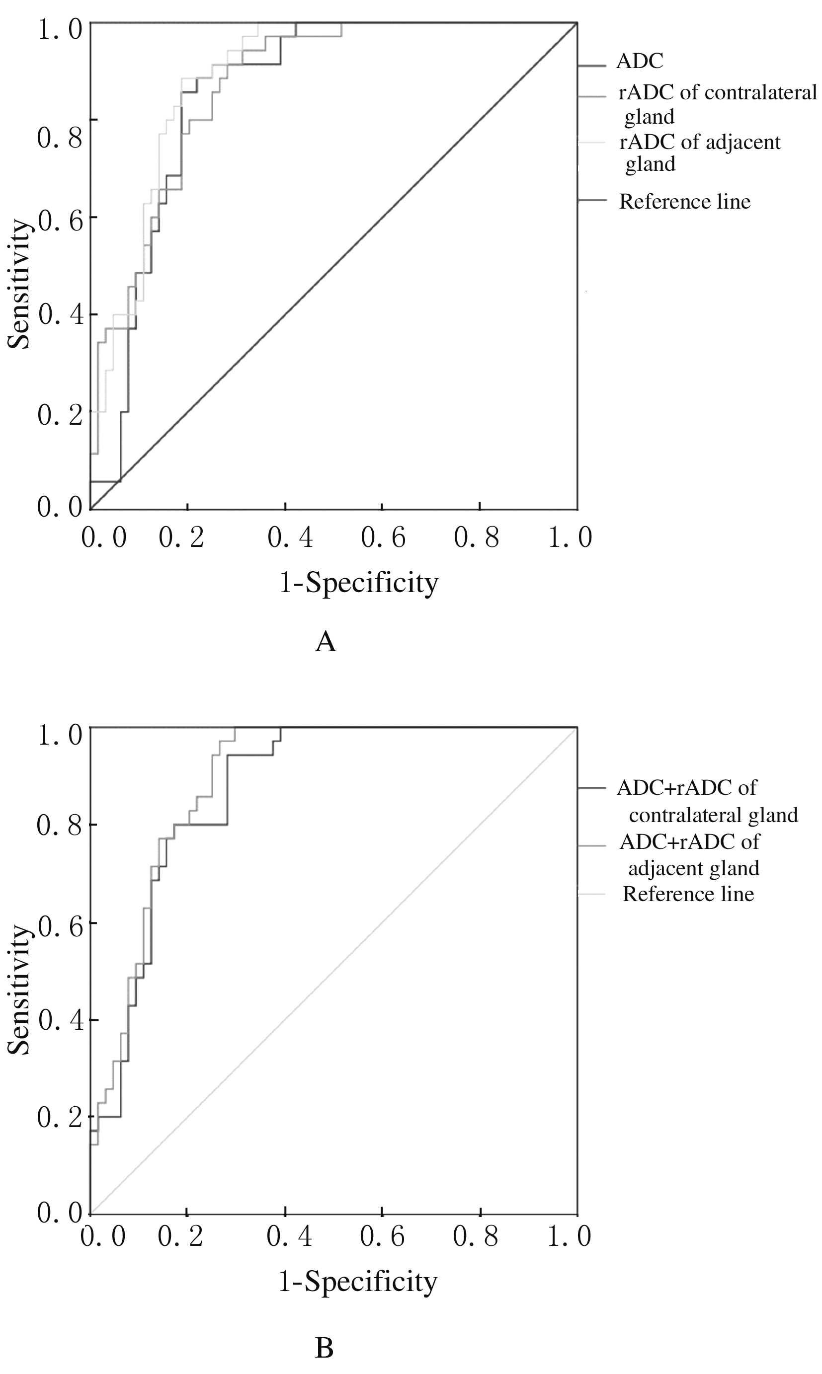
  • Received:2020-03-25 Online:2020-11-28 Published:2021-01-27
  • Contact: Jun DING E-mail:dingjun@jlu.edu.cn

摘要: 目的

探讨腮腺良恶性肿瘤的磁共振弥散加权成像(DWI)表观扩散系数(ADC)和表观扩散系数比值(rADC)在鉴别腮腺良恶性肿瘤中的诊断价值。

方法

选取经病理证实的腮腺肿瘤患者84例,根据患者术后病理类型分为腮腺良性肿瘤组和恶性肿瘤组,患者术前行常规MRI及DWI序列扫描,HE染色观察不同病理分型腮腺肿瘤组织病理形态表现,测定腮腺良恶性肿瘤及其临近腺体和腮腺良恶性肿瘤及其对侧腺体的ADC,计算相应rADC。

结果

腮腺良性肿瘤60例,恶性肿瘤24例;良性肿瘤多边界清晰,混合瘤和腺淋巴瘤是最常见的类型。T2WI显示病灶呈等、低信号;恶性肿瘤以黏液表皮样癌较为多见,形态不规则,边界模糊,信号欠均匀,常伴周围结构的侵犯及颈部淋巴结的增大。与腮腺良性肿瘤比较,腮腺恶性肿瘤的ADC降低(P<0.05),腮腺恶性肿瘤对侧腺体rADC和邻近腺体rADC均降低(P<0.05)。受试者工作特征(ROC)曲线检测,ADC、对侧腺体rADC和邻近腺体rADC曲线下面积(AUC)分别为0.859、0.874和0.894;多项指标联合并计算AUC结果显示ADC与对侧腺体rADC、ADC与邻近腺体rADC的 AUC分别为0.874和0.894。

结论

DWI的ADC和rADC能够为腮腺良恶性肿瘤的诊断及鉴别诊断提供参考依据,具有重要的临床指导意义。

关键词: 腮腺肿瘤, 磁共振成像, 弥散加权成像, 表观扩散系数, 表现扩散系数比值

Abstract: Objective

To investigate the diagnostic value of apparent diffusion coefficient (ADC) and apparent diffusion coefficient ratio (rADC)of diffusion weighted imaging(DWI) in the differentiation of benign and malignant parotid tumors.

Methods

Eighty-four patients with parotid gland tumor confirmed by pathology were selected. According to the postoperative pathological types, the patients were divided into benign tumor group and malignant tumor group.The patients received preoperative routine MRI and DWI sequence scanning.HE staining was used to detect the pathomorphology of benign and malignant parotid gland tumors and their parotid gland tumor tissue with different pathological types,and the ADC of benign and malignat parotid gland tumors and their adjacent glands of and contralateral glands of were measured, respectively.The corresponding rADC were calculated.

Results

There were 60 cases of benign parotid gland tumor and 24 cases of malignant parotid gland tumor;the benign tumor had the clear multi-boundary, and the mixed tumor and the adenomas were the most common types;the T2WI results showed the isostatic and low-signal lesions; the malignant parotid gland tumor was more common in mucoepidermoid carcinoma with irregular morphology, blurred boundaries and uneven signals, usually accompanied by invasion of peripheral structures and enlargement of cervical lymph nodes. Compared with benign parotid gland tumor,the ADC of parotid gland malignant tumor was decreased(P<0.05),and the rADC of contralateral glands and adjacent glands of parotid malignant tumor were decreased (P<0.05);the ROC curve detection results showed that the area under the curves (AUC) of ADC, rADC of contralateral,glands, and rADC of adjacent glands were 0.859, 0.874,and 0.894, respectively.The AUC of ADC and contralateral glands and the AUC of ADC and adjacent glands were 0.874 and 0.894 calculated by combined multiple indexes.

Conclusions

The ADC and rADC of DWI can provide the reference for the diagnosis and differential diagnosis of benign and malignant parotid gland tumors, which has important clinical significance.

Key words: parotid gland tumor, magnetic resonance imaging, diffusion weighted imaging, apparent diffusion coefficient, apparent diffusion coefficient ratio

中图分类号: 

  • R739.87