吉林大学学报(医学版) ›› 2022, Vol. 48 ›› Issue (2): 277-283.doi: 10.13481/j.1671-587X.20220202

• 基础研究 • 上一篇    下一篇

不同发育期小鼠卵巢组织中Wnt5a蛋白表达水平及其对卵母细胞自噬的影响

马亚博1,2,原筱潭1,2,谢贤国1,2,刘新峰1,2,徐金瑞1,2,杨易1,2()   

  1. 1.宁夏大学西部特色生物资源保护与利用教育部重点实验室,宁夏 银川 750021
    2.宁夏大学 生命科学学院微生物与分子生物学系,宁夏 银川 750021
  • 收稿日期:2021-06-28 出版日期:2022-03-28 发布日期:2022-05-10
  • 通讯作者: 杨易 E-mail:yangyi@nxu.edu.cn
  • 作者简介:马亚博(1999-),女,宁夏回族自治区固原市人,在读硕士研究生,主要从事动物生物学方面的研究。
  • 基金资助:
    国家自然科学基金项目(31960712);国家重点研发计划项目(2018YFC1003701)

Expression levels of Wnt5a protein in ovary tissue of mice at different development stages and its effect on oocyte autophagy

Yabo MA1,2,Xiaotan YUAN1,2,Xianguo XIE1,2,Xinfeng LIU1,2,Jinrui XU1,2,Yi YANG1,2()   

  1. 1.Key Laboratory of Conservation and Utilization of Western Characteristic Biological Resources,Ministry of Education,Ningxia University,Yinchuan 750021,China
    2.Department of Microbiology and Molecular Biology,College of Life Sciences,Ningxia University,Yinchuan 750021,China
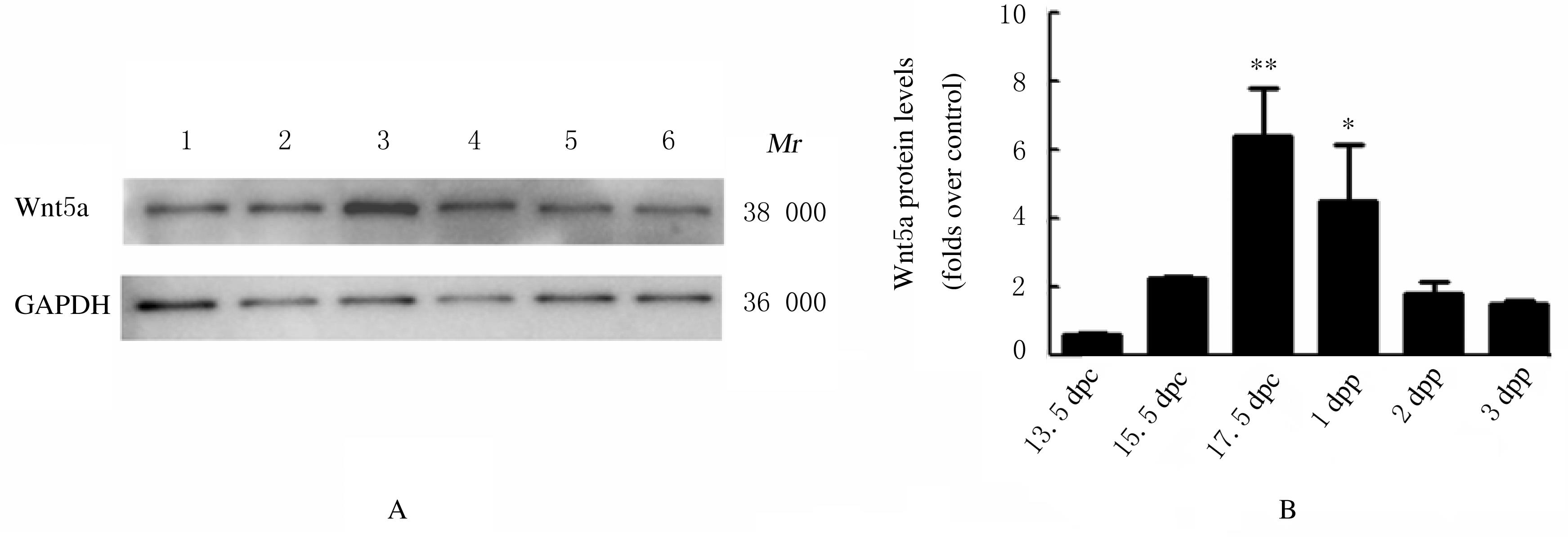
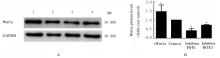
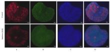
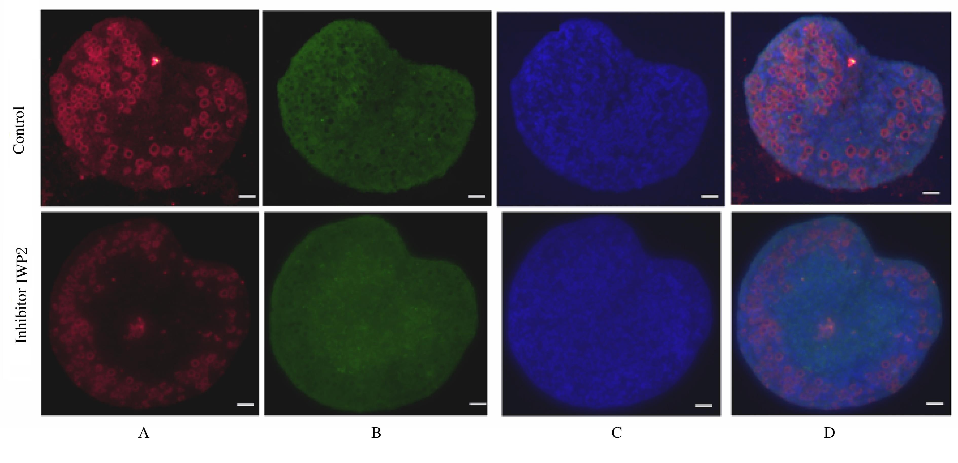
  • Received:2021-06-28 Online:2022-03-28 Published:2022-05-10
  • Contact: Yi YANG E-mail:yangyi@nxu.edu.cn

摘要: 目的

探讨不同发育期小鼠卵巢组织中Wnt5a蛋白表达水平,并阐明Wnt5a对卵母细胞自噬的影响。

方法

收集不同发育期小鼠卵巢组织,采用Western blotting法检测Wnt5a蛋白在小鼠围产期卵巢组织中的表达水平,选取新生1 d(1 dpp)小鼠卵巢组织进行免疫荧光染色,将卵母细胞胞质标记物DDX4和颗粒细胞标记物FOXL2(红色)分别与Wnt5a(绿色)和细胞核标记物Hoechst(蓝色)共染后,观察卵母细胞形态和数量。体外培养胚胎期17.5 d(17.5 dpc)小鼠卵巢组织,分为对照组、1 mol·L-1过表达Wnt5a组(rWnt5a组)、1 mol·L-1抑制剂IWP2组和1 mol·L-1抑制剂BOX5组,采用Western blotting法检测各组小鼠卵巢组织中Wnt5a、微管相关蛋白1轻链3(LC3)和P62蛋白表达水平,免疫荧光法观察卵巢卵母细胞数量。

结果

Wnt5a在不同发育阶段小鼠卵巢组织中均有表达,与13.5 dpc组比较,17.5 dpc和1 dpp组Wnt5a蛋白表达水平均明显升高(P<0.05),荧光定位发现目的蛋白Wnt5a与标记物DDX4和FOXL2有重叠部分。Western blotting法检测,与对照组比较,rWnt5a组Wnt5a蛋白表达水平明显升高(P<0.01),抑制剂IWP2组Wnt5a和LC3蛋白表达水平明显降低(P<0.01),抑制剂BOX5组Wnt5a蛋白表达水平明显升高(P<0.01);与rWnt5a组比较,抑制剂IWP2组LC3蛋白表达水平明显降低(P<0.01),P62蛋白表达水平明显升高(P<0.01)。免疫荧光染色,与对照组比较,抑制剂IWP2组卵巢中卵母细胞数目减少。

结论

Wnt5a可能通过影响小鼠卵巢内自噬水平调控卵母细胞的存活。

关键词: Wnt5a, 卵巢组织, 卵母细胞, 颗粒细胞, 自噬

Abstract: Objective

To investigate the expression levels of Wnt5a protein in ovary tissue of the mice at different development stages, and to clarify its effect on oocyte autophagy.

Methods

The ovary tissue of mice at different developmental stages were collected, and Western blotting method was used to detect the expression levels of Wnt5a protein in perinatal ovary tissue of the mice. The ovarian tissue of the 1 d after partum (1 dpp) mice were selected for immunofluorescence staining. The oocyte cytoplasmic marker DDX4 and granulosa cell marker FOXL2 (red) were co-stained with Wnt5a (green) and nuclear marker Hoechst (blue), respectively,and the morphology and the number of oocytes were observed. The ovary tissue of the mice at 17.5 d of embryonic period(17.5 dpc)were divided into control group, 1 mol·L-1 overexpressing Wnt5a group (rWnt5a group), 1 mol·L-1 inhibitor IWP2 group and 1 mol·L-1 inhibitor BOX5 group. The expression levels of Wnt5a, LC3 and P62 proteins in ovary tissue of the mice in various groups were detected by Western blotting method, and the number of ovarian oocytes was observed by immunofluorescence method.

Results

Wnt5a was expressed in ovary tissue of the mice at different developmental stages. Compared with control group, the expression levels of Wnt5a protein in 17.5 dpc and 1 dpp groups were significantly increased (P<0.05). The fluorescence localization revealed that the target protein Wnt5a overlapped with the markers DDX4 and FOXL2. The Western blotting results showed that compared with control group, the expression level of Wnt5a protein in rWnt5a group was significantly increased (P<0.01), the expression levels of Wnt5a and LC3 proteins in inhibitor IWP2 group were significantly decreased (P<0.01), and the expression level of Wnt5a protein in inhibitor BOX5 group was significantly increased (P<0.01). Compared with rWnt5a group, the expression level of LC3 protein in inhibitor IWP2 group was significantly decreased(P<0.01),and the expression level of P62 protein was significantly increased (P<0.01). The immunofluorescence staining results showed that the number of oocytes in IWP2 inhibitor group was reduced compared with control group.

Conclusion

Wnt5a may regulate the oocyte survival by affecting the level of autophagy in ovary of the mice.

Key words: Wnt5a, Ovary tissue, Oocyte, Granulosa cell, Autophagy

中图分类号: 

  • Q955